
More Related Content
What's hot
What's hot (20)
Similar to Advances in CBCT in cleft patients
Similar to Advances in CBCT in cleft patients (20)
Recently uploaded
Recently uploaded (20)
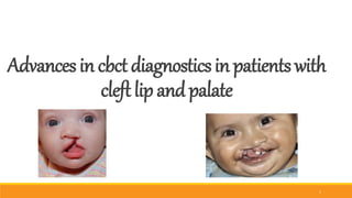
Advances in CBCT in cleft patients
- 3. Introduced in 1998 CRANIOFACIAL IMAGING IN 21st CENTUARY Evolutionary process of conventional CT’s CBCT 3
- 4. Godfrey Hounsfield Nobel prize in Medicine, 1979 Allan Cormack COMPUTED TOMOGRAPHY 4
- 5. 5
- 6. NewTom 9000 / NewTom 3G i-CAT 6
- 7. CBCT in orthodontics: assessment of treatment outcomes and indications for its use Kapila et al ,Dentomaxillofacial Radiology (2015) 44, 20140282 7
- 8. Dose AS LOW AS RESONABLY ACHEIVABLE 8
- 9. 9
- 10. C B C T vs PANORAMIC radiograph Panoramic Provides a distorted and magnified image Shows image layer view only Structures are superimposed CBCT Provides an undistorted image Cross-sectional (bucco-lingual), axial, coronal, sagittal, and panoramic views Structures can be separated 10
- 12. CBCT in orthodontics: assessment of treatment outcomes and indications for its use Kapila et al ,Dentomaxillofacial Radiology (2015) 44, 20140282 ITERATIVE CLOSEST POINT 12
- 13. CBCT in orthodontics: assessment of treatment outcomes and indications for its use Kapila et al ,Dentomaxillofacial Radiology (2015) 44, 20140282 SHAPE CORRESPONDENCE 13
- 14. CBCT in orthodontics: assessment of treatment outcomes and indications for its use Kapila et al ,Dentomaxillofacial Radiology (2015) 44, 20140282 IMPACTION 14
- 15. CBCT in orthodontics: assessment of treatment outcomes and indications for its use Kapila et al ,Dentomaxillofacial Radiology (2015) 44, 20140282 IMPACTION 15
- 16. CBCT in orthodontics: assessment of treatment outcomes and indications for its use Kapila et al ,Dentomaxillofacial Radiology (2015) 44, 20140282 SUPERNUMERARY TEETH 16
- 17. CBCT in orthodontics: assessment of treatment outcomes and indications for its use Kapila et al ,Dentomaxillofacial Radiology (2015) 44, 20140282 ALVEOLAR BOUNDARY CONDITION 17
- 18. CBCT in orthodontics: assessment of treatment outcomes and indications for its use Kapila et al ,Dentomaxillofacial Radiology (2015) 44, 20140282 ALVEOLAR BOUNDARY CONDITION- transposition 18
- 19. TMJ 19
- 20. CBCT in orthodontics: assessment of treatment outcomes and indications for its use Kapila et al ,Dentomaxillofacial Radiology (2015) 44, 20140282 AIRWAY MORPHOLOGY 20
- 21. CLEFT LIP AND PALATE CBCT in orthodontics: assessment of treatment outcomes and indications for its use Kapila et al ,Dentomaxillofacial Radiology (2015) 44, 20140282 21
- 22. CLEFT LIP AND PALATE 22
- 23. 23
- 24. 24
- 25. Internationally Approved classification:- Classification of Cleft of the Lip, Alveolus and Palate: MILLARD 1976 (Based on embryological principles) Group 1: Cleft of anterior (primary) palate. Lip Alveolus Group 2: Cleft of anterior and posterior (primary and secondary) palate: Lip Alveolus Hard Palate Group 3: Cleft of posterior (secondary) palate: Hard palate Soft palate 25
- 26. VEAU’S CLASSIFICATION (1931) Group I - Cleft of soft palate only Group II - Cleft of hard and soft palate, extending no further than the incisive foramen thus involving the secondary palate alone. 26
- 27. Group III - Complete unilateral cleft of soft and hard palate, lip and alveolar ridge Group IV - Complete bilateral cleft of soft and hard palate, lip and alveolar ridge on both sides. 27
- 28. Kernahans striped Y logo classification. 28
- 30. LAHSHAL Classification L - A - H - S - H - A - L - Lip Alveolus Hard palate Soft palate Hard palate Alveolus Lip 30
- 31. Overview of orthodontic care for children with cleft lip and palate, 1915-2015 Katherine et al,Am J Orthod Dentofacial Orthop 2015;148:543-56 31
- 32. Pre surgical Orthopedics 1-4 weeks Repositioning palatal segment facilitates lip repair Lip closure 8 to 12 weeks May be preceded by preliminary lip adhesion as an alternative to presurgical orthopedics Palate closure 18 to 24 months Closing only the soft palate initially an alternative, but one stage closure of the hard and soft palate is the usual procedure 32
- 33. Speech therapy 6 to 11 years Articulation errors often develop as child tries to compensate for cleft Early orthodontics 7 to 8 years Usually incisor alignment and maxillary transverse expansion Alveolar grafting 6 to 10 years Needed before permanent canines erupt; timing determined by stage and sequence of dental development 33
- 34. Comprehensive orthodontics 11 to 14 years Class III elastics often very helpful Orthognathic surgery 17 to 19 years Maxillary advancement, perhaps combined with mandibular set-back; not done until growth completed except in rare instances of severe psychosocial impact; needed infrequently Fixed prosthodontics 17 to 19 years Replacement of missing lateral incisors: comprehensive treatment only after growth completed 34
- 35. CBCT IN CLEFT LIP AND PALATE 35
- 36. Assessment of Alveolar Bone Morphology 36
- 37. Alveolar Bone Morphology in Patients With Bilateral Complete Cleft Lip and Palate in the Mixed Dentition: CBCT Evaluation Garib et al, Cleft Palate–Craniofacial Journal, March 2012, Vol. 49 No. 2 Alveolar Bone Morphology in Patients With Bilateral Complete Cleft Lip and Palate 37
- 38. Alveolar Bone Morphology in Patients With Bilateral Complete Cleft Lip and Palate in the Mixed Dentition: CBCT Evaluation Garib et al, Cleft Palate–Craniofacial Journal, March 2012, Vol. 49 No. 2 38
- 39. Alveolar Bone Morphology in Patients With Bilateral Complete Cleft Lip and Palate in the Mixed Dentition: CBCT Evaluation Garib et al, Cleft Palate–Craniofacial Journal, March 2012, Vol. 49 No. 2 39
- 40. Alveolar Bone Morphology in Patients With Bilateral Complete Cleft Lip and Palate in the Mixed Dentition: CBCT Evaluation Garib et al, Cleft Palate–Craniofacial Journal, March 2012, Vol. 49 No. 2 40
- 41. Alveolar Bone Morphology in Patients With Bilateral Complete Cleft Lip and Palate in the Mixed Dentition: CBCT Evaluation Garib et al, Cleft Palate–Craniofacial Journal, March 2012, Vol. 49 No. 2 41
- 42. Assessment of the alveolar bone support of patients with unilateral cleft lip and palate: A cone-beam computed tomography study Esra et al, Angle Orthod. 2015;85:1003–1008 Assessment of the alveolar bone support of patients with unilateral cleft lip and palate 42
- 43. Assessment of the alveolar bone support of patients with unilateral cleft lip and palate: A cone-beam computed tomography study Esra et al, Angle Orthod. 2015;85:1003–1008 43
- 44. Assessment of the alveolar bone support of patients with unilateral cleft lip and palate: A cone-beam computed tomography study Esra et al, Angle Orthod. 2015;85:1003–1008 44
- 45. Use of Cone Beam Computed Tomography to Volumetrically Assess Alveolar Cleft Defects— Preliminary Results Fasial et al, J Oral Maxillofac Surg 70:188-191, 2012 Volumetric Assessment of Alveolar Cleft Defects Cleft height 45
- 46. Cleft width Use of Cone Beam Computed Tomography to Volumetrically Assess Alveolar Cleft Defects—Preliminary Results Fasial et al, J Oral Maxillofac Surg 70:188-191, 2012 46
- 47. Cleft length Use of Cone Beam Computed Tomography to Volumetrically Assess Alveolar Cleft Defects—Preliminary Results Fasial et al, J Oral Maxillofac Surg 70:188-191, 2012 47
- 48. Assessment of secondary alveolar bone grafting 48
- 49. Volumetric Assessment of Secondary Alveolar Bone Grafting Using Cone Beam Computed Tomography Oberoi et al, Cleft Palate–Craniofacial Journal, September 2009, Vol. 46 No. 5 Volumetric Assessment of Secondary Alveolar Bone Grafting 49
- 50. Pre and one year after grafting Volumetric Assessment of Secondary Alveolar Bone Grafting Using Cone Beam Computed Tomography Oberoi et al, Cleft Palate–Craniofacial Journal, September 2009, Vol. 46 No. 5 50
- 51. Application of Limited CBCT to Clinical Assessment of Alveolar Bone Grafting: A Preliminary Report HAMADA et al, Cleft Palate–Craniofacial Journal, March 2005, Vol. 42 No. 2 Assessment of Alveolar Bone Grafting 51
- 52. Application of Limited CBCT to Clinical Assessment of Alveolar Bone Grafting: A Preliminary Report HAMADA et al, Cleft Palate–Craniofacial Journal, March 2005, Vol. 42 No. 2 52
- 53. Application of Limited CBCT to Clinical Assessment of Alveolar Bone Grafting: A Preliminary Report HAMADA et al, Cleft Palate–Craniofacial Journal, March 2005, Vol. 42 No. 2 53
- 55. Assessment of Bone Resorption After Secondary Alveolar Bone Grafting Using Three-Dimensional Computed Tomography: A Three-Year Study Matthias et al, Cleft Palate–Craniofacial Journal, March 2007, Vol. 44 No. 2 Assessment of Bone Resorption After Secondary Alveolar Bone Grafting 55
- 56. Assessment of alveolar bone grafting CBCT in the assessment of alveolar bone grafting in children with unilateral cleft lip and palate Anni et al, European Journal of Orthodontics 36 (2014) 603–611 56
- 57. CBCT in the assessment of alveolar bone grafting in children with unilateral cleft lip and palate Anni et al, European Journal of Orthodontics 36 (2014) 603–611 57
- 58. fair labiopalatal bone support CBCT in the assessment of alveolar bone grafting in children with unilateral cleft lip and palate Anni et al, European Journal of Orthodontics 36 (2014) 603–611 58
- 59. Good labiopalatal bone support CBCT in the assessment of alveolar bone grafting in children with unilateral cleft lip and palate Anni et al, European Journal of Orthodontics 36 (2014) 603–611 59
- 60. Nasal floor CBCT in the assessment of alveolar bone grafting in children with unilateral cleft lip and palate Anni et al, European Journal of Orthodontics 36 (2014) 603–611 60
- 61. Comparison between multislice and cone-beam computerized tomography in the volumetric assessment of cleft palate Marco et al, Oral Surg Oral Med Oral Pathol Oral Radiol Endod 2011;112:249-257 Comparison between multislice CT and CBCT in the volumetric assessment of cleft palate 61
- 62. Comparison between multislice and cone-beam computerized tomography in the volumetric assessment of cleft palate Marco et al, Oral Surg Oral Med Oral Pathol Oral Radiol Endod 2011;112:249-257 62
- 63. Comparison between multislice and cone-beam computerized tomography in the volumetric assessment of cleft palate Marco et al, Oral Surg Oral Med Oral Pathol Oral Radiol Endod 2011;112:249-257 63
- 64. Comparison between multislice and cone-beam computerized tomography in the volumetric assessment of cleft palate Marco et al, Oral Surg Oral Med Oral Pathol Oral Radiol Endod 2011;112:249-257 64
- 65. Comparison between multislice and cone-beam computerized tomography in the volumetric assessment of cleft palate Marco et al, Oral Surg Oral Med Oral Pathol Oral Radiol Endod 2011;112:249-257 65
- 66. Evaluation of donor site 66
- 67. Evaluation of the anterior mandibular donor site one year after secondary reconstruction of an alveolar cleft:3-dimensional analysis using CBCT Bilsen et at, British Journal of Oral and Maxillofacial Surgery 53 (2015) 719–724 Evaluation of the anterior mandibular donor site 67
- 68. Evaluation of the anterior mandibular donor site one year after secondary reconstruction of an alveolar cleft:3-dimensional analysis using CBCT Bilsen et at, British Journal of Oral and Maxillofacial Surgery 53 (2015) 719–724 68
- 69. Regeneration of bone Evaluation of the anterior mandibular donor site one year after secondary reconstruction of an alveolar cleft:3-dimensional analysis using CBCT Bilsen et at, British Journal of Oral and Maxillofacial Surgery 53 (2015) 719–724 69
- 71. Maxillary Dental Anomalies in Patients with Cleft Lip and Palate: A Cone Beam Computed Tomography Study Buyuk et al, The Journal of Clinical Pediatric Dentistry Volume 39, Number 2/2015 Maxillary Dental Anomalies 71
- 72. Tooth Lengths of the Permanent Upper Incisors in Patients With Cleft Lip and Palate Determined With CBCT Zhou et al, The Cleft Palate-Craniofacial Journal 50(1) pp. 88–95 January 2013 Tooth Lengths of the Permanent Upper Incisors 72
- 73. Tooth Lengths of the Permanent Upper Incisors in Patients With Cleft Lip and Palate Determined With CBCT Zhou et al, The Cleft Palate-Craniofacial Journal 50(1) pp. 88–95 January 2013 73
- 74. Three-Dimensional Assessment of the Eruption Path of the Canine in Individuals With Bone-Grafted Alveolar Clefts Using CBCT Oberoi et al, Cleft Palate–Craniofacial Journal, September 2010, Vol. 47 No. 5 Eruption Path of the Canine 74
- 75. Incidental findings on cone beam computed tomography scans in cleft lip and palate patients Mette et al, Clin Oral Invest (2014) 18:1237–1244 Incidental findings on cone beam computed tomography scans 75
- 76. Incidental findings on cone beam computed tomography scans in cleft lip and palate patients Mette et al, Clin Oral Invest (2014) 18:1237–1244 76
- 77. Incidental findings on cone beam computed tomography scans in cleft lip and palate patients Mette et al, Clin Oral Invest (2014) 18:1237–1244 77
- 78. Evaluation of facial soft tissue 78
- 79. Facial soft-tissue thickness in patients affected by bilateral cleft lip and palate: A retrospective CBCT study Mevlut et al, Am J Orthod Dentofacial Orthop 2014;146:573-8 SNA , SNB , ANB , SN-MP , Co- A (mm), CoGn (mm), U1-SN, and IMPA Facial soft-tissue thickness 79
- 80. Glabella (G-G0), Nasion (N-N0), Rhinion (rhi-rhi0), Subnasale (a-sn), Labrale superior (pr-ls), Stomion (u1-sto), Labrale inferior (id-li), Labiomental (b-labm), Pogonion (pog-pog0), Gnathion (gn-gn0). Facial soft-tissue thickness in patients affected by bilateral cleft lip and palate: A retrospective CBCT study Mevlut et al, Am J Orthod Dentofacial Orthop 2014;146:573-8 80
- 81. Facial soft-tissue thickness in patients affected by bilateral cleft lip and palate: A retrospective CBCT study Mevlut et al, Am J Orthod Dentofacial Orthop 2014;146:573-8 81
- 83. Three-dimensional evaluation of midfacial asymmetry in patients with nonsyndromic unilateral cleft lip and palate by CBCT Park et al, Korean J Orthod 2013;43(3):113-119 Evaluation of midfacial asymmetry 83
- 84. 84
- 85. CBCT Assessment of Lower Facial Asymmetry in Unilateral Cleft Lip and Palate and Non-Cleft Patients with Class III Skeletal Relationship Lin et al, PLOS ONE | DOI:10.1371/journal.pone.0130235 August 3, 2015 Lower Facial Asymmetry 85
- 86. 86
- 87. 87
- 89. Three Dimensional Assessment of the Pharyngeal Airway in Individuals with Non-Syndromic Cleft Lip and Palate Oberoi et al, PLoS ONE August 2012 | Volume 7 | Issue 8 89
- 91. Dentoskeletal effects of 3 maxillary expanders in patients with clefts: A cone-beam computed tomography study Daniel e al, Am J Orthod Dentofacial Orthop 2014;146:73-81 Hyrax 91
- 92. Fan-type 92
- 94. 94
- 95. CONCLUSION 95
- 96. REFERENCES • CBCT in orthodontics: assessment of treatment outcomes and indications for its use Kapila et al ,Dentomaxillofacial Radiology (2015) 44, 20140282 • Overview of orthodontic care for children with cleft lip and palate, 1915-2015 Katherine et al,Am J Orthod Dentofacial Orthop 2015;148:543-56 • Alveolar Bone Morphology in Patients With Bilateral Complete Cleft Lip and Palate in the Mixed Dentition: CBCT Evaluation Garib et al, Cleft Palate–Craniofacial Journal, March 2012, Vol. 49 No. 2 • Assessment of the alveolar bone support of patients with unilateral cleft lip and palate: A cone-beam computed tomography study Esra et al, Angle Orthod. 2015;85:1003–1008 • Use of Cone Beam Computed Tomography to Volumetrically Assess Alveolar Cleft Defects—Preliminary Results Fasial et al, J Oral Maxillofac Surg 70:188-191, 2012 • Comparison between multislice and cone-beam computerized tomography in the volumetric assessment of cleft palate Marco et al, Oral Surg Oral Med Oral Pathol Oral Radiol Endod 2011;112:249-257 96
- 97. • Volumetric Assessment of Secondary Alveolar Bone Grafting Using Cone Beam Computed Tomography Oberoi et al, Cleft Palate–Craniofacial Journal, September 2009, Vol. 46 No. 5 • Evaluation of the anterior mandibular donor site one year after secondary reconstruction of an alveolar cleft:3-dimensional analysis using cone-beam computed tomography Bilsen et at, British Journal of Oral and Maxillofacial Surgery 53 (2015) 719–724 • Application of Limited CBCT to Clinical Assessment of Alveolar Bone Grafting: A Preliminary Report HAMADA et al, Cleft Palate–Craniofacial Journal, March 2005, Vol. 42 No. 2 • Assessment of Bone Resorption After Secondary Alveolar Bone Grafting Using Three-Dimensional Computed Tomography: A Three-Year Study Matthias et al, Cleft Palate–Craniofacial Journal, March 2007, Vol. 44 No. 2 • Tooth Lengths of the Permanent Upper Incisors in Patients With Cleft Lip and Palate Determined With Cone Beam Computed Tomography Zhou et al, The Cleft Palate-Craniofacial Journal 50(1) pp. 88–95 January 2013 • Incidental findings on cone beam computed tomography scans in cleft lip and palate patients Mette et al, Clin Oral Invest (2014) 18:1237–1244 • Three Dimensional Assessment of the Pharyngeal Airway in Individuals with Non-Syndromic Cleft Lip and Palate Oberoi et al, PLoS ONE August 2012 | Volume 7 | Issue 8 97
- 98. • CBCT in the assessment of alveolar bone grafting in children with unilateral cleft lip and palate Anni et al, European Journal of Orthodontics 36 (2014) 603–611 • Maxillary Dental Anomalies in Patients with Cleft Lip and Palate: A Cone Beam Computed Tomography Study Buyuk et al, The Journal of Clinical Pediatric Dentistry Volume 39, Number 2/2015 • Three-Dimensional Assessment of the Eruption Path of the Canine in Individuals With Bone-Grafted Alveolar Clefts Using CBCT Oberoi et al, Cleft Palate–Craniofacial Journal, September 2010, Vol. 47 No. 5 • Facial soft-tissue thickness in patients affected by bilateral cleft lip and palate: A retrospective cone-beam computed tomography study Mevlut et al, Am J Orthod Dentofacial Orthop 2014;146:573-8 • Three-dimensional evaluation of midfacial asymmetry in patients with nonsyndromic unilateral cleft lip and palate by cone-beam computed Tomography Park et al, Korean J Orthod 2013;43(3):113-119 • Cone-Beam Computed Tomography Assessment of Lower Facial Asymmetry in Unilateral Cleft Lip and Palate and Non-Cleft Patients with Class III Skeletal Relationship Lin et al, PLOS ONE | DOI:10.1371/journal.pone.0130235 August 3, 2015 98
- 99. • CLEFT LIP AND PALATE :DIEGO.F.WYSZYNSKI • Cone Beam Computed Tomography in orthodontics:Sunil S Kapila • Dentoskeletal effects of 3 maxillary expanders in patients with clefts: A cone-beam computed tomography study Daniel e al, Am J Orthod Dentofacial Orthop 2014;146:73-81 99
- 100. 100