• Functions andcharacteristics of plasma proteins
• Measurement of plasma proteins and diagnosis of
diseases
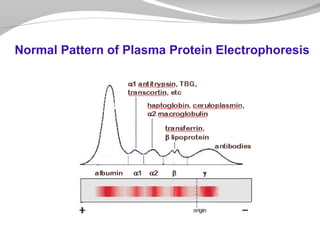
• Electrophoretic patterns of plasma proteins
• Acute phase proteins
Overview:
3.
Plasma contains >300different proteins
Many pathological conditions affect
level of pps
Mostly synthesized in the liver
Some are produced in other sites
A normal adult contains ~70 g/L of pps
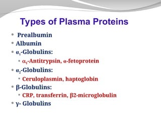
Plasma Proteins (pps)
4.
• Transport (Albumin,prealbumin, globulins)
• Maintain plasma oncotic pressure (Albumin)
• Defense (Immunoglobulins and
complement)
• Clotting and fibrinolysis (Thrombin and
plasmin)
Functions of pps
5.
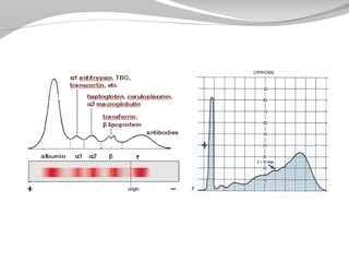
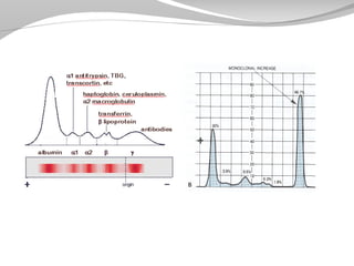
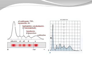
A) Quantitative measurementof a specific protein:
Chemical or immunological reactions
B) Semiquantitative measurement by
electrophoresis:
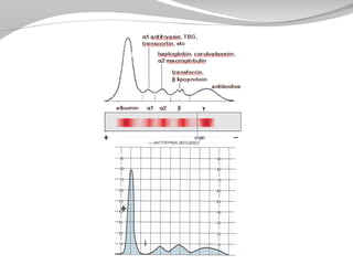
Proteins are separated by their electrical charge in
electrophoresis
Five separate bands of proteins are observed
These bands change in disease
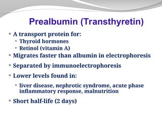
Measurement of Plasma Proteins
A transportprotein for:
Thyroid hormones
Retinol (vitamin A)
Migrates faster than albumin in electrophoresis
Separated by immunoelectrophoresis
Lower levels found in:
liver disease, nephrotic syndrome, acute phase
inflammatory response, malnutrition
Short half-life (2 days)
Prealbumin (Transthyretin)
9.
Most abundantplasma protein (~40 g/L) in
normal adult
Synthesized in the liver as preproalbumin and
secreted as albumin
Half-life in plasma: 20 days
Decreases rapidly in injury, infection and
surgery
Albumin
10.
Functions
• Maintains oncoticpressure:
– The osmotic pressure exerted by
plasma proteins that pulls water into
the circulatory system
– Maintains fluid distribution in and
outside cells and plasma volume
• 80% of plasma oncotic pressure is
maintained by albumin
11.
Functions
• A non-specificcarrier of
– hormones, calcium, free fatty acids, drugs,
etc.
• Tissue cells can take up albumin by
pinocytosis where it is hydrolyzed to
amino acids
• Useful in treatment of liver diseases,
hemorrhage, shock and burns
12.
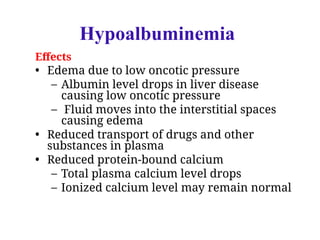
Hypoalbuminemia
• Causes
– Decreasedalbumin synthesis (liver
cirrhosis, malnutrition)
– Increased losses of albumin
• Increased catabolism in infections
• Excessive excretion by the kidneys
(nephrotic syndrome)
• Excessive loss in bowel
• Severe burns (plasma loss in the absence of
skin barrier)
13.
Effects
• Edema dueto low oncotic pressure
– Albumin level drops in liver disease
causing low oncotic pressure
– Fluid moves into the interstitial spaces
causing edema
• Reduced transport of drugs and other
substances in plasma
• Reduced protein-bound calcium
– Total plasma calcium level drops
– Ionized calcium level may remain normal
Hypoalbuminemia
14.
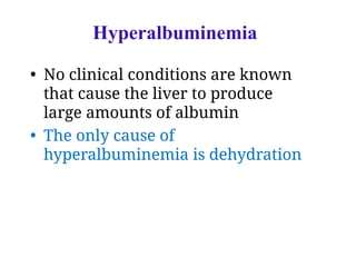
Hyperalbuminemia
• No clinicalconditions are known
that cause the liver to produce
large amounts of albumin
• The only cause of
hyperalbuminemia is dehydration
15.
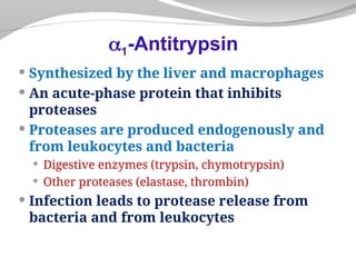
Synthesized bythe liver and macrophages
An acute-phase protein that inhibits
proteases
Proteases are produced endogenously and
from leukocytes and bacteria
Digestive enzymes (trypsin, chymotrypsin)
Other proteases (elastase, thrombin)
Infection leads to protease release from
bacteria and from leukocytes
1-Antitrypsin
17.
Over 30types are known
The most common is M type
Genetic deficiency of 1-Antitrypsin
Synthesis of the defective 1-Antitrypsin
occurs in the liver but it cannot secrete the
protein
1-Antitrypsin accumulates in hepatocytes
and is deficient in plasma
Types of 1-Antitrypsin
18.
Neonatal jaundicewith evidence of cholestasis
Childhood liver cirrhosis
Pulmonary emphysema in young adults
Laboratory Diagnosis
Lack of 1-globulin band in protein
electrophoresis
Quantitative measurement of 1-Antitrypsin by:
Radial immunodiffusion, isoelectric focusing or
nephelometry
Clinical Consequences of 1-Antitrypsin
Deficiency
19.
Synthesized inthe developing embryo and
fetus by the parenchymal cells of the liver
AFP levels decrease gradually during intra-
uterine life and reach adult levels at birth
Function is unknown but it may protect fetus
from immunologic attack by the mother
No known physiological function in adults
-Fetoprotein (AFP)
20.
Elevated maternalAFP levels are associated
with:
Neural tube defect, anencephaly
Decreased maternal AFP levels are associated
with:
Increased risk of Down’s syndrome
AFP is a tumor marker for:
Hepatoma and testicular cancer
-Fetoprotein (AFP)
21.
Synthesized bythe liver
Contains >90% of serum copper
An oxidoreductase that inactivates ROS
causing tissue damage in acute phase
response
Important for iron absorption from the
intestine
Wilson’s disease:
Due to low plasma levels of ceruloplasmin
Copper is accumulated in the liver and brain
Ceruloplasmin
22.
Synthesized bythe liver
Binds to free hemoglobin to form
complexes that are metabolized in the RES
Limits iron losses by preventing Hb loss
from kidneys
Plasma level decreases during hemolysis
Haptoglobin
23.
A majoriron-transport protein in plasma
30% saturated with iron
Plasma level drops in:
Malnutrition, liver disease, inflammation,
malignancy
Iron deficiency results in increased hepatic
synthesis
A negative acute phase protein
Transferrin
24.
A componentof human leukocyte antigen
(HLA)
Present on the surface of lymphocytes and
most nucleated cells
Filtered by the renal glomeruli due to its small
size but most (>99%) is reabsorbed
Elevated serum levels are found in
Impaired kidney function
Overproduction in disease
May be a tumor marker for:
Leukemia, lymphomas, multiple myeloma
2–Microglobulin
25.
An acute-phaseprotein synthesized by the liver
Important for phagocytosis
High plasma levels are found in many
inflammatory conditions such as rheumatoid
arthritis
A marker for ischemic heart disease
C-Reactive Protein (CRP)
26.
May resultfrom stimulation of
B cells (Polyclonal hypergammaglobulinemia)
Monoclonal proliferation (Paraproteinemia)
Polyclonal hypergammaglobulinemia:
Stimulation of many clones of B cells produce a
wide range of antibodies
-globulin band appears large in electophoresis
Clinical conditions: acute and chronic
infections, autoimmune diseases, chronic liver
diseases
Hypergammaglobulinemia
28.
Monoclonal
Hypergammaglobulinemia
Proliferation ofa single B-cell clone
produces a single type of Ig
Appears as a separate dense band
(paraprotein or M band) in electrophoresis
Paraproteins are characteristic of
malignant B-cell proliferation
Clinical condition: multiple myeloma
30.
Plasma proteinlevels increase in:
Infection, inflammation , malignancy, trauma,
surgery
These proteins are called acute phase
reactants
Synthesized due to body’s response to injury
Examples: 1-Antitypsin, haptoglobin,
ceruloplasmin, fibrinogen, c-reactive protein
Positive Acute Phase Proteins
31.
Mediators causethese proteins to increase
after injury
Mediators: Cytokines (IL-1, IL-6), tumor
necrosis factors and , interferons, platelet
activating factor
Functions:
1. Bind to polysaccharides in bacterial walls
2. Activate complement system
3. Stimulate phagocytosis
Positive Acute Phase Proteins
33.
These proteinsdecrease in inflammation
Albumin, prealbumin, transferrin
Mediated by inflammatory response via
cytokines and hormones
Synthesis of these proteins decrease to save
amino acids for positive acute phase proteins
Negative Acute Phase Proteins
34.
summery
summery
• Functions andcharacteristics of plasma proteins
• Measurement of plasma proteins and diagnosis of
diseases
• Electrophoretic patterns of plasma proteins
• Acute phase proteins