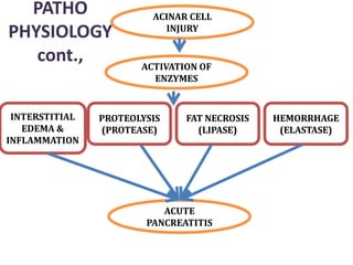
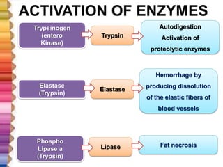
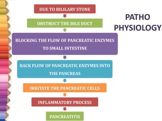
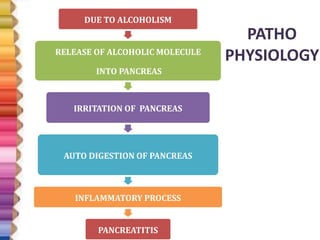
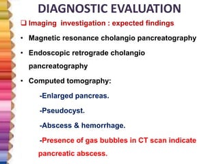
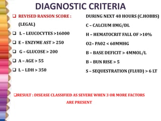
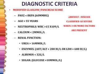
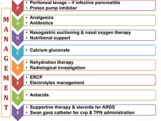
MS.MUTHU RAJATHI, M.SC (N)
ASST., PROFESSOR
DEPARTMENT OF MEDICAL SURGICAL NURSING
GANGA INSTITUTE OF HEALTH SCIENCES
COIMBATORE
PANCREATITIS
INTRODUCTION
• Acute emergency condition of gastro intestinal
system.
• Requires immediate medical attention.
• Higher incidence found in south India .
• Inflammatory process in pancreas due to
cholelitheasis.
SIGNIFICANT ANATOMY &
PHYSIOLOGY
SIGNIFICANT ANATOMY &
PHYSIOLOGY
• Subsances into the intestiine.
• Enzymes – amylase, peptase,
ribonuclease, lipase
EXOCRINE
PANCREAS
• Substances into the blood stream.
• Hormones – insulin, glucagon,
somatostatin, pancreatic
polypeptide
ENDOCRINE
PANCREAS
SIGNIFICANT ANATOMY &
PHYSIOLOGY
CHEMOTRYPSIN
TRYPSIN
ELASTASE
LIPASE
AMYLASE
COLLAGENASE
AMINOPEPTIDASE
1. Neturalizes HCL
2. Digestion of CHO,
lipids, proteins,
polysaccharides
SIGNIFICANT ANATOMY & PHYSIOLOGY
ENDOCRINE PANCREAS
BETA CELLSALPHA CELLS
POLYPEPTIDE
CELLS
GAMMA CELLS
INSULINGLUCAGON
PANCREATIC
PEPTIDE
SOMATOSTATIN
•LOWERING
THE BLOOD
GLUCOSE
LEVEL
•STORAGE OF
GLYCOGEN
•BREAKDOWN
OF GLYCOGEN
FROM LIVER &
MUSCLES
•INCREASES
BLOOD
GLUCOSE LEVEL
•SELF REGULATION
OF PANCREATIC
SECRETION &
EFFECTS THE
HEPATIC GYLCOGEN
LEVELS.
•INHIBITION &
RELEASE OF GI
HORMONES
DEFINITION
• Is an acute inflammation, where the activated
pancreatic enzymes leak into the pancreas and
begins the process of auto digestion of pancreas.
• An acute inflammatory process of the pancreas ,
degree of inflammation varies from mild edema to
severe necrosis.
TISSUE
INJURY
MILD
EDEMA
SEVERE
NECROSIS
DEFINITION
• CLINICAL DEFINITION : An acute
inflammation, presenting with
abdominal pain usually associated with
raised pancreatic enzymes due to
pancreatic inflammation.
• PATHOLOGICAL DEFINITION : Reversible
pancreatic parenchymal injury
associated with inflammatory process.
INCIDENCE AND PREVALENCE
 Region : Highest incidence in South India – 114-200/
100000 population
 Sex : male to female ratio – 4.6:1
 Age : 19 -45 years
CLASSIFICATION
PANCREATITIS
CHRONIC –
PROLONGED
DISORDER
ACUTE – REVERSIBLE
DAMAGE WITH ACUTE
SYMPTOMS
CLASSIFICATION
ACCORDING TO
DURATION
EARLY – WITHIN A
WEEK
LATE – MORE THAN
TWO WEEKS
ACCORDING TO
SEVERITY
MILD – NO
ORGAN FAILURE
MODERATE –
ORGAN FAILURE
LESS THAN 48 HRS
SEVERE – ORGAN
FAILURE LONGER
THAN 48 HRS
ACCORDING TO
DISEASE PROCESS
EDEMATOUS - NECROTIZING
CAUSES AND RISK FACTORS
 I : idiopathic
 G: gallstone obstruction .(common)
 E: ethanol
 T: trauma – injury to the pancreatic tissue
 S: chronic use of steroids
 M: mumps. Viral infections
 A: alcoholism (common) or autoimmune disorder
 S: scorpion bits.
 H: hyperlipidemia.
 E: endoscopic retrograde cholangio
 Pancreatography - procedure
 D: drugs: thiazide, azathioprine.
I
G
E
T
S
M
A
S
H
E
D
• Gall stone, amupullary tumour,
pancreatic cancer, ERCP, trauma
MECHANICAL
• Alcoholism, hyperlipidemia,
drugs,, malnutrition, azotemia
METABOLIC
• Mumps, coxsackie B, snake bite,
scorpion bite, ascares
INFECTIVE
• Cystic fibrosis, auto immune
disorder
GENETIC
• Shock , hyperthermia, athero
embolism, vasculitis
VASCULAR
• 70% due to microlithiasis
IDIOPATHIC
C
A
U
S
E
S
CAUSES AND RISK FACTORS
70%
20%
10%
BILIARY OBSTRUCTION
ALCOHOLIC
IDIOPATHIC
LESS COMMON CAUSES AND RISK FACTORS
Trauma (postsurgical, abdominal)
Viral infections (mumps, coxsackievirus hiv)
Penetrating duodenal ulcer
Cysts
Idiopathic
Abscesses
Cystic fibrosis
Kaposi’s sarcoma
Metabolic disorders
Vascular diseases
Post operative gi surgery
DUE TO ANY CAUSE
BILIARY DUCT
OBSTRUCTION
OTHER CAUSES
IMPAIRED METABOLIC
TRANSPORT
CHOLELITHIASIS TRAUMA
ALCOHOL
VIRUS
METABOLIC INJURY
INTERSTITIAL
EDEMA
IMPAIRED
BLOOD FLOW
ISCHEMIA
ACINAR CELL
INJURY
INTRACELLULAR RELEASE OF PROENZYMES
AND LYSOSOMAL HYDROXYLASE
INTRACELLULAR ACTIVATION OF ENZYMES
PATHO
PHYSIOLOGY
ACINAR CELL
INJURY
INTERSTITIAL
EDEMA &
INFLAMMATION
PROTEOLYSIS
(PROTEASE)
FAT NECROSIS
(LIPASE)
HEMORRHAGE
(ELASTASE)
ACUTE
PANCREATITIS
ACTIVATION OF
ENZYMES
PATHO
PHYSIOLOGY
cont.,
ACTIVATION OF ENZYMES
Trypsinogen
(entero
Kinase)
Phospho
Lipase a
(Trypsin)
Elastase
(Trypsin)
Trypsin
Elastase
Lipase
Autodigestion
Activation of
proteolytic enzymes
Hemorrhage by
producing dissolution
of the elastic fibers of
blood vessels
Fat necrosis
PATHO
PHYSIOLOGY
DUE TO BILILARY STONE
OBSTRUCT THE BILE DUCT
BLOCKING THE FLOW OF PANCREATIC ENZYMES
TO SMALL INTESTINE
BACK FLOW OF PANCREATIC ENZYMES INTO
THE PANCREAS
IRRITATE THE PANCREATIC CELLS
INFLAMMATORY PROCESS
PANCREATITIS
PATHO
PHYSIOLOGY
DUE TO ALCOHOLISM
RELEASE OF ALCOHOLIC MOLECULE
INTO PANCREAS
IRRITATION OF PANCREAS
AUTO DIGESTION OF PANCREAS
INFLAMMATORY PROCESS
PANCREATITIS
STAGES
• May stimulate production of
digestive enzymes
• Increases sensitivity to hormone
cholecystokinin
• Stimulates production of
pancreatic enzymes
ALCOHOLIC
PANCREATITITS
• Mild and self-limiting
EDEMATOUS
PANCREATITIS
• Degree of necrosis correlates with
severity of manifestations
NECROTIZING
PANCREATITIS
CLINICAL MANIFESTATIONS
 Cardinal symptom : abdominal pain
 Site: mid epigastrium , left upper quadrant radiating to back.
 Onset: quickly, generally following substantial meal.
 Severity: frequently severe, reaching maximum intensity
within minutes rather than hours.
 Nature: “boring through”, “knifing” (illimitable agony)
 Duration: hours-days
 Course: constant , piercing type
 Aggravating factor: food/alcohol intake, walking, lying
supine
 Alleviating factor : sitting or leaning /stooping forward
position
CLINICAL MANIFESTATIONS
 Nausea, frequent and effortless vomiting
 Anorexia
 Diarrhea
 Hiccups
 Fever - low grade, seen in infective pancreatitis
 Weakness
 Leukocytosis
 Hypotension & tachycardia
 Jaundice
 Tenderness in epigastric
 Guarding of abdominal muscles.
 Mild abdominal distention
CLINICAL SIGNS –
GREY TURNER’S SIGN
• Bruising of the flanks, (the part of the
body between the last rib and the top
of the hip). The bruising appears as a
blue discoloration, and is a sign of
retroperitoneal hemorrhage, or
bleeding behind the peritoneum,
which is a lining of the abdominal
cavity
CLINICAL SIGNS –
CULLEN SIGN
Superficial edema and
bruising in the
subcutaneous fatty tissue
around the umbilicus.
CLINICAL SIGNS –
FOX SIGN
Fox's sign is a clinical sign in
which bruising is seen over the
inguinal ligament. It occurs in
patients with retroperitoneal
bleeding, usually due to acute
haemorrhagic pancreatitis.
DIAGNOSTIC EVALUATION
• History collection
– Personal habits
– Family history of pancreatitis
– History of hematological disorder
– Dietary pattern
– Present medical illness
• Physical examination
– Rigidity of abdomen
– Guarding pain
– Abdominal distention
– Pallor
– Clinical signs of pancreatitis
DIAGNOSTIC EVALUATION
 Lab investigation : expected findings
 Serum amylase – three times higher than normal elevated
for 48-72hrs
 Serum lipase – three times higher than normal : elevated for
7-14 days.
 WBC (15000-30000) - Leukocytosis
 Increased glucose level
 Increased BUN & CRP level
 Decreased albumin.
 Decreased calcium in serum.
 Increased AST, Bilirubin, Alkaline Phosphate
 ABG show hypoxia.
DIAGNOSTIC EVALUATION
 Imaging investigation : expected findings
• Ultrasonagraphy:
-Gall stones.
-Bilary obstruction.
-Pseudocyst.
• X-ray of abdomen:
-Gall stones.
-Sentinel loop: Air filled small intestine in the LUQ.
-Colon cut off sign: gas-filled segment of transverse colon
abruptly ending at the area of pancreatic inflammation.
DIAGNOSTIC EVALUATION
 Imaging investigation : expected findings
• Magnetic resonance cholangio pancreatography
• Endoscopic retrograde cholangio
pancreatography
• Computed tomography:
-Enlarged pancreas.
-Pseudocyst.
-Abscess & hemorrhage.
-Presence of gas bubbles in CT scan indicate
pancreatic abscess.
DIAGNOSTIC CRITERIA
 REVISED RANSON SCORE :
(LEGAL)
 L – LEUCOCYTES >16000
 E – ENZYME AST > 250
 G – GLUCOSE > 200
 A – AGE > 55
 L – LDH > 350
DURING NEXT 48 HOURS (C.HOBBS)
C – CALCIUM 8MG/DL
H – HEMATOCRIT FALL OF >10%
O2– PA02 < 60MMHG
B – BASE DEFICIT > 4MMOL/L
B – BUN RISE > 5
S – SEQUESTRATION (FLUID) > 6 LT
RESULT : DISEASE CLASSIFIED AS SEVERE WHEN 3 OR MORE FACTORS
ARE PRESENT
DIAGNOSTIC CRITERIA
• MODIFIED GLASGOW/PANCREAS SCORE
– PAO2 < 8KPA (60MMHG)
– AGE > 55 YEARS
– NEUTROPHILS: WBC >15 X109/L
– CALCIUM < 2MMOL/L
– RENAL FUNCTION:
• UREA > 16MMOL/L
• ENZYMES: (AST/ALT > 200 IU/L OR LDH > 600 IU/L)
• ALBUMIN < 32G/L
• SUGAR: (GLUCOSE >10MMOL/L)
RESULT : DISEASE
CLASSIFIED AS SEVERE
WHEN 3 OR MORE FACTORS
ARE PRESENT
P
• Peritoneal lavage – if infective pancreatitis
• Proton pump inhibitor
A
• Analgesics
• Antibiotics
N
• Nasogastric suctioning & nasal oxygen therapy
• Nutritional support
C
• Calcium gluconate
R
• Rehydration therapy
• Radiological investigation
E
• ERCP
• Electrolytes management
A
• Antacids
S
• Supportive therapy & steroids for ARDS
• Swan ganz catheter for cvp & TPN administration
M
A
N
A
G
E
M
E
N
T
MANAGEMENT - MILD
• Determine and treat the etiology
• Nil per mouth till the vomiting subsides
• Complete bed rest
• Fluid resuscitation : 4 pints per day
• Administer analgesics : inj.Tramadol 50mg tds
• Avoid morphine.
MANAGEMENT - SEVERE
1. Admit in intensive care unit
2. Complete bed rest
3. High flow oxygen administration.
4. Nil per oral
5. Continuous naso gastric tube suctioning
6. Aggressive IV fluid replacement
7. Pethidine (75-100 mg) for 3-4 h to control pain
8. IV saline.
9. Inj. Dopamine ( if shock not respond to the fluid replacement)
10. Calcium gluconate IV only if there are hypocalcemia.
11. Fresh frozen plasma for coagulopathy.
12. Serum albumin should be given.
13. Administer insulin.
MANAGEMENT - SEVERE
1. Parenteral nutrition if there are sever pancreatitis &
illeus for 7-10 days
2. Antibiotics: inj . cefuroxime 1.5g IV 8h.
3. ERCP : If sever pancreatitis result from gall stone
in biliary duct.
 Treatment of pseudo cyst:
 NPO & suctioning
PHARMACOLOGICAL MANAGEMENT
–IV morphine
–Nitroglycerin or papaverine
–Antispasmodics
–Carbonic anhydrase inhibitor
–Antacids
–Histamine (h2) receptor
NUTRITIONAL MANAGEMENT
• After free of pain, normal vitals, bowel sounds return
• Begin with clear liquid diet
– Very few calories
– Low residue food in liquid form to minimize amount of food to
be digested in the intestines
• Next step up to full liquid diet
– All liquids added so some protein and fat are available
• Next step up to small meals, low fat, low cholesterol, low
triglyceride
• Avoid alcohol, eat small meals
SURGICAL MANAGEMENT
TO TREAT THE
ETIOLOGY
CHOLE
CYSTECTOMY
ERCP
PANCREATICO
JEJUNOSTOMY
TO TREAT THE
COMPLICATIONS
ROUX EN Y
CYSTO
JEJUNOSTOMY
PANCREATIC
RESECTION
PANCREATIC
DEBRIDE
MENT
SURGICAL MANAGEMENT
SURGICAL MANAGEMENT - ERCP
SURGICAL MANAGEMENT -
PANCREATICOJEJUNOSTOMY
SURGICAL MANAGEMENT –
ROUX EN Y CYSTOJEJUNOSTOMY
SURGICAL MANAGEMENT –
PANCREATIC RESECTION
PANCREATIC DEBRIDEMENT –
NECROSECTOMY OF PANCREAS
COMPLICATIONS
COMPLICATIONS
NURSING MANAGEMENT
• Nursing assessment :
• Health history
– Biliary tract disease
– Alcohol use
– Abdominal trauma
– Duodenal ulcers
– Infection
• Metabolic disorders
• Medication usage
– Thiazides, estrogens,
corticosteroids, nsaids
• Surgical procedures
• Nausea/vomiting
• Dyspnea
• Severe pain
NURSING MANAGEMENT
• Nursing diagnosis:
– Acute pain related to inflammatory process as
evidenced by pain scale score
– Fluid volume deficit related to vomiting as evidenced by
dryness of skin & hypotension
– Imbalanced nutrition less than body requirement related
to nil per oral secondary to abdominal pain as
evidenced by verbalization.
– Impaired peripheral tissue perfusion related to capillary
bleeding as evidenced by cullen sign
– Risk for shock related to progressive fluid loss.
NURSING MANAGEMENT
• Nursing intervention:
– Monitor vital signs
– Iv fluids
– Observe for side effects of medications
– Assess respiratory function
– Pain assessment and management
• Frequent position changes
– Side-lying with head end of bed elevated 45
degrees
– Knees up to abdomen
NURSING MANAGEMENT
• Nursing intervention:
– Fluid/electrolyte balance
• Blood glucose monitoring
• Monitor for signs of hypocalcemia
– Tetany (jerking, irritability, twitching)
– Numbness around lips/fingers
– Positive chvostek or trousseau sign
• Monitor for hypomagnesemia
– Ng tube care
– Frequent oral/nasal care
– Observe for signs of infection
– Wound care
– Observe for paralytic ileus, renal failure, mental changes
NURSING MANAGEMENT
• Nursing intervention: ambulatory care
– Physical therapy
– Counseling regarding abstinence from alcohol, caffeine,
and smoking
– Assessment of narcotic addiction
– Dietary teaching
• High-carbohydrate, low-fat diet
– Patient/family teaching
• Signs of infection, high blood glucose, steatorrhea
• Medications/diet
SOURCES
• LEWIS , TEXTBOOK OF MEDICAL SURGICAL
NURSING
• JOYCE.M. BLACK., TEXTBOOK OF MEDICAL
SURGICAL NURSING
• BRUNNER & SUDDHARTH’S, TEXTBOOK OF
MEDICAL SURGICAL NURSING
• WWW. NHS.UK.COM
• WWW. PANCREAPEDIA.COM
ACUTE PANCREATITIS

ACUTE PANCREATITIS

  • 1.
    MS.MUTHU RAJATHI, M.SC(N) ASST., PROFESSOR DEPARTMENT OF MEDICAL SURGICAL NURSING GANGA INSTITUTE OF HEALTH SCIENCES COIMBATORE
  • 2.
  • 3.
    INTRODUCTION • Acute emergencycondition of gastro intestinal system. • Requires immediate medical attention. • Higher incidence found in south India . • Inflammatory process in pancreas due to cholelitheasis.
  • 4.
  • 5.
    SIGNIFICANT ANATOMY & PHYSIOLOGY •Subsances into the intestiine. • Enzymes – amylase, peptase, ribonuclease, lipase EXOCRINE PANCREAS • Substances into the blood stream. • Hormones – insulin, glucagon, somatostatin, pancreatic polypeptide ENDOCRINE PANCREAS
  • 6.
  • 7.
    SIGNIFICANT ANATOMY &PHYSIOLOGY ENDOCRINE PANCREAS BETA CELLSALPHA CELLS POLYPEPTIDE CELLS GAMMA CELLS INSULINGLUCAGON PANCREATIC PEPTIDE SOMATOSTATIN •LOWERING THE BLOOD GLUCOSE LEVEL •STORAGE OF GLYCOGEN •BREAKDOWN OF GLYCOGEN FROM LIVER & MUSCLES •INCREASES BLOOD GLUCOSE LEVEL •SELF REGULATION OF PANCREATIC SECRETION & EFFECTS THE HEPATIC GYLCOGEN LEVELS. •INHIBITION & RELEASE OF GI HORMONES
  • 8.
    DEFINITION • Is anacute inflammation, where the activated pancreatic enzymes leak into the pancreas and begins the process of auto digestion of pancreas. • An acute inflammatory process of the pancreas , degree of inflammation varies from mild edema to severe necrosis. TISSUE INJURY MILD EDEMA SEVERE NECROSIS
  • 9.
    DEFINITION • CLINICAL DEFINITION: An acute inflammation, presenting with abdominal pain usually associated with raised pancreatic enzymes due to pancreatic inflammation. • PATHOLOGICAL DEFINITION : Reversible pancreatic parenchymal injury associated with inflammatory process.
  • 10.
    INCIDENCE AND PREVALENCE Region : Highest incidence in South India – 114-200/ 100000 population  Sex : male to female ratio – 4.6:1  Age : 19 -45 years
  • 11.
  • 12.
    CLASSIFICATION ACCORDING TO DURATION EARLY –WITHIN A WEEK LATE – MORE THAN TWO WEEKS ACCORDING TO SEVERITY MILD – NO ORGAN FAILURE MODERATE – ORGAN FAILURE LESS THAN 48 HRS SEVERE – ORGAN FAILURE LONGER THAN 48 HRS ACCORDING TO DISEASE PROCESS EDEMATOUS - NECROTIZING
  • 13.
    CAUSES AND RISKFACTORS  I : idiopathic  G: gallstone obstruction .(common)  E: ethanol  T: trauma – injury to the pancreatic tissue  S: chronic use of steroids  M: mumps. Viral infections  A: alcoholism (common) or autoimmune disorder  S: scorpion bits.  H: hyperlipidemia.  E: endoscopic retrograde cholangio  Pancreatography - procedure  D: drugs: thiazide, azathioprine. I G E T S M A S H E D
  • 14.
    • Gall stone,amupullary tumour, pancreatic cancer, ERCP, trauma MECHANICAL • Alcoholism, hyperlipidemia, drugs,, malnutrition, azotemia METABOLIC • Mumps, coxsackie B, snake bite, scorpion bite, ascares INFECTIVE • Cystic fibrosis, auto immune disorder GENETIC • Shock , hyperthermia, athero embolism, vasculitis VASCULAR • 70% due to microlithiasis IDIOPATHIC C A U S E S
  • 15.
    CAUSES AND RISKFACTORS 70% 20% 10% BILIARY OBSTRUCTION ALCOHOLIC IDIOPATHIC
  • 16.
    LESS COMMON CAUSESAND RISK FACTORS Trauma (postsurgical, abdominal) Viral infections (mumps, coxsackievirus hiv) Penetrating duodenal ulcer Cysts Idiopathic Abscesses Cystic fibrosis Kaposi’s sarcoma Metabolic disorders Vascular diseases Post operative gi surgery
  • 17.
    DUE TO ANYCAUSE BILIARY DUCT OBSTRUCTION OTHER CAUSES IMPAIRED METABOLIC TRANSPORT CHOLELITHIASIS TRAUMA ALCOHOL VIRUS METABOLIC INJURY INTERSTITIAL EDEMA IMPAIRED BLOOD FLOW ISCHEMIA ACINAR CELL INJURY INTRACELLULAR RELEASE OF PROENZYMES AND LYSOSOMAL HYDROXYLASE INTRACELLULAR ACTIVATION OF ENZYMES PATHO PHYSIOLOGY
  • 18.
    ACINAR CELL INJURY INTERSTITIAL EDEMA & INFLAMMATION PROTEOLYSIS (PROTEASE) FATNECROSIS (LIPASE) HEMORRHAGE (ELASTASE) ACUTE PANCREATITIS ACTIVATION OF ENZYMES PATHO PHYSIOLOGY cont.,
  • 19.
    ACTIVATION OF ENZYMES Trypsinogen (entero Kinase) Phospho Lipasea (Trypsin) Elastase (Trypsin) Trypsin Elastase Lipase Autodigestion Activation of proteolytic enzymes Hemorrhage by producing dissolution of the elastic fibers of blood vessels Fat necrosis
  • 20.
    PATHO PHYSIOLOGY DUE TO BILILARYSTONE OBSTRUCT THE BILE DUCT BLOCKING THE FLOW OF PANCREATIC ENZYMES TO SMALL INTESTINE BACK FLOW OF PANCREATIC ENZYMES INTO THE PANCREAS IRRITATE THE PANCREATIC CELLS INFLAMMATORY PROCESS PANCREATITIS
  • 21.
    PATHO PHYSIOLOGY DUE TO ALCOHOLISM RELEASEOF ALCOHOLIC MOLECULE INTO PANCREAS IRRITATION OF PANCREAS AUTO DIGESTION OF PANCREAS INFLAMMATORY PROCESS PANCREATITIS
  • 22.
    STAGES • May stimulateproduction of digestive enzymes • Increases sensitivity to hormone cholecystokinin • Stimulates production of pancreatic enzymes ALCOHOLIC PANCREATITITS • Mild and self-limiting EDEMATOUS PANCREATITIS • Degree of necrosis correlates with severity of manifestations NECROTIZING PANCREATITIS
  • 23.
    CLINICAL MANIFESTATIONS  Cardinalsymptom : abdominal pain  Site: mid epigastrium , left upper quadrant radiating to back.  Onset: quickly, generally following substantial meal.  Severity: frequently severe, reaching maximum intensity within minutes rather than hours.  Nature: “boring through”, “knifing” (illimitable agony)  Duration: hours-days  Course: constant , piercing type  Aggravating factor: food/alcohol intake, walking, lying supine  Alleviating factor : sitting or leaning /stooping forward position
  • 24.
    CLINICAL MANIFESTATIONS  Nausea,frequent and effortless vomiting  Anorexia  Diarrhea  Hiccups  Fever - low grade, seen in infective pancreatitis  Weakness  Leukocytosis  Hypotension & tachycardia  Jaundice  Tenderness in epigastric  Guarding of abdominal muscles.  Mild abdominal distention
  • 25.
    CLINICAL SIGNS – GREYTURNER’S SIGN • Bruising of the flanks, (the part of the body between the last rib and the top of the hip). The bruising appears as a blue discoloration, and is a sign of retroperitoneal hemorrhage, or bleeding behind the peritoneum, which is a lining of the abdominal cavity
  • 26.
    CLINICAL SIGNS – CULLENSIGN Superficial edema and bruising in the subcutaneous fatty tissue around the umbilicus.
  • 27.
    CLINICAL SIGNS – FOXSIGN Fox's sign is a clinical sign in which bruising is seen over the inguinal ligament. It occurs in patients with retroperitoneal bleeding, usually due to acute haemorrhagic pancreatitis.
  • 28.
    DIAGNOSTIC EVALUATION • Historycollection – Personal habits – Family history of pancreatitis – History of hematological disorder – Dietary pattern – Present medical illness • Physical examination – Rigidity of abdomen – Guarding pain – Abdominal distention – Pallor – Clinical signs of pancreatitis
  • 29.
    DIAGNOSTIC EVALUATION  Labinvestigation : expected findings  Serum amylase – three times higher than normal elevated for 48-72hrs  Serum lipase – three times higher than normal : elevated for 7-14 days.  WBC (15000-30000) - Leukocytosis  Increased glucose level  Increased BUN & CRP level  Decreased albumin.  Decreased calcium in serum.  Increased AST, Bilirubin, Alkaline Phosphate  ABG show hypoxia.
  • 30.
    DIAGNOSTIC EVALUATION  Imaginginvestigation : expected findings • Ultrasonagraphy: -Gall stones. -Bilary obstruction. -Pseudocyst. • X-ray of abdomen: -Gall stones. -Sentinel loop: Air filled small intestine in the LUQ. -Colon cut off sign: gas-filled segment of transverse colon abruptly ending at the area of pancreatic inflammation.
  • 31.
    DIAGNOSTIC EVALUATION  Imaginginvestigation : expected findings • Magnetic resonance cholangio pancreatography • Endoscopic retrograde cholangio pancreatography • Computed tomography: -Enlarged pancreas. -Pseudocyst. -Abscess & hemorrhage. -Presence of gas bubbles in CT scan indicate pancreatic abscess.
  • 32.
    DIAGNOSTIC CRITERIA  REVISEDRANSON SCORE : (LEGAL)  L – LEUCOCYTES >16000  E – ENZYME AST > 250  G – GLUCOSE > 200  A – AGE > 55  L – LDH > 350 DURING NEXT 48 HOURS (C.HOBBS) C – CALCIUM 8MG/DL H – HEMATOCRIT FALL OF >10% O2– PA02 < 60MMHG B – BASE DEFICIT > 4MMOL/L B – BUN RISE > 5 S – SEQUESTRATION (FLUID) > 6 LT RESULT : DISEASE CLASSIFIED AS SEVERE WHEN 3 OR MORE FACTORS ARE PRESENT
  • 33.
    DIAGNOSTIC CRITERIA • MODIFIEDGLASGOW/PANCREAS SCORE – PAO2 < 8KPA (60MMHG) – AGE > 55 YEARS – NEUTROPHILS: WBC >15 X109/L – CALCIUM < 2MMOL/L – RENAL FUNCTION: • UREA > 16MMOL/L • ENZYMES: (AST/ALT > 200 IU/L OR LDH > 600 IU/L) • ALBUMIN < 32G/L • SUGAR: (GLUCOSE >10MMOL/L) RESULT : DISEASE CLASSIFIED AS SEVERE WHEN 3 OR MORE FACTORS ARE PRESENT
  • 34.
    P • Peritoneal lavage– if infective pancreatitis • Proton pump inhibitor A • Analgesics • Antibiotics N • Nasogastric suctioning & nasal oxygen therapy • Nutritional support C • Calcium gluconate R • Rehydration therapy • Radiological investigation E • ERCP • Electrolytes management A • Antacids S • Supportive therapy & steroids for ARDS • Swan ganz catheter for cvp & TPN administration M A N A G E M E N T
  • 35.
    MANAGEMENT - MILD •Determine and treat the etiology • Nil per mouth till the vomiting subsides • Complete bed rest • Fluid resuscitation : 4 pints per day • Administer analgesics : inj.Tramadol 50mg tds • Avoid morphine.
  • 36.
    MANAGEMENT - SEVERE 1.Admit in intensive care unit 2. Complete bed rest 3. High flow oxygen administration. 4. Nil per oral 5. Continuous naso gastric tube suctioning 6. Aggressive IV fluid replacement 7. Pethidine (75-100 mg) for 3-4 h to control pain 8. IV saline. 9. Inj. Dopamine ( if shock not respond to the fluid replacement) 10. Calcium gluconate IV only if there are hypocalcemia. 11. Fresh frozen plasma for coagulopathy. 12. Serum albumin should be given. 13. Administer insulin.
  • 37.
    MANAGEMENT - SEVERE 1.Parenteral nutrition if there are sever pancreatitis & illeus for 7-10 days 2. Antibiotics: inj . cefuroxime 1.5g IV 8h. 3. ERCP : If sever pancreatitis result from gall stone in biliary duct.  Treatment of pseudo cyst:  NPO & suctioning
  • 38.
    PHARMACOLOGICAL MANAGEMENT –IV morphine –Nitroglycerinor papaverine –Antispasmodics –Carbonic anhydrase inhibitor –Antacids –Histamine (h2) receptor
  • 39.
    NUTRITIONAL MANAGEMENT • Afterfree of pain, normal vitals, bowel sounds return • Begin with clear liquid diet – Very few calories – Low residue food in liquid form to minimize amount of food to be digested in the intestines • Next step up to full liquid diet – All liquids added so some protein and fat are available • Next step up to small meals, low fat, low cholesterol, low triglyceride • Avoid alcohol, eat small meals
  • 40.
    SURGICAL MANAGEMENT TO TREATTHE ETIOLOGY CHOLE CYSTECTOMY ERCP PANCREATICO JEJUNOSTOMY TO TREAT THE COMPLICATIONS ROUX EN Y CYSTO JEJUNOSTOMY PANCREATIC RESECTION PANCREATIC DEBRIDE MENT
  • 41.
  • 42.
  • 43.
  • 44.
    SURGICAL MANAGEMENT – ROUXEN Y CYSTOJEJUNOSTOMY
  • 45.
  • 46.
  • 47.
  • 48.
  • 49.
    NURSING MANAGEMENT • Nursingassessment : • Health history – Biliary tract disease – Alcohol use – Abdominal trauma – Duodenal ulcers – Infection • Metabolic disorders • Medication usage – Thiazides, estrogens, corticosteroids, nsaids • Surgical procedures • Nausea/vomiting • Dyspnea • Severe pain
  • 50.
    NURSING MANAGEMENT • Nursingdiagnosis: – Acute pain related to inflammatory process as evidenced by pain scale score – Fluid volume deficit related to vomiting as evidenced by dryness of skin & hypotension – Imbalanced nutrition less than body requirement related to nil per oral secondary to abdominal pain as evidenced by verbalization. – Impaired peripheral tissue perfusion related to capillary bleeding as evidenced by cullen sign – Risk for shock related to progressive fluid loss.
  • 51.
    NURSING MANAGEMENT • Nursingintervention: – Monitor vital signs – Iv fluids – Observe for side effects of medications – Assess respiratory function – Pain assessment and management • Frequent position changes – Side-lying with head end of bed elevated 45 degrees – Knees up to abdomen
  • 52.
    NURSING MANAGEMENT • Nursingintervention: – Fluid/electrolyte balance • Blood glucose monitoring • Monitor for signs of hypocalcemia – Tetany (jerking, irritability, twitching) – Numbness around lips/fingers – Positive chvostek or trousseau sign • Monitor for hypomagnesemia – Ng tube care – Frequent oral/nasal care – Observe for signs of infection – Wound care – Observe for paralytic ileus, renal failure, mental changes
  • 53.
    NURSING MANAGEMENT • Nursingintervention: ambulatory care – Physical therapy – Counseling regarding abstinence from alcohol, caffeine, and smoking – Assessment of narcotic addiction – Dietary teaching • High-carbohydrate, low-fat diet – Patient/family teaching • Signs of infection, high blood glucose, steatorrhea • Medications/diet
  • 54.
    SOURCES • LEWIS ,TEXTBOOK OF MEDICAL SURGICAL NURSING • JOYCE.M. BLACK., TEXTBOOK OF MEDICAL SURGICAL NURSING • BRUNNER & SUDDHARTH’S, TEXTBOOK OF MEDICAL SURGICAL NURSING • WWW. NHS.UK.COM • WWW. PANCREAPEDIA.COM