This document provides information about a book titled "ACSM's Resources for Clinical Exercise Physiology: Musculoskeletal, Neuromuscular, Neoplastic, Immunologic, and Hematologic Conditions, Second Edition". It lists the senior editors and section editors of the book. It also provides a preface that describes the purpose and organization of the book, which is to serve as a resource for exercise professionals working with patients who have chronic diseases beyond just cardiovascular and pulmonary conditions. It aims to complement existing ACSM guidelines and provides information on epidemiology, pathophysiology, testing, exercise prescription, and case studies for various medical conditions.


![Acquisitions Editor: Emily Lupash
Managing Editor: Andrea M. Klingler
Marketing Manager: Christen D. Murphy
Project Manager: Debra Schiff
Designer: Doug Smock
Production Services: Aptara, Inc.
ACSM Publication Committee Chair: Jeffrey L. Roitman, EdD, FACSM
ACSM Group Publisher: D. Mark Robertson
Copyright © 2010 and 2002 American College of Sports Medicine
351 West Camden Street 530 Walnut Street
Baltimore, MD 21201 Philadelphia, PA 19106
All rights reserved. This book is protected by copyright. No part of this book may be reproduced or transmitted in any form or by any means,
including as photocopies or scanned-in or other electronic copies, or utilized by any information storage and retrieval system without written
permission from the copyright owner, except for brief quotations embodied in critical articles and reviews.
The publisher is not responsible (as a matter of product liability, negligence, or otherwise) for any injury resulting from any material contained
herein. This publication contains information relating to general principles of medical care that should not be construed as specific instructions
for individual patients. Manufacturers’ product information and package inserts should be reviewed for current information, including
contraindications, dosages, and precautions.
Printed in China
Library of Congress Cataloging-in-Publication Data
ACSM's resources for clinical exercise physiology : musculoskeletal,
neuromuscular, neoplastic, immunologic, and hematologic conditions /
American College of Sports Medicine. – 2nd ed.
p. ; cm.
Includes bibliographical references and index.
ISBN 978-0-7817-6870-2 (alk. paper)
1. Exercise therapy. I. American College of Sports Medicine. II.
Title: Resources for clinical exercise physiology.
[DNLM: 1. Exercise Therapy–Practice Guideline. 2.
Exercise–physiology–Practice Guideline. WB 541 A1875 2010]
RM725.A34 2010
615.8'2--dc22
2008047172
The publishers have made every effort to trace the copyright holders for borrowed material. If they have inadvertently overlooked any, they will be pleased to make
the necessary arrangements at the first opportunity.
To purchase additional copies of this book, call our customer service department at (800) 638-3030 or fax orders to (301) 223-2320. International
customers should call (301) 223-2300.
Visit Lippincott Williams & Wilkins on the Internet: http://www.lww.com. Lippincott Williams & Wilkins customer service representatives are available from
8:30 am to 6:00 pm, EST.
For more information concerning American College of Sports Medicine Certification and suggested preparatory materials, call (800) 486-5643 or visit the
American College of Sports Medicine web site www.acsm.org.
9 8 7 6 5 4 3 2 1
LWBK191-4034G-FM_i-xii.qxd 6/11/08 6:02 pm Page iv Aptara (PPG-Quark)](https://image.slidesharecdn.com/acsmsresourcesforclinicalexercisephysiology-230410064150-fb83bfa2/85/ACSM_s-Resources-for-Clinical-Exercise-Physiology-pdf-6-320.jpg)


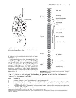
![deficits compared with right-hemisphere lesions. The
motor impairment from stroke usually results in hemiple-
gia (paralysis) or hemiparesis (weakness). When damage
occurs to the descending neural pathways, an abnormal
regulation of spinal motor neurons results in adverse
changes in postural and stretch reflexes and difficulty
with voluntary movement. Deficits in motor control can
involve muscle weakness, abnormal synergistic organiza-
tion of movements, impaired regulation of force, de-
creased reaction times, abnormal muscle tone, and loss of
active range of motion (ROM) (51).
EXERCISE TESTING AND
SCREENING CRITERIA
SCREENING PROTOCOL
Before conducting a graded exercise test, persons with
stroke should be screened by their primary care physi-
cian. Although no published guidelines exist on the type
of screening that should be conducted before a graded ex-
ercise test, taking the utmost precaution is critical be-
cause many stroke survivors are older and have cardio-
vascular disease. The screening should include a fasting
blood draw, resting electrocardiogram (ECG), resting
heart rate, resting blood pressure (standing, seated,
supine), and basal temperature. To be approved for peak
V
.
O2 testing, participants’ blood screening tests (i.e., com-
plete blood count [CBC], enzymes, protein levels) should
be within normal limits. If the preliminary blood work is
acceptable, the participant can be scheduled for testing.
Participants who successfully complete the graded exer-
cise test can be recommended for an exercise program.
Individuals who have adverse cardiovascular changes
during exercise testing should be advised for further
follow-up and may need to begin an exercise program in
a closely supervised setting, such as cardiac rehabilitation.
EXERCISE TESTING
Peak Oxygen Uptake (Cardiorespiratory Fitness)
Cycle ergometer testing with stroke survivors is consid-
ered safe and feasible when a medical prescreening is per-
formed and the participant’s exercise response is closely
monitored to minimize risk (52). Determination of aero-
bic exercise capacity is an important component for de-
veloping appropriate exercise programs and evaluating
the effectiveness of the programming (52). Because of a
significant loss of muscle function resulting from hemi-
paresis or hemiplegia, stroke survivors have a severely re-
duced maximal or peak oxygen uptake (37,52). Because
many stroke survivors also have cardiovascular comor-
bidity (e.g., hypertension, CAD), graded exercise tests
are often symptom-limited, thus not allowing the person
to achieve high peak capacities. Additionally, limited
data are available on the test-retest reliability of peak ex-
ercise testing among stroke survivors and it is recom-
mended that at least one preliminary testing trial be per-
formed to minimize the possible practice effect (53). In
the postacute stage, a symptom-limited graded exercise
test (peak V
.
O2) can be performed on a stationary bike or
treadmill, or in persons with severe hemiplegia, with an
arm ergometer. Although it may be necessary to perform
the exercise test with an arm ergometer, performance will
be limited because of the limited amount of muscle re-
cruitment and a greater strain on the cardiac system per
unit of peripheral muscle mass recruited (53). Typi-
cally, arm-cranking yields a peak V
.
O2 30%–35% less than
treadmill performance. The two most preferred exercise
modes are the stationary cycle and treadmill. For individ-
uals with balance difficulties or severe hemiplegia where
walking on a treadmill is difficult or not possible, the sta-
tionary cycle is preferred because it eliminates the risk of
falls.
Ramp Cycle Protocol
There are several different testing protocols that have
been used with persons with stroke. Rimmer et al. (43)
measured peak V
.
O2 in 35 stroke survivors using a ramp
cycle ergometer testing protocol. Participants began cy-
cling at a workload of 20 W at a target cadence of 60 rev-
olutions per minute (rpm) and increased by 10 W every
minute until maximal effort was achieved. Heart rate and
blood pressure were recorded every 2 minutes. Tests were
terminated if one of the following criteria was observed:
(a) respiratory exchange ratio (RER) 1.1, (b) peak heart
rate within 10 beat per min1
of age-predicted maximal
value, (c) abnormal blood pressure or ECG response, or
(d) unable to continue pedaling above 50 rpm. Potempa
et al. (22) used a similar ramp cycle ergometer protocol
with stroke survivors. Testing began at 10 W and in-
creased 10 W each minute until maximal effort was at-
tained.
Macko et al. (26) performed several exercise testing
and training studies with stroke survivors using a tread-
mill protocol. They recommended that a treadmill test at
0% incline be conducted before the maximal exercise test
to assess gait safety and walking velocity. As a safety
measure, individual wore a gait support belt. Participants
who successfully completed at least 3 consecutive min-
utes of treamill walking at .22 m/s or faster (0.5 mph)
were allowed to have a maximal exercise stress. After a
15-minute rest, the participants performed a constant ve-
locity, progressively graded treadmill test to volitional fa-
tigue or peak effort and were continuously monitored
(ECG and vital signs) during testing.
Tang et al. (52) conducted exercise testing on stroke
survivors who were in the subacute (3 months) stage of
recovery. They used a semirecumbent cycle ergometer
(Biodex) to perform the exercise test. The ramp protocol
CHAPTER 1 Stroke 5
LWBK191-4034G-C01_01-18.qxd 6/11/08 5:50 pm Page 5 Aptara (PPG-Quark)](https://image.slidesharecdn.com/acsmsresourcesforclinicalexercisephysiology-230410064150-fb83bfa2/85/ACSM_s-Resources-for-Clinical-Exercise-Physiology-pdf-19-320.jpg)
![CHAPTER 1 Stroke 15
Smoking history is part of the risk factor profile that
accompanies stroke. In this case, her pulmonary
function may be a limiting factor during exercise. An in-
quiry regarding her pulmonary function is important, es-
pecially if she is short of breath.
Pharmacologic issues: Be aware of reduced patient
arousal because of the somnolence side effects of her
medications (e.g., muscle relaxant [cyclobenzaprine],
antiseizure medications [carbamazepine]). Metoprolol is
a -blocker that reduces maximal heart rate and may
limit her exercise endurance.
Nerve conduction studies and needle electromyography test-
ing were used to discover the slowed median nerve con-
duction across the wrist called carpal tunnel syndrome,
explaining the numbness and pain in the nonhemiplegic
left hand. Electrical stimulation by a recording device
measures the speed and size (amplitude) of nerve
conduction to determine if the nerve is healthy or
injured. She has both a nerve pinch (i.e., carpal tunnel
syndrome) and diabetes, which will also show abnormal
results on nerve conduction study. The needle form of
electromyography is also used to assess for loss of motor
nerve function or muscle disease by observing the electri-
cal muscle membrane and muscle contraction potentials
heard and viewed on a screen.
CASE 3
JP: 72-year-old man suffered a cerebrovascular accident
2 years before his visit. He was a depressed gentleman, a
retired successful businessman, and he had a history of
participation in competitive sports. His chief complaint
was lack of improvement from a left hemiparesis after
the stroke and lack of energy. Extensive medical work-up
uncovered sleep apnea and a home airway (CPAP)
device was implemented through an affiliated sleep lab-
oratory. He needed help in bathing, dressing, and for
community walks greater than household distances (i.e.,
50 feet). He could eat, but not cut his own food. A
wheelchair was used for community mobility pushed
usually by his wife. His wife would state that he had sev-
eral falls at home while ambulating, none with any
injury. Medications: Dilantin, Coumadin, Synthroid,
Prozac, Zocor, MVI, Digoxin. The physical examination
noted a tall slender man with clear speech and commu-
nication, but with wandering of thoughts during his re-
sponses and slowed responses. He showed a left facial
hemiparesis. He could repeat five of seven numbers in
forward order, but was unable to repeat them in reverse
order. He also would tend to avoid looking over to his
left side and would not pay attention to his left arm
hanging off of the wheelchair. He displayed minimal
movement of the proximal scapula-related muscles and
no voluntary motor response in the more distal upper
extremity. The lower extremity strength was graded fair in
the proximal muscle of the hip and knee and poor in
more distal muscles of the ankle and foot. Sensation was
partially decreased on the left side, with sensory
extinction on simultaneous (left and right tested at the
same time) light touch. Grafesthesia was also impaired
but present on the left side. Reflexes were decreased on
the left side, with a positive Babinski sign on the left and
right sides. Cranial deficits were noted in facial muscles
on the left side. No aphasia was present. He needed sev-
eral attempts to arise from a sitting to a stand position.
However, with a quad cane he did ambulate at less than
1 mph with no deviations at that speed on noncarpeted
surface. He had a foot drop and would have difficulty
clearing his toe during swing phase of gait. He entered
the clinical exercise physiology program to improve both
safety in ambulation and focus on details as they related
to a home exercise program. The goal was also to facili-
tate maximal use of the left upper extremity and attention
to his left side. Throughout the program he displayed in-
terruption in ambulation, reaching tasks, and therapeutic
exercises, accompanied by random conversations about
issues that concerned him. He would also periodically
bump his left side in the doorway or other obstacles. The
program was adjusted to raise his awareness by having
him navigate various obstacle courses and objects to
reach another point. He was noted to not bump into
these obstacles when asked specifically to walk around
them. He also gradually responded to verbal cues to the
left side. Resistance exercise for the right side involved
lifting the left extremities. Electrical muscle stimulation
pulsed to turn on every 5 seconds for 5 seconds duration
was used to cue muscles of the left shoulder, and left hip-
and knee-related muscles. No gait training was done on
the treadmill, because his speed of ambulation was
initially 0.6 mph and never faster than 1.5 mph. He was
successful in the cessation of any falls at home. His speed
of ambulation increased, and he could discontinue his
left plastic ankle foot orthosis without a foot drop at the
end of 6 months of the program, on an every-other-week
basis. His left upper extremity became functional for arm
swing during ambulation. He was successfully connected
with psychological support care, as well as support coun-
seling for him and his wife.
Problems to Consider
One-sided neglect is a problem most common to individu-
als who have had a stroke to the right (or dominant side)
hemisphere with left hemiplegia. They will ignore the left
side of the body, often as if it did not exist. Setting up
safe challenges that requires the individual to become
aware of that side, such as an obstacle course, is one
form of cuing to that side. Other strategies are to use the
recognized extremities to find and use the neglected side
in bilateral two-extremity exercises. Biofeedback, such as
electrical stimulation with activities, can be useful.
Sensory extinction is the absence of recognition of a sen-
sory stimulus to one extremity when both are touched.
For example, if both extremities recognize a sensory
CHAPTER 1 Stroke 15
LWBK191-4034G-C01_01-18.qxd 6/11/08 5:50 pm Page 15 Aptara (PPG-Quark)](https://image.slidesharecdn.com/acsmsresourcesforclinicalexercisephysiology-230410064150-fb83bfa2/85/ACSM_s-Resources-for-Clinical-Exercise-Physiology-pdf-29-320.jpg)

![34
Multiple Sclerosis
3
C
HA
P
TER
EPIDEMIOLOGY/ETIOLOGY
Multiple sclerosis (MS) is a common neurologic disease
that affects women at a ratetwo to three times greater
than men (1). It is estimated that approximately
400,000 individuals in the United States have MS, with
a worldwide estimate of 2.5 million (1). Studies of mi-
grating populations have indicated that where a person
resides in relation to the equator before the age of 15 ap-
pears to determine the likelihood of developing MS (2).
The incidence of MS is nearly 3/100,000 in temperate
zones, and below 1/100,000 in tropical areas (3). The
initiation of MS, either propensity for the disease or the
disease itself, begins in childhood. A variety of mild
viral infections, such as measles and upper-respiratory
infections, are thought to be etiological (4). A threefold
increase in the incidence of exacerbations of MS is seen
following an upper-respiratory infection (5,6). Exacer-
bation rate is reduced during pregnancy and is increased
threefold in the postpartum period up to approximately
3 months (7). The onset of MS usually occurs between
the ages of 20 and 40; however, it is often possible to ob-
tain a history of transient neurologic deficits, such as
numbness of an extremity, weakness, blurring of vision,
and diplopia, in childhood or adolescence before the de-
velopment of more persistent neurologic deficits. The
latter often lead to a definitive diagnosis (8,9). It is pos-
sible that viruses causing upper-respiratory infections
may be responsible for sensitizing the brain to subse-
quent autoimmune insult, producing inflammatory de-
myelination.
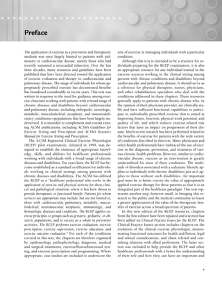
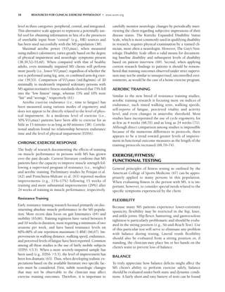
Several distinct courses of the disease are now recog-
nized, as well as the prevalence rate associated with each
type: Relapsing-remitting MS (RRMS; 85%), primary pro-
gressive MS (PPMS; 10%), and progressive relapsing MS
(PRMS; 5%). After an initial period of RRMS, many de-
velop a secondary-progressive (SPMS) disease course,
characterized by a more steady decline in function with
or without flare-ups and remissions. Of the 85% of those
initially diagnosed with RRMS, more than 50% will go
onto to develop SPMS within 10 years. Furthermore, 90%
of patients with RRMS will develop SPMS within 25
years. These statistics, however, are based on data
collected before the widespread use of newer disease-
modifying agents that may delay or reduce the progression
of RRMS to SPMS. An overview of the course of each pat-
tern and related disability is presented in Figure 3.1 (10).
For decades scientists have speculated about a “ge-
netic predisposition” for MS; however, only recently has
the presence of two specific genes been identified that
could be related to the increased susceptability (11). Pa-
tients who have a definite diagnosis of MS are more
likely to have a variety of other illnesses of an autoim-
mune nature, such as systemic lupus erythematosus
(SLE), rheumatoid arthritis (RA), polymyositis, myas-
thenia gravis, and so forth (12). Studies have shown that
if a first-degree relative has MS, there is a 12- to 20-fold
increase in the likelihood of having MS (13). In monozy-
gotic twins there is a 33% increase in the incidence of
MS, whereas in dizygotic twins the incidence is about
8%, or that found in the normal population (14,15).
PATHOPHYSIOLOGY
Multiple sclerosis is a disease of the central nervous sys-
tem in which there are multiple areas of inflammatory de-
myelination with a predilection for distribution around
the ventricles and vascular spaces. Multiple mechanisms
are involved in producing damage to central nervous sys-
tem myelin, as well as axons (16,17). An immune reac-
tion to myelin (myelin basic protein [MBP]) and myelin
oligodendrocyte glycoprotein (MOG) occurs. Activated T
cells attach to the endothelium of capillaries within the
brain and migrate into the brain parenchyma, where acti-
vated macrophages attack and digest myelin. A number
of cytokines, including tumor necrosis factor (TNF), and
interferons, as well as IgG, are involved in the immune at-
tack. B cells produce IgG directed at MOG. There is in-
creased production of IgG and an increased prevalence of
specific IgG moieties, some of which represent antiviral
IgG. Recent studies of total brain N-acetylaspartate (NAA)-
to-creatine ratios have provided evidence for loss of axons,
as well as evidence for membrane damage (demyelination)
as an increase in choline-to-creatine ratio (18).
Lesions representing focal areas of inflammatory de-
myelination can be present in the cerebral hemispheres,
brainstem, and spinal cord. For a definite diagnosis to be
established, two or more areas of demyelination (white
matter lesions) must be established. Furthermore, there
must be two or more remissions of neurologic deficits.
LWBK191-4034G-C03_34-43.qxd 06/11/2008 10:00 AM Page 34](https://image.slidesharecdn.com/acsmsresourcesforclinicalexercisephysiology-230410064150-fb83bfa2/85/ACSM_s-Resources-for-Clinical-Exercise-Physiology-pdf-48-320.jpg)
![36 RESOURCES FOR CLINICAL EXERCISE PHYSIOLOGY • www.acsm.org
early diagnosed patients complain of urinary urgency and
frequency (24,25).
Sexual dysfunction is common, with loss of sensation,
lack of the ability to have an orgasm, and impotence
being very common (26). Adynamia (i.e., a loss of
strength or vigor) of the colon is also common with most
patients experiencing severe constipation (25). Some pa-
tients require partial colectomy secondary to obstipation
(intractable constipation).
A variety of skin sensations and loss of sensation
occur, most commonly vibration sense loss in both feet
with position sense preserved until vibration sense loss is
severe. Dysesthesias, characterized as abnormal sensa-
tions produced by touching or stroking the skin, are also
very common. These sensory disturbances do not occur
in a distribution characteristic of involvement of a pe-
ripheral nerve. Abnormal sensation over the trunk, par-
ticularly a bandlike sensation around the abdomen or
chest, is also characteristic. Most patients, however, do
not complain of severe pain in the extremities, but this
does sometimes occur (27,28).
Cognitive difficulties develop along with cranial,
motor, and sensory symptoms (29–31). The patient may
complain of an inability to function in the workplace
when he or she is required to monitor two or more activi-
ties at the same time. Attentiveness is decreased so that
the patient may be unable to register information accu-
rately in memory. Emotional lability is also common in as-
sociation with subfrontal demyelination, which produces
a pseudobulbar palsy. Studies have shown that cognitive
deficit is greater in individuals with prominent signs of
pseudobulbar palsy or excessive emotional lability (32).
MEDICAL AND SURGICAL TREATMENTS
Exacerbations of MS are usually treated with high-dose
adrenocortical steroids customarily administered intra-
venously at a dose of 1000 mg/day for 3 to 5 days, fol-
lowed by a prednisone taper over approximately 6 weeks
(22,23). It has been demonstrated that patients with
monosymptomatic MS initially treated with methylpred-
nisolone defer the development of more typical MS when
they are treated with methylprednisolone (22). Develop-
ment of two or more deficits, marked weakness and inco-
ordination, loss of sensation in both lower extremities, or
all of those are indication for treatment with adrenocorti-
cal steroids.
Prophylactic treatments include interferon beta-1a
(Rebif, Avonex) and -1b (Betaseron), which reduce lym-
phocytic invasion of the brain, induce a suppressor im-
mune reaction, provide an antiviral action, reduce the
number of exacerbations over time, and preserve brain
mass (32–34). Glatiramer (copolymer I or Copaxone), a
peptide consisting of four amino acids—glycine, alanine,
lycine, and tyrosine—act by inducing immune tolerance to
myelin basic protein (35). This is also effective in reducing
the number of exacerbations and preserving brain mass
over time. Chemotherapy, such as methotrexate and mi-
toxantrone (Novantrone), are also recommended for pa-
tients with chronic progressive disease (36,37).
Symptomatic management includes the treatment of
neurogenic bladder with anticholinergic medications,
such as oxybutynin, for urinary frequency and urgency.
Regimens for treating obstipation include psyllium
preparations, laxatives, suppositories, and physical activ-
ity. Spasticity, particularly flexor and extensor spasms, is
treated with either -aminobutyric acid (GABA-b-ergic)
compound (baclofen), which increases spinal inhibition,
or Tizanidine, which increases supraspinal inhibitors of
spinal reflex activities (38,39).
DIAGNOSTIC TECHNIQUES
If the patient’s history is highly suggestive of MS, then an
MRI of the brain, often combined with imaging of the
spinal cord are obtained. Sometimes, to establish the ex-
istence of another lesion characteristic of MS, evoked po-
tentials of the visual systems (visual evoked potential
[VEP]), the brainstem, or somatosensory (SSEP) systems
are obtained. In MS, there is a marked delay in conduc-
tion of the action potential. Evaluation of the spinal fluid
during an exacerbation early in the disease may reveal a
mild pleocytosis of usually less than 100 cells consisting
predominantly of lymphocytes and a mild elevation of
protein, usually less than 100 mg%. An increase in IgG
synthesis and decreased variability of the different moi-
eties of IgG are also characteristic. In the first few months
of the disease, spinal fluid findings may be normal, but
repeat examination a year or so later may reveal increased
IgG synthesis and OCBs. Early definitive diagnosis is im-
portant so that prophylactic treatment can be instituted
to prevent injury to the central nervous system (40).
CLINICAL EXERCISE PHYSIOLOGY
ACUTE EXERCISE RESPONSE
Studies have shown that persons with MS have a lower
maximal aerobic capacity than the average age- and gen-
der-matched, nondisabled adult without MS (38,39). Fur-
thermore, maximal aerobic capacity appears to be in-
versely related to level of disability (39) as measured on
the Kurtzke Expanded Disability Status Scale (EDSS)
(40). Individuals with a higher EDSS score, indicative of
more neurologic impairment as derived from a clinical ex-
amination, have a lower maximal and submaximal aerobic
exercise capacity. Despite the variability in peformance,
one common effect of acute exercise in individuals with
MS is an overwhelming sense of fatigue during postexer-
cise recovery. No scientific evidence, however, suggests
that the postexercise fatigue is reflective of an exacerba-
tion of existing or new MS symptom. Furthermore, recent
LWBK191-4034G-C03_34-43.qxd 06/11/2008 10:00 AM Page 36](https://image.slidesharecdn.com/acsmsresourcesforclinicalexercisephysiology-230410064150-fb83bfa2/85/ACSM_s-Resources-for-Clinical-Exercise-Physiology-pdf-50-320.jpg)
![CHAPTER 3 Multiple Sclerosis 37
evidence shows that the level of self-reported postexercise
fatigue as measured by the Modified Fatigue Impact Scale
(MFIS) can be reduced following training (40,41).
To understand basic physiologic responses to an acute
bout of exercise, it is helpful to use the two directions
commonly taken in the literature: (a) as it relates to mus-
cle performance (i.e., strength and endurance) and (b)
cardiovascular responses (i.e., heart rate, blood pressure,
oxygen utilization).
MUSCLE PERFORMANCE
In the absence of documented spasticity or use of anti-
spasmodic drugs, several studies have reported that mus-
cle endurance for persons with MS during a sustained
isometric contraction (e.g., 30% of maximal voluntary
contraction [MVC]) is similar to that of a nondisabled,
healthy adult (42). Although these findings are in direct
conflict with other studies of acute muscle response
(43–47), it is important to consider that in none of the
these other studies was the presence of spasticity or the
use of antispasmodic drugs controlled in the design. Both
Chen et al. (46) and Ponichtera et al. (44) hypothesized
that spasticity of antagonistic muscles may decrease con-
centric agonist force production. In contrast, antagonists
are not stretched during an eccentric contraction,
whereas the agonist receives additional stretch to facili-
tate its contraction. Ponichtera et al. (44) suggested that,
although spasticity in 44% of their sample may have con-
tributed to a significantly lower force production during
concentric knee extension, force production during ec-
centric knee extension for the MS patients was normal.
Thus, spasticity and or co-contraction of opposing mus-
cle groups may be one factor that can reduce concentric
muscle performance in this population.
To believe that spasticity is the sole contributor to dif-
ferences in muscle performance observed between MS
and non-MS persons would be naïve. Other contributors
to observed differences have been attributed to conduc-
tion block of demyelinated fibers (48), reduced muscle
metabolic responses during voluntary exercise (49),
muscle weakness owing to fiber atrophy (50,51), as well
as sensory deficits, which have been discussed previously.
Maximal muscle force during isokinetic exercise at var-
ious velocities has also been shown to be consistently
lower for persons with MS when matched to controls
(43–46). Furthermore, maximal aerobic power (POmax)
using leg cycling or combination leg–arm cycling proto-
cols have shown that most persons with MS are likely to
generate 20%–68% less power (38,39) than healthy indi-
viduals. More recently, similar findings have confirmed
that persons with MS have lower POmax than healthy,
sedentary adults. Performance during arm cranking and
combined arm–leg cycling shows similar findings: 31%
lower during arm cranking and 24% lower during com-
bined arm–leg cycling (39). However, when disability
level (i.e., EDSS) is taken into account and power output
is expressed in terms of body weight (watts/kilogram), it
appears that individuals with an EDSS of 4.0 or less (i.e.,
fully ambulatory) are not significantly weaker than able-
bodied, matched controls (52). Thus, documentation of
disability level in both research and clinical practice is im-
portant to understanding the potential work capacity in
this population and to setting realistic therapeutic goals.
ACUTE CARDIORESPIRATORY RESPONSES
Physiologic responses to an acute bout of submaximal aer-
obic exercise appear to be normal for many persons with
MS. Heart rate (HR), blood pressure (BP), and oxygen up-
take (V̇O2), and minute ventilation (VE) have been shown
to increase in a linear fashion to increments in workload
(38,39). Normal metabolic and cardiovascular responses
are consistent over a wide range of impairment levels (i.e.,
EDSS). In contrast, when HR response to incremental ex-
ercise is examined in the context of metabolic cost (i.e.,
oxygen pulse-V̇O2/HR), Tantucci et al. (53) reported a sig-
nificantly lower oxygen pulse for those with MS compared
with healthy controls. These findings are consistent during
both submaximal and maximal aerobic exercise. A higher
HR at a given V̇O2 might imply that stroke volume is in-
sufficient to support the metabolic demand. However, in a
case study by Vaz Fragoso and associates (54), right and
left ventricular ejection fraction were recorded at rest
and at anaerobic threshold showed normal cardiac output
and O2 saturation levels; implicating an abnormality in
peripheral O2 distribution or utilization. A reduction in O2
distribution could be related to a diminished sympathetic
outflow to arterial smooth muscle. Cardiovascular auto-
nomic dysfuction, both sympathetic and parasympathetic,
has been well-documented in the MS population (55–58).
A deficiency in O2 utilization is suggestive of peripheral
muscle pathology, which has already been supported in
earlier research findings (49–51). Future research should
focus on the question of peripheral issues, such as oxygen
extraction during exercise. Furthermore, whether oxygen
pulse (i.e, reduced HR) can be improved with increased
stroke volume following training is certainly an important
question to be answered.
From a clinical perspective, using HR as an index of ex-
ercise intensity presents a twofold problem. First, if oxygen
pulse is significantly lower in this population, then absolute
workloads will need to be lower during training. With ex-
ercise intensity being lower, smaller absolute gains in aero-
bic capacity are probable. Second, in the presence of dimin-
shed cardioacceleration, the application of either the
Karvonen method or the standard practice of calculating
HRmax as 220–age, from which a training HR is calculated,
should be done so with caution. Percieved exertion scales
would be better suited for this population with the use of
the Category-Ratio Rating of Perceived Exertion (59) possi-
bly being the best choice. This scales uses perceived stress
LWBK191-4034G-C03_34-43.qxd 06/11/2008 10:00 AM Page 37](https://image.slidesharecdn.com/acsmsresourcesforclinicalexercisephysiology-230410064150-fb83bfa2/85/ACSM_s-Resources-for-Clinical-Exercise-Physiology-pdf-51-320.jpg)
![REFERENCES
1. Web site home page-National Mulitple Sclerosis Society [Internet].
New York, NY. National MS Society; [cited 2007]. Avaiblable from:
http://www.nmms.org.
2. Rosati G. Descriptive epidemiology of MS in Europe in the 1980’s:
A critical overview. Ann Neurol 1994;36:5164–5174.
3. Weinshenker BG. Epidemiology of MS. Neurol Clin 1996;14:291–308.
4. Sibley WA, Bamford CR, Clark K. Clinical viral infections and MS.
Lancet 1985;1:1313–1315.
5. Panitch HS. Influence of infection on exacerbation of MS. Ann Neu-
rol 1994;36:525–528.
6. Korn-Labetzki I, Khana E, Cooper G, et al. Activity of MS during
pregnancy and puerperium. Ann Neurol 1984;16:229–231.
7. Carriere, W, Baskerville J, Ebers, GC. The natural history of MS: A ge-
ographically based study. Applications to planning and interpretation
of clinical and therapuetic trials. Brain 1991;114:1057–1067.
8. Wynn DR, Rodriguez M, O’Fallon M, Kurland LT. A reappraisal of
the epidemiology of MS in Olmstead County, Minnesota. Neurology
1990;40:780–786.
9. Lublin FD, Reingold SC. Defining the clinical course of multiple
sclerosis: Results of an international survey. National Multiple Scle-
rosis Society (USA) Advisory Committee on Clinical Trials of New
Agents in Multiple Sclerosis. Neurology 1996;46:907–911.
10. The International Multiple Sclerosis Genetics Consortium. Risk
alleles for multiple sclerosis identified by a genomewide study.
N Engl J Med [Internet]. 2007 [cited 2007, July 29. Available from:
http://www.nejm.com.
11. Haegert DG, Marrosu MG. Genetic susceptibility to MS. Ann Neurol
1994;36:2S04–S210.
12. Mumford CJ, Wood NW, Kellar-Wood H, Thorpe JW, Miller DH,
Compston, DA. The British Isles survey of MS in twins. Neurology
1994;44:11–15.
13. Sadovnick AD, Armstrong H, Rice GP
, et al. A population-based
study of MS in twins: Update. Ann Neurol 1993; 33:281–285.
14. Prineas JW. Pathology of MS. In: Cook SD, ed. Handbook of MS.
New York: Marcel Dekker. 1990:187–218.
15. Sobel RA. The pathology of MS. Neurol Clin 1995;13:1–21.
16. Waxman SG. Pathophysiology of MS. In: Coo DS, ed. Handbook of
MS. New York: Marcel Dekker. 1990:219–249.
17. Prineas JW, Barnanrd RD, Revesz T, Kwon EE, Sharer L, Cho E-S.
MS: Pathology of recurrent lesions. Brain 1993;116:681–693.
18. Gonen O, Patalace I, Babb JS, et al. Total brain N-acetylaspartate, a
new measure of disease load in MS. Neurology 2000;54:15–19.
19. Poser CM, Paty DW, Scheinberg L, et al. New diagnostic criteria for
MS: Guidelines for research protocols. Ann Neurol 1983;13:227–231.
20. Krupp LB, Alvarez LA, LaRocca NG, et al. Fatigue in MS. Arch Neu-
rol 1988;45:435–437.
21. Freal JE, Kraft GH, Coryell JK. Symptomatic fatigue in MS. Arch
Phys Med Rehabil 1984;65:165–168.
22. Thompson AJ, Kennard C, Swash M, et al. Relative efficacy of in-
travenous methylprednisolone and ACTH in the treatment of acute
relapse in MS. Neurology 1989;39:696–971.
23. Beck RW, Cleary PA, Anderson MM, et al. A randomized controlled
trial of corticosteroids in the treatment of acute optic neuritis. N
Engl J Med 1992;326:581–588.
24. Blaivas JG. Management of bladder dysfunction in MS. Neurology
1980;30:12–18.
25. Bradley WE, Logothetis JL, Timm GW. Cystometric and sphincter
abnormalities in MS. Neurology 1973;23:1131–1139.
26. Valleroy ML, Kraft G. Sexual dysfunction in MS. Arch Phys Med Re-
habil 1984;65:125–128.
27. Svendsen, KB, Jensen, TS, Hansen, HJ, Bach, FW. Sensory function
and quality of life in patients with multiple sclerosis and pain. Pain
2005;114:473–481.
28. Osterberg, A, Boivie, J, Thuomas, K-A. Central pain in multiple
sclerosis—prevelance and clinical characteristics. Eur J Pain 2005;
9:531–542.
29. Herholz, K. Cognitive dysfunction and emotional-behavioural
changes in MS: The potential of positron emission tomography.
J Neurol Sci 2006;245(1–2);9–12.
30. The IFNB MS study group and the University of British Columbia
MS/MRI analysis group. Interferon beta-1b in the treatment of MS:
Final outcome of the randomized controlled trial. Neurology
1995;45:1277–1285.
31. Gold R, Rieckmann P
, Chang P
, Abdalla J; the PRISMS Study Group.
The long-term safety and tolerability of high-dose interferon -1a
in relapsing-remitting multiple sclerosis: 4-year data from the
PRISMS study. Eur J Neurol 2005;12:649–656.
32. Johnson KP
, Brooks BR, Cohen JA, et al. Copolymer-1 reduces relapse
rate and improves disability in relapsing-remitting MS: Results of a
phase III multicenter double-blind placebo-controlled trial. Neurol-
ogy 1995;45:1268–1276.
33. Thompson AJ, Noseworthy JH. New treatments for MS: Clinical
perspective. Curr Opin Neurol 1996;9:187–198.
34. Fidler JM, DeJoy SQ, Smith FR 3rd, Gibbons JJ Jr. Selective im-
munomodulation by the antineoplastic agent mitoxantrone. Non-
specific adherent suppressor cells derived from mitoxantrone-
treated mice. J Immunol 1986;136:2747–2754.
35. Katz R. Management of spasticity. Am J Phys Med Rehabil 1988;
67:108–116.
36. Nance DW, Sheremata WA, Lynch SG, et al. Relationships of the an-
tispasticity effect of Tizanidine to plasma concentration in patients
with MS. Arch Neurol 1997;54:731–736.
37. Rudick RA, Goodman A, Herndon RM, Panitch HS. Selecting re-
lapsing-remitting MS patients for treatment: The care for early
treatment. J Neuroimmunol 1999;98:22–28.
38. Ponichtera-Mulcare JA, Glaser RM, Mathews T, Camaione, DN.
Maximal aerobic exercise in persons with MS. Clin Kinesiol 1983;
46(4):12–21.
39. Ponichtera-Mulcare JA, Mathews T, Glaser RM, Mathrews T Gupta
SC. Maximal aerobic exercise of individuals with MS using three
modes of ergometery. Clin Kinesiol 1995;49:4–13.
40. White LJ, McCoy SC, Castellano V, Guiterrez G, Stevens JE, Walter
GA, Vandenborne K. Resistance training improves strength and
functional capacity in persons with multiple sclerosis. Mult Scler
2004;10:668–674.
41. Surakka J, Romberg A, Ruutiainen J, Aunola S, Virtanen A, Karppi
SL, Maentaka K. Effects of aerobic and strength exercise on motor
fatigue in men and women with multiple sclerosis: A randomized
controlled trial. Clin Rehabil 2004;18(7):737–746.
counter extensor tone bias. Standing hip and knee
flexion to improve toe clearance during swing phase of
gait. Lateral pull-downs and seated rows to increase
strength of postural extensor muscles to reduce effects
of prolonged sitting and improve antigravity muscle con-
trol during functional activities.
Flexibility
• Stretching of pectoralis major or minor and illiopsoas
to improve trunk and hip extension.
• Stretching of gastocnemius or soleus to improve ankle
dorsiflexion. Perform two repetitions holding each
stretch for 60 seconds twice a day.
42 RESOURCES FOR CLINICAL EXERCISE PHYSIOLOGY • www.acsm.org
LWBK191-4034G-C03_34-43.qxd 06/11/2008 10:00 AM Page 42](https://image.slidesharecdn.com/acsmsresourcesforclinicalexercisephysiology-230410064150-fb83bfa2/85/ACSM_s-Resources-for-Clinical-Exercise-Physiology-pdf-56-320.jpg)
![CHAPTER 3 Multiple Sclerosis 43
42. Ng AV, Dao HT, Miller RG, Gelina DF
, Kent-Braun JA. Blunted pres-
sor and intramuscular metabolic responses to voluntary isometric
exercise in MS. J Appl Physiol 2000;88:871–880.
43. Lambert CP
, Archer RL, Evans WJ. Muscle strength and fatigue dur-
ing isokinetic exercise in individuals with multiple sclerosis. Med
Sci Sport Exerc 2001;33(10):1613–1619.
44. Ponichtera JA, Rodgers MM, Glaser RM, Mathews, T. Concentric
and eccentric isokinetic lower extremity strength in persons with
MS. J Orthop Sport Phys Ther 1988;16(3):114–122.
45. Armstrong LE, Winant DM, Swasey PR, Seidle ME, Carter AL,
Gehlsen GM. Using isokinetic dynamometry to test ambulatory pa-
tients with MS. Phys Ther 1983;63:1274–1279.
46. Chen W-Y, Peirson FM, Burnett CN. Force-time measurements of
knee muscle function in MS. Phys Ther 1987;67:934–940.
47. Rice CL, Volmer TL, Bigland-Ritchie B. Neuromuscular responses
of patients with MS. Muscle Nerve 1992;15:1123–1132.
48. McDonald WI, Sears TA. Effect of a demyelinating lesion on con-
duction in the central nervous system studied in single nerve fibers.
J Physiol (London) 1970;207:53–54P
.
49. Kent-Braun JA, Sharma KR, Miller RG, Weiner MW. Postexercise
phosphocreatine resysntheis is slowed in multiple sclerosis. Muscle
Nerve 1994;17(8):835–841.
50. Kent-Braun JA, Sharma KR, Weiner MW, Miller RG. Effects of exer-
cise on muscle activation and metabolism in MS. Muscle Nerve
1994;17(10):1162–1169.
51. Sharma KR, Kent-Braun J, Mynhier MA, Weiner MW, Miller RG.
Evidence of an abnormal intramuscular component of fatigue in
MS. Muscle Nerve 1995;18(12):1403–1411.
52. Mulcare JA, Webb P
, Mathews T, Gupta, SC. Sweat response in per-
sons with multiple sclerosis during submaximal aerobic exercise.
International Journal of MS Care 2001;3(4):26–33.
53. Tantucci, C Massucci M, Piperno R, Grassi V, Sorbini CA. Energy
cost of exercise in MS patients with low degree of disability. Mult
Scler 1996; 2(3):161–167.
54. Vaz Fragoso C, Wirz D, Mashman J. Establishing a physiological
basis to multiple sclerosis-related fatigue: A case report. Arch Phys
Med Rehabil 1995;76:583–586.
55. Linden D, Diehl RR, Kretzschmar A, Berlit P
. Autonomic evaluation
by means of standard tests and power spectral analysis in multiple
sclerosis. Muscle Nerve 1997;20(7):809–814.
56. Flachenecker P
, Wolf A, Krauser M, Hartung HP
, Reiners K. Cardio-
vascular autonomic dysfunction in multiple sclerosis: Correlation
with orthostatic intolerance. J Neurol 1999;246(7):578–586.
57. Bonnett M, Mulcare J, Mathews T, Gupta SA, Ahmed N, Yeragani V.
Heart rate and QT interval variability in multiple sclerosis: Evi-
dence for decreased sympathetic activity. J Neurol Sci [Turkish]
2006;23(4):248–256.
58. Pepin EB, Hicks RW, Spencer MK, Tan ZC, Jackson CGR. Pressor
response to isometric exercise in patients with multiple sclerosis.
Med Sci Sports Exerc 1996;23:656–660.
59. Noble BJ, Roberton RJ. Perceived Exertion. Champaign, IL: Human
Kinetics; 1996.
60. Pariser G, Madras D, Weiss E. J Outcomes of an aquatic exercise
program including aerobic capacity, lactate threshold, and fatigue
in two individuals with multiple sclerosis. Neur Phys Ther 2006;
30(2):82–90.
61. Nieman DC. Fitness and Sports Medicine: An Introduction. Palo Alto,
CA: Bull Publishing Company. 1990:500.
62. Petajan JH, Gappmaier E, White AT, Spencer JK, Mino L, Hicks RW.
Impact of aerobic training on fitness and quality of life in MS. Ann
Neurol 1996;34:432–441.
63. Ponichtera-Mulcare JA, Mathews T, Barrett PJ, Gupta SC. Change
in aerobic fitness of patients with MS during 6-month training pro-
gram. Sports Medicine, Training, and Rehabilitation 1997;7:265–272.
64. Gutierrez GM, Chow JW, Tillman MD, McCoy SC, Castellano V,
White J. Resistance training improves gait kinematics in persons
with multiple sclerosis. Arch Phys Med Rehabil 2005;86:1824–1929.
65. White LJ, McCoy SC, Castellano V
, Guiterrez G, Stevens JE, Walter GA,
Vandenborne K. Resistance training improves strength and functional
capacity in persons with multiple sclerosis. Mult Scler 2004;10:668–674
66. Taylor NF
, Dodd KJ, Prasad D, Denisenko S. Progressive resistance
exercise for people with multiple sclerosis. Disabil Rehabil 2006;
28(18):1119–1126.
67. American College of Sports Medicine. Progression models in resist-
ance training for healhty adults. Med Sci Sports Exerc 2002;34(2):
364–380.
68. van den Berg M, Dawes H, Wade DT, Newman M, Burridge J, Izadi
H, Sackley CM. Treadmill training for individuals with multiple
sclerosis: A pilot randomised trial. J Neurol Neurosurg Psychiatry
2006;77:531–533.
69. Hoogervorst ELJ, Eikelenboom MJ, Uitdehaag BMJ, Polman CH.
One year changes in disability in multiple sclerosis: Neurlogical ex-
amination compared with patient self report. J Neurol Neurosurg
Psych 2003;74(4):439–442.
70. Mostert S, Kesselring J. Effect of a short-term exercise training pro-
gram on aerobic fitness, fatigue, health perception and activity level
of subjects with multiple sclerosis. Mult Scler 2002;8:61–168.
71. Romberg A, Virtanen A, Ruutiainen J. Long-term exercise improves
functional impairment but not quality of life in multiple sclerosis.
J Neurol 2005;252:839–845.
72. Kileff J, Ashburn A. A pilot study of the effect of aerobic exercise on
people with moderate disabilty multiple sclerosis. Clin Rehabil
2005;19:165–169.
73. Newman MA, Dawes H, van den Berg M, Wade DT, Burridge J, Izadi
H. Can aerobic treadmill training reduce the effort of walking and
fatigue in people with multiple sclerosis: A pilot study. Mult Scler
2007;13:113–119.
74. Giesser B, Beres-Jones J, Budovitch A, Herlihy E, Harkema S. Loco-
motor training using body weight support on a treadmill improves
mobility in persons with multiple sclerosis: A pilot study. Mult Scler
2007;13:224–231.
75. Berg KO, Wood-Dauphine SL, Williams JI, Maki B. Measuring bal-
ance in the elderly: Validation of an instrument. Can J Public Health
1992;83[Suppl 2]:S7–S11.
76. Jackson KJ, Mulcare JA, Donahoe-Fillmore B, Fritz HI, Rodgers
MM. Home balance training intervention for people with multiple
sclerosis. Int J MS Care 2007;9:111–117.
77. Gutierrez GM, Chow JW, Tillman MD, McCoy SC, Castellano V,
White LJ. Resistance training improves gait kinematics in persons
with multiple sclerosis. Arch Phys Med Rehabil 2005;86:1824–1829.
78. White LJ, McCoy SC, Castellano V, Guiterrez G, Stevens JE, Walter
GA, Vandenborne K. Resistance training improves strength and
functional capacity in persons with multiple sclerosis. Mult Scler
2004;10:668–674.
79. Stuifbergen A. Health promoting behaviors and quality of life
among individuals with MS. Scholarly Inquiry for Nursing Practice
1995;9:31–50.
80. Stuifbergen A, Becker H. Predictors of health promoting lifestyles in
persons with disabilities. Res Nurs Health 1994;17:3–13.
81. Ng AV, Kent-Braun J. Quantification of lower physical activity in
persons with MS. Med Sci Sports Exerc 1997;29(4):517–523.
82. Rimmer JH, Rubin SS, Braddock D. Barriers to exercise in African
American women with physical disabilities. Arch Phys Med Rehabil
2000;81(2):182–188.
83. Abramson S, Stein J, Schaufele M, et al. Personal exercise habits and
counseling practices of primary care physicians: A national survey.
Clin J Sport Med 2000;10(1):40–48.
84. Mulcare JA, Webb P
, Mathew T, Gupta SC. The effect of body cool-
ing on the aerobic endurance of persons with MS following a
3-month aerobic training program. Med Sci Sports Exerc 1997;
29(5):S83.
85. White AT, Wilson TE, Petajan JH. Effect of pre-exercise cooling on
physical function and fatigue in MS patients. Med Sci Sports Exerc
1997;29(5):S83.
LWBK191-4034G-C03_34-43.qxd 06/11/2008 10:00 AM Page 43](https://image.slidesharecdn.com/acsmsresourcesforclinicalexercisephysiology-230410064150-fb83bfa2/85/ACSM_s-Resources-for-Clinical-Exercise-Physiology-pdf-57-320.jpg)
![CHAPTER 4 Parkinson’s Disease 45
neurological syndrome displaying similar motor symp-
toms as IPD, but the cause of damage to the nigrostriatal
system has been identified (40). This secondary classifi-
cation includes postencephalitic, drug-induced, toxic,
traumatic, metabolic, and neoplastic causes (40,54). A
third classification includes parkinsonism due to multi-
ple system degenerations or atrophies and has also been
labeled parkinsonism-plus syndromes (40). This classifi-
cation includes striatonigral and pallidonigral degenera-
tions, olivopontocerebellar atrophy, progressive supranu-
clear palsy, and Shy-Drager syndrome (40,64). This
category also includes various degenerative diseases and
disorders of the nervous system that are inherited, all of
which can present with or cause parkinsonianlike symp-
toms (54). Approximately 10% of all patients with
parkinsonism have “secondary” parkinsonism, and 15%
of all patients seen in specialized clinics are diagnosed
with multiple system degenerations (54).
For purposes of this chapter, emphasis will be placed
on IPD. As much as 75% to 90% of parkinsonian syn-
dromes are thought to be IPD (54,116). Within this clas-
sification, distinct clinical pictures occur with the de-
scriptive subgroups labeled depending on which
authority one reads (40,54,64).
Clinical symptoms for IPD may be categorized within
three subgroups (64): (i) tremor predominant; (ii) pos-
tural instability-gait difficulty (PIGD), and (iii) akinetic-
rigidity predominant. In addition, there are differentiat-
ing features dependent upon age of onset, mental status,
and clinical course of the disease. The age of onset is typ-
ically broken down into juvenile, younger than 40 years,
between 40 and 75 years, or older than 75 years. Classifi-
cation of mental status is dependent upon dementia
being present or absent. The clinical course of the disease
can be classified as benign, progressive, or malignant.
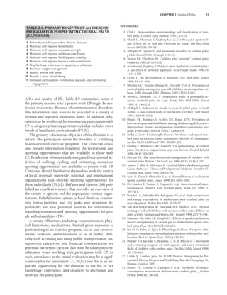
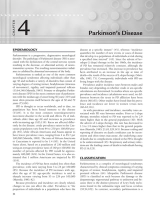
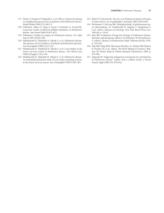
As a means to classify the severity of the disease, one of
the historical scales, which continues to be used by neu-
rologists today, is the Hoehn and Yahr Staging Scale (50).
This original scale stages the progression or severity of the
disease from I to V. The scale is based upon symptoms
being unilateral or bilateral (I), one’s impairment of bal-
ance (II), one’s functional capability in relation to normal
activities (III), employment status (IV), and level of inde-
pendence (V). In the original version, the descriptives
under each stage lack congruity and allow for extreme sub-
jectivity when trying to rate an individual (Table 4.1). Al-
though modified scales have been developed that are more
congruent and less ambiguous (such as the United Parkin-
son’s Disease Rating Scale [32]), they still allow for subjec-
tivity when rating an individual. The latter scale is fre-
quently used clinically and in research as a means of
classifying patients according to the severity of their dis-
ease. Despite its measurement limitations, when reference
is made to a particular stage these scales, permit some con-
cept of where an individual is in the possible progression
of the disease.
PATHOPHYSIOLOGY
As stated previously, IPD is a neurodegenerative process
that can result in movement disorders. In addition, these
symptoms of dysfunctional movement are often accompa-
nied by nonmotor abnormalities, such as cognitive
changes and mood disturbances. The anatomical struc-
ture within the central nervous system known to be a pri-
mary area affected by the disease is the basal ganglia. Col-
lectively, the basal ganglia are thought to control the more
complex aspects of motor planning. In addition, parts of
the thalamus and reticular formation work in close associ-
ation with the above structures and are, therefore, consid-
ered to be part of the basal ganglia system for motor con-
trol. Furthermore, the basal ganglia is anatomically linked
to other parts of the brain that control not only motor and
sensory programs, but cognitive and motivational aspects
of the human body and psyche as well. Therefore, any dis-
ease of the basal ganglia can result in various movement
disorders as well as nonmotor abnormalities.
Although it is not the only area affected by the disease,
the structure within the basal ganglia most vulnerable to
the pathological process of IPD is the substantia nigra.
Widespread destruction of the pigmented neurons in the
substantia nigra pars compacta is associated with IPD,
and as a result of this destruction, the nigrostriatal tract
degenerates. The degeneration of the dopaminergic
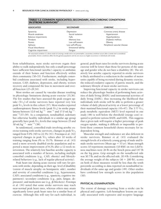
TABLE 4.1. HOEHN AND YAHR STAGING OF
PARKINSON’S DISEASE
Stage 1
1. Signs and symptoms on one side only
2. Symptoms mild
3. Symptoms inconvenient but not disabling
4. Usually presents with tremor of one limb
5. Friends have noticed changes in posture, locomotion, and facial
expression
Stage 2
1. Symptoms are bilateral
2. Minimal disability
3. Posture and gait affected
Stage 3
1. Significant slowing of body movements
2. Early impairment of equilibrium on walking or standing
3. Generalized dysfunction that is moderately severe
Stage 4
1. Severe symptoms
2. Can still walk to a limited extent
3. Rigidity and bradykinesia
4. No longer able to live alone
5. Tremor may be less than earlier stages
Stage 5
1. Cachectic stage
2. Invalidism complete
3. Cannot stand or walk
4. Requires constant nursing care
LWBK191-4034G-C04_44-57.qxd 06/11/2008 10:00 AM Page 45](https://image.slidesharecdn.com/acsmsresourcesforclinicalexercisephysiology-230410064150-fb83bfa2/85/ACSM_s-Resources-for-Clinical-Exercise-Physiology-pdf-59-320.jpg)
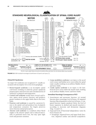
![CHAPTER 5 Spinal Cord Dysfunction 61
innervation, vasomotor paralysis, susceptibility to
hypotension and autonomic dysreflexia, impaired
cough.
• Paraplegia: impaired lower body function.
3. Spastic or flaccid bladder requires catheterization or
urinary collection system for emptying and manage-
ment; risk of urinary incontinence.
4. Bowel constipation; risk of bowel incontinence.
5. Bone demineralization or osteopenia; risk of fracture.
Secondary Conditions and SCI
Secondary medical conditions may further compromise the
health and function of persons with SCI as they age. These
conditions generally increase with time since SCI. The most
prevalent secondary conditions are chronic pain, problem-
atic spasticity, depression, obesity, urinary tract infections,
and pressure sores (9–12). Many years of dependence on
the upper extremities for daily activities (wheelchair or
crutch use, and transfers) makes the shoulders, elbows, and
wrists susceptible to overuse injury, tendon inflammation,
joint degeneration, and pain. Severe spasticity can cause
joint contractures and loss of range of motion. Paralysis of
abdominal (expiratory) musculature impairs cough and in-
creases susceptibility to respiratory infections. Frequent
bladder infections and use of antibiotics can lead to kidney
damage and systemic infection. Inactivity, dyslipidemia,
insulin resistance, and hypertension put long-surviving
persons with SCD at risk for cardio- and cerebrovascular
disease and metabolic disease (13,14).
Exercise-related Consequences of SCI
Spinal cord injury can result in two major exercise-related
problems: (a) reduced ability to perform large-muscle-
group aerobic exercise voluntarily (i.e., without using
functional electrical stimulation leg cycle ergometry [FES-
LCE] with paralyzed leg muscles), and (b) the inability to
stimulate the autonomic and cardiovascular systems to
support higher rates of aerobic metabolism (15,16).
Therefore, catecholamine production by the adrenal
medullae, skeletal muscle venous pump, and thermoregu-
lation (17) may be impaired, which restricts exercise car-
diac output (CO) to subnormal levels. Hopman et al. (18)
examined the properties of the venous vasculature in the
lower extremities in persons with paraplegia. Compared
with non-SCI subjects, they noted lower venous distensi-
bility and capacity and higher venous flow resistance.
They attributed these to vascular adaptations to inactivity
and muscle atrophy rather than the effect of an inoperable
leg muscle pump and sympathetic denervation.
Common secondary complications during exercise,
especially in persons with tetraplegia, may include lim-
ited positive cardiac chronotropy and inotropy, excessive
venous pooling, venous atrophy, orthostatic and exercise
hypotension, exercise intolerance, and autonomic dysre-
flexia. This latter condition is a syndrome resulting from
mass activation of autonomic reflexes causing extreme
hypertension, headache, bradycardia, flushing, goose-
flesh, unusual sweating, shivering, or nasal congestion.
Tetraplegia usually results in a sedentary lifestyle with
profound deconditioning of many physiologic systems.
This exacerbates mobility impairment, bone demineral-
ization, myocardial and skeletal muscle atrophy, and
changes in body composition, such as decreased lean
body mass, body water content, blood volume, and in-
creased percentage of body fat (19).
SPINA BIFIDA
Immediate Neurologic Consequences of SB
Infants with SB generally have surgery within 24 hours of
birth to close the spinal malformation to minimize the risk
of infection and prevent further neurologic damage. About
80% of SB affects the lumbosacral nerve roots (1), resulting
in damage to the lumbar or sacral segments of the cauda
equina from L1 to S4. As with SCI at the same neurologic
level, SB usually results in sensorimotor and autonomic
impairment to the legs, pelvic organs (bladder, bowels, and
sexual organs), or both. The exact neurologic level and
completeness of injury determines the degree of impair-
ment (Figs. 5.2 and 5.3). Also as with SCI, the ASIA Im-
pairment Scale in Table 5.1 can be used to grade the degree
of impairment or completeness in SB at a given level (6,7).
A frequent complication of SB is “hydrocephalus” (an
abnormal accumulation of cerebrospinal fluid (CSF) in
the cavities of the brain, which can lead to increased in-
tracranial pressure and progressive enlargement of the
head, convulsion, and mental disability) occurring in
about 90% of individuals with SB (1). SB impairs proper
absorption and drainage of CSF and allows excessive
accumulation of CSF in the ventricles of the brain. If
ineffectively treated or left untreated, hydrocephalus
compresses the brain and causes brain damage and per-
manent cognitive impairment and learning disabilities.
Most people with SB have a plastic shunt implanted to
drain CSF from the ventricles of the brain under the skin
into the chest or abdomen. Shunts will fail if they become
obstructed; people with SB typically have their failed
shunt replaced twice in their lifetime.
Presence of the Arnold-Chiari malformation, a dis-
placement of the cerebellar tonsils and the medulla
through the foramen magnum, may result in compres-
sion of the brainstem and cerebellum (1). Symptoms in
children or adolescents may include neck pain, changes
in sensorimotor function, or problems with swallowing,
speech, or breathing. The only treatment is surgical de-
compression. Some may exhibit cerebellar dysfunction
with dysmetria, fine motor incoordination, tremors, nys-
tagmus, and ataxic gait. These individuals may experi-
ence difficulty with fine motor skills.
Spinal cord function must be closely monitored
in people with SB. Changes in muscle tone or strength,
LWBK191-4034G-C05_58-78.qxd 06/11/2008 10:02 AM Page 61](https://image.slidesharecdn.com/acsmsresourcesforclinicalexercisephysiology-230410064150-fb83bfa2/85/ACSM_s-Resources-for-Clinical-Exercise-Physiology-pdf-75-320.jpg)
![CHAPTER 5 Spinal Cord Dysfunction 67
in adults with SCI and SB. Both programs involved home-
based exercises to stretch anterior shoulder musculature
and to strengthen posterior shoulder musculature.
Mixed Fitness Training
Hicks et al. (75) conducted a randomized controlled trial
of mixed fitness training (aerobic and resistance exercises
for 9 months, twice weekly) with a diverse group of 23
men and women (ages 19–65 years, SCI levels C4-L1,
ASIA A-D). The training intervention primarily used ACE,
pulley and free weights, and accessible Equalizer weight
machines. Compared with an educational intervention
control group (n 13), the exercise training group signif-
icantly increased submaximal ACE power output by 81%)
and 1-RM strength in upper body muscle groups (19%–
34%). The training group also reported less pain, stress,
and depression after training and scored higher than the
control group on indices of satisfaction with physical func-
tion, perceived health, and quality of life.
Body Weight Supported Treadmill Training (BWSTT)
Early animal studies examining recovery of hindlimb step-
ping after complete low thoracic SCI have evolved into
human trials. An experimental training method for persons
with SCI involves manual positioning of the legs by thera-
pists or robotic devices while subjects perform limb-loaded
stepping movements on a treadmill with their body weight
partially suspended by a harness, with or without electrical
stimulation of paralyzed muscles. Several reports describe
subjects with complete and incomplete SCI recovering ther-
apeutic or functional levels of ambulation (76,77). Nash
et al. (78) demonstrated in a case report that robotically as-
sisted BWSTT can induce slight acute increases in metabolic
rate (by 2.4 metabolic equivalents [METs]) and HR (by 17
bpm). The potential for BWSTT to improve fitness and
health in selected SCI individuals is largely undocumented.
Dobkin et al. (79) reported a recent clinical trial to compare
the functional outcomes of BWSTT with conventional am-
bulation training and concluded no differences between the
interventions in persons with incomplete SCI.
Spina Bifida
Arm Ergometry
Ekblom and Lundberg (80) trained 10 adolescents (7 SB
and 3 SCI, mean age 17 years, 6 female and 4 male) with
wheelchair exercise (30 min/session, 2–3 sessions/wk,
6 weeks). Although V
.
O2peak (1.1 L/min) did not change,
POpeak increased by 5.5 W (10%) to 60 W.
Resistance Training
Although no resistance exercise training studies are pub-
lished that utilized adults with only SB, a few small-scale
studies have documented training responses of children
or adolescents with SB. Andrade et al. (25) found that a
10-week exercise program significantly increased cardio-
vascular fitness, isometric muscle strength, and self-con-
cept in eight children with SB compared with control chil-
dren. Also, O’Connell and Barnhart (81) resistance-trained
three children (ages 4, 5, and 16) with thoracic SB. Train-
ing consisted of seven upper body exercises using free
weights: 30 min/session, 3 sets 6-repetition maximum
(RM), 3 sessions/wk for 9 weeks. All children improved 6-
RM muscular strength by 70%–300%, 50-m dash time by
20%, and 12-min wheelchair propulsion distance by 29%.
Thus, similar to youth without SCD, resistance training
improves strength and general fitness.
PHYSICAL EXAMINATION
To design a program for the participant with SCD ade-
quately, a systematic neurologic examination of the sen-
sory and motor function is required. A well-defined se-
quence is provided in the International Standards for
Neurological and Functional Classification of Spinal Cord
Injury (6). Beyond motor and sensory evaluation, atten-
tion must be paid to joint range of motion (ROM), spas-
ticity status, and skin integrity. Programs will need to be
tailored depending on contracture status, severe spasticity
or flaccidity, and the presence of open pressure sores in
weight-bearing areas. Some people with SCI require com-
prehensive pain management for chronic dysesthetic,
spinal, or upper-extremity overuse syndromes. These and
other functional tests (82,83) may include joint flexibility
or ROM; manual muscle testing to determine muscle im-
balance and risk of contracture; testing of reflexes, muscle
tone, and spasticity; equipment evaluation (wheelchair
and cushion, assistive and orthotic devices); home evalu-
ation for accessibility and modification; and psychological
evaluation to promote adjustment or coping and to assess
or control depression and substance abuse. Because of
susceptibility to pressure sores, people with SCD also
need to perform frequent inspections of insensitive
weight-bearing skin areas to assess skin integrity.
MEDICAL AND SURGICAL TREATMENTS
Multiple medical, nursing, and allied health professional
services are utilized during SCD rehabilitation (84).
Shortly after injury, neurosurgery, orthopedic surgery, or
both are usually necessary to stabilize spinal fractures
and dislocations. Internal fixation devices and fusion
(rodding, plating, screws, bone grafts) are often neces-
sary to accomplish this after traumatic SCI. The instru-
mentation and postsurgical healing must be adequate to
withstand exercise demands. External spinal orthoses,
such as halo, and other spinal orthoses are common for
several weeks after surgery to stabilize the healing spine.
Other orthopedic injuries often acquired during trau-
matic SCI include limb fracture and closed head injury.
LWBK191-4034G-C05_58-78.qxd 06/11/2008 10:02 AM Page 67](https://image.slidesharecdn.com/acsmsresourcesforclinicalexercisephysiology-230410064150-fb83bfa2/85/ACSM_s-Resources-for-Clinical-Exercise-Physiology-pdf-81-320.jpg)
![CHAPTER 5 Spinal Cord Dysfunction 69
antithrombics (anticoagulation, e.g., warfarin), and an-
tibiotics (e.g., Bactrim). Neurogenic bladder treatment
may require alpha-blocking agents that induce hypoten-
sion, especially in persons with tetraplegia (86). Persons
with a history of deep venous thrombosis may be taking
warfarin, which leads to easy bruisability. Aging persons
with SCD are at risk for cardiovascular and metabolic dis-
ease and may take medications for hypertension, diabetes,
dyslipidemia, dysrhythmia, and congestive heart failure.
DIAGNOSTIC PROCEDURES
The physician will judge the necessity and extent of initial
diagnostic procedures that will depend on the prospective
participant’s documented medical history and physical ex-
amination. If FES-LCE exercise will be utilized in exercise
programming, the participant’s file should include baseline
radiographs (including plain x-rays, scans for osseous tis-
sue, and magnetic resonance imaging [MRI] for soft tis-
sues) showing adequacy of spinal alignment and integrity
of internal stabilization. Baseline pulmonary function tests
are desirable for those with tetraplegia or tetraparesis. The
nature and extent of the changes in ventilatory function
and cough depend to a great extent on level of neurological
injury or dysfunction. If the client is middle-aged or older
or if sufficient coronary risk factors exist, electrocardio-
grams (ECG) and myocardial perfusion tests should be ob-
tained as baseline evaluations of the participant’s cardiac
status. Also, laboratory analysis of baseline hematologic
and metabolic status would be useful, including a complete
blood count, electrolytes, renal indices, thyroid and liver
function, lipid panel, fasting blood sugar, and glucose toler-
ance. Urodynamic evaluation is necessary to assess bladder
responses to filling and emptying (e.g., voiding cys-
tourethrogram). Finally, because osteoporosis is common
below the level of injury in SCD and, with immobilization
in other conditions, consider evaluation of bone mineral
status (bone densitometry) as part of the initial workup.
EXERCISE/FITNESS/FUNCTIONAL
TESTING
GUIDELINES
During rehabilitation of persons with SCD, functional
testing takes priority over physiologic testing to promote
functional independence at the fastest rate possible. For
example, rehabilitation goals usually include independent
mobility via weight-bearing or wheelchair ambulation,
transfers, and self-care with or without assistive devices.
The fitness requirements of these tasks are specific to the
functional tasks themselves. The cardiovascular and
metabolic demands of walking and wheelchair ambula-
tion are the greatest of all functional tasks, hence, the im-
portance of exercise tolerance and capacity during reha-
bilitation. Aerobic fitness is necessary for long-distance
mobility, some recreational activities, competitive sports,
and long-term cardiovascular health. Neuromuscular co-
ordination and skill, balance and stability, and muscular
strength and endurance are necessary to various degrees
for safe standing, ambulation, transfers, driving, and other
self-care activities.
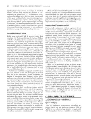
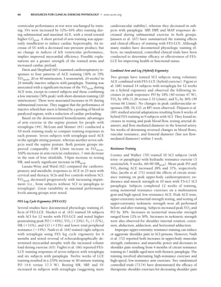
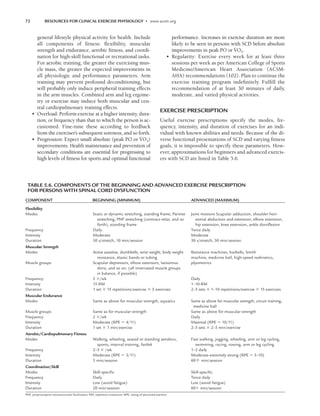
Table 5.5 lists relative and absolute contraindications
for cardiovascular exercise testing of persons with SCD.
These are the same as for people without disabilities and
include several disability-specific conditions.
Advice from the person with SCD concerning exercise
modes and proper positioning or strapping is often use-
ful. Adapt the exercise equipment, as needed, and pro-
vide for the following special needs (87):
TABLE 5.5. DISABILITY-SPECIFIC RELATIVE AND ABSOLUTE CONTRAINDICATIONS FOR EXERCISE
TESTING OF PERSONS WITH SPINAL CORD INJURY (SCI) AND SPINA BIFIDA (SB)
SCI
RELATIVE Tetraplegia Paraplegia SB
Asymptomatic hypotension X
Muscle and joint discomfort X X X
ABSOLUTE
Autonomic dysreflexia X
Severe or infected skin pressure sore on weight-bearing skin areas X X X
Symptomatic hypotension (dizziness, nausea, palor, extreme fatigue, X
visual disturbance, confusion)
Illness caused by acute urinary tract infection X X X
Uncontrolled spasticity or pain X X X
Unstable fracture X X X
Uncontrolled hot humid environments X
Inability to safely seat and stabilize the person on well-cushioned or padded X X X
ergometers or equipment
Insufficient range of motion to perform exercise task X X
X, special relevance to SCI or SB.
LWBK191-4034G-C05_58-78.qxd 06/11/2008 10:02 AM Page 69](https://image.slidesharecdn.com/acsmsresourcesforclinicalexercisephysiology-230410064150-fb83bfa2/85/ACSM_s-Resources-for-Clinical-Exercise-Physiology-pdf-83-320.jpg)
![76 RESOURCES FOR CLINICAL EXERCISE PHYSIOLOGY • www.acsm.org
REFERENCES
1. Nelson MR, Rott EJ. Spina bifida. In: Grabois M, Garrison SJ, Hart
KA, et al., eds. Physical Medicine and Rehabilitation: The Complete
Approach. Malden, MA: Blackwell Science; 2000:1414–1432.
2. Cardenas DD, Burns SP
, Chan L. Rehabilitation of spinal cord in-
jury. In: Grabois M, Garrison SJ, Hart KA, et al, eds. Physical Med-
icine and Rehabilitation: The Complete Approach. Malden, MA:
Blackwell Science; 2000:1305–1324.
3. Yarkony GM, Chen D. Rehabilitation of patients with spinal cord
injuries. In: Braddom RJ, ed. Physical Medicine and Rehabilitation.
Philadelphia: W.B. Saunders; 1996:1149–1179.
4. Mallory B. Autonomic dysfunction in spinal cord disease. In: Lin
VW, ed. Spinal Cord Medicine: Principles and Practice. New York:
Demos; 2003:477–500.
5. Goshgarian HG. Anatomy and function of the spinal cord. In: Lin
VW, ed. Spinal Cord Medicine: Principles and Practice. New York:
Demos; 2003:15–34.
6. ASIA. International Standards for Neurological Classification of
SCI. Atlanta: American Spinal Injury Association; 2002.
7. ASIA. Standard neurological classification of spinal cord injury.
Atlanta: American Spinal Injury Association; 2006. (accessed
online, 2-17-08: http://www.asia-spinalinjury.org/publications/
2006_Classif_worksheet.pdf)
8. Consortium for Spinal Cord Medicine. Outcomes following trau-
matic spinal cord injury: Clinical practice guidelines for healthcare
professions. Washington, DC: Paralyzed Veterans of America; 1999.
9. Furhrer MJ. Rehabilitation and research training center in com-
munity-oriented services for persons with spinal cord injury: A
progress report. Houston, TX: The Baylor College of Medicine and
The Institute for Rehabilitation Research; 1991.
10. Whiteneck GG. Learning from recent empirical investigations. In:
Whiteneck GG, Charlifue SW, Gerhart KS, et al., eds. Aging with
Spinal Cord Injury. New York: Demos Publications; 1993.
11. Anson CA, Shepherd C. Incidence of secondary complications in
spinal cord injury. Int J Rehabil Res 1996;19:55–66.
12. Johnson RL, Gerhart KA, McCray J, et al. Secondary conditions
following spinal cord injury in a population-based sample. Spinal
Cord 1998;36:45–50.
13. Bauman WA, Spungen AM. Metabolic changes in persons after
spinal cord injury. Phys Med Rehabil Clin N Am 2000;11:109–140.
14. Washburn RA, Figoni SF
. Physical activity and chronic cardiovas-
cular disease prevention: A comprehensive literature review. Top
Spinal Cord Injury Rehabil 1998;3:16–32.
15. Davis GM. Exercise capacity of individuals with paraplegia. Med
Sci Sports Exerc 1993;25:423–432.
16. Figoni SF
. Exercise responses and quadriplegia. Med Sci Sports
Exerc 1993;25:433–441.
17. Price MJ. Thermoregulation during exercise in individuals with
spinal cord injuries. Sports Med 2006;36:863–879.
18. Hopman MTE, Nommensen E, van Asten WNJC, et al. Properties
of the venous vascular system in the lower extremities on individ-
uals with parapelgia. Paraplegia 1994;32:810–816.
19. Bauman WA. Endocrinology and metabolism after spinal cord in-
jury. In: Kirshblum S, Campagnolo DI, DeLisa JA, eds. Spinal Cord
Medicine. Philadelphia, PA: Lippincott Williams Wilkins; 2002:
164–180.
20. Kelly LE. Spinal cord disabilities. In: Winnick JP
, ed. Adapted Phys-
ical Education and Sport. 4th ed. Champaign, IL: Human Kinetics;
2005:275–306.
21. Hart AL, Malone TR, English T. Shoulder function and rehabilita-
tion implications for the wheelchair athlete. Top Spinal Cord Injury
Rehabil 1998;3:50–65.
22. Winnick JP
, Short FX. The physical fitness of youngsters with spinal
neuromuscular conditions. Adapted Phys Activ Q 1984;1:37–51.
23. Shephard RJ. Fitness in Special Populations. Champaign, IL: Hu-
man Kinetics, 1990.
24. Winnick JP
, Short FX. The Brockport Physical Fitness Test Manual.
Champaign, IL: Human Kinetics; 1999.
25. Andrade CK, Kramer J, Garber M, et al. Changes in self-concept, car-
diovascular endurance and muscular strength of children with spina
bifida aged 8 to 13 years in response to a 10-week physical-activity
programme: A pilot study. Child Care Health Dev 1991;17:183–196.
26. Morrison SA, Melton-Rogers SL, Hooker SP
. Changes in physical
capacity and physical strain in persons with acute spinal cord in-
jury. Top Spinal Cord Injury Rehabil 1997;3:1–15.
27. Hjeltnes N, Jansen T. Physical endurance capacity, functional sta-
tus and medical complications in spinal cord injured subjects with
long-standing lesions. Paraplegia 1990;428–432.
28. Stewart MW, Melton-Rogers SL, Morrison S, et al. The measure-
ment properties of fitness measures and health status for persons
with spinal cord injuries. Arch Phys Med Rehabil 2000;81:394–400.
29. Raymond J, Davis GM, Fahey A, et al. Oxygen uptake and heart rate
responses during arm vs combined arm/electrically stimulated leg
exercise in people with paraplegia. Spinal Cord 1997;35:680–685.
30. Haisma JA, van der Woude LH, Stam HJ, Bergen MP
, Sluis TA,
Bussmann JB. Physical capacity in wheelchair-dependent persons
with a spinal cord injury: A critical review of the literature. Spinal
Cord 2006;44(11):642–652.
31. Glaser RM. Exercise and locomotion for the spinal cord injured.
Exerc Sports Sci Rev 1985;13:263–303.
b. Lateral pull-downs (pulleys)
c. Rowing (pulleys)
d. Incline press (against light machine weights)
e. Bench press (weight machine)
Day 1: Assessments: Determine 10-RM for each
exercise, and perform Wingate anaerobic power test
(maximal work performed during maximal-effort, high-
intensity arm-cranking for 30 seconds).
Day 2: Training: 2 sets 10 repetitions at 90%
10-RM
Progress according to principles of progressive resis-
tive exercise and principles of overload, progression,
specificity, regularity, and so forth.
4. Aerobic Exercise: Choose two to three acceptable ac-
cessible available modes (e.g., arm-crank ergometry):
a. Find workload (20–30 W) that elicits RPE
4/11 for at least 5 minutes or until arm muscle
fatigue or dizziness.
b. Perform at least two assessments on different days
to establish training baselines. Example:
1) Maximal work performed during 6-minute period
2) ACE field test
c. Progress over time to 30–60 minutes per workout.
d. Vary direction of cranking (forward versus backward).
e. Vary intensities and durations of workouts (i.e.,
shorter durations with higher intensity [intervals]
versus long, slow distance).
f. Watch videos or find partner to combat boredom
of arm ergometry.
g. Repeat assessments every 2 weeks.
LWBK191-4034G-C05_58-78.qxd 06/11/2008 10:02 AM Page 76](https://image.slidesharecdn.com/acsmsresourcesforclinicalexercisephysiology-230410064150-fb83bfa2/85/ACSM_s-Resources-for-Clinical-Exercise-Physiology-pdf-90-320.jpg)
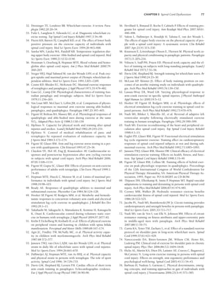
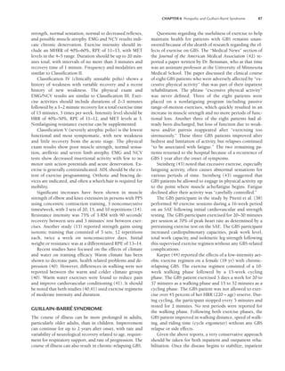
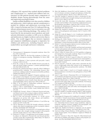
![91
EPIDEMIOLOGY AND
PATHOPHYSIOLOGY
Myopathies encompass a wide range of disorders that can
be broadly considered as either congenital, inherited, or
acquired. The congenital forms include the muscular dy-
strophies, congenital myopathies, channelopathies, and
metabolic myopathies (Table 7.1). The acquired my-
opathies include endocrine myopathies, inflammatory
myopathies, toxic myopathies (including drugs), and pri-
mary infectious myopathies (Table 7.2). The incidence
and prevalence of muscle disorders varies widely, with
myotonic dystrophy and Duchenne dystrophy (DMD)
ocurring in about 1 of 3,500 live births (DMD only in
men), whereas other disorders, such as MELAS 3271 (mi-
tochondrial myopathy), is present in only about 40 peo-
ple in the world (10,42).
Most of the genetically based myopathies are caused
by mutations in structural or metabolism linked proteins.
These can be categorized as autosomal dominant (AD),
autosomal recessive (AR), X-linked recessive (XR), ma-
ternally inherited (MI, mitochondrial disorders), or spo-
radic spontaneous mutations. In general, the dystrophies
often result from abnormalities in the cytoskeleton (e.g.,
dystrophin in DMD), whereas the metabolic myopathies
are caused by mutations in the energy transduction en-
zymes (e.g., carnitine palmitoyl transferase deficiency
[CPT-2]). Some exceptions to this general rule exist,
such as calpain-3 deficiency (a proteolytic enzyme) seen
in a form of AR limb girdle dystrophy. The inheritance
pattern and gene product of many of the inherited my-
opathies are presented in Table 7.3.
The acquired myopathies encountered in North Amer-
ica are usually inflammatory, endocrine, or toxic. Acute
viral myopathies, which are frequently encountered with
a wide spectrum of severity, are most commonly caused
by influenza or coxsackie viruses. The most common
toxic myopathies are those caused by lipid-lowering
agents, primarily the statins (hydroxymethylglutaryl-
coenzyme A [HMG-CoA]-reductase inhibitors) (36).
Fungal and bacterial myopathies rarely occur in first-
world countries, but should be considered in recent im-
migrants or those with travel histories to developing
countries (Table 7.2).
SYMPTOMS AND FUNCTIONAL
CONSEQUENCES
Progressive muscle weakness is the most common symp-
tom of structural myopathies, and episodic fatigability is
the most common symptom of the metabolic myopathies
and channelopathies. Most of the congenital myopathies
or dystrophies start with weakness in the proximal mus-
cles that eventually spreads more distally. Notable excep-
tions are fascioscapulohumeral dystrophy (FSHD), where
facial weakness may be a prominent and early sign, my-
opathies with distal weakness including the “distal my-
opathies,” and myotonic muscular dystrophy. In general,
the disorders of fat metabolism and mitochondrial cy-
topathies present with impairment in endurance-type ac-
tivities, whereas glycogen storage diseases present with
symptoms during higher-intensity muscle contractions.
A common outcome from prolonged weakness is joint
contracture and secondary bony abnormalities, including
osteoarthritis, osteoporosis, and scoliosis. Contractures
are most commonly seen at the shoulders, elbows, an-
kles, knees, and hips. Several of the myopathies can also
affect cardiac muscle with conduction block (e.g., myo-
tonic dystrophy) and cardiomyopathy (dystrophinopa-
thy, Emery-Dreifuss) (Table 7.4).
In general, because most myopathies initially affect
proximal limb muscles, early in the course of the disease
individuals with these conditions will often experience
difficultly arising from a low chair, climbing stairs, or ris-
ing from a kneeling position or from the ground (e.g.,
after a fall). As weakness progresses, gait disturbance and
limitations in functional mobility become more problem-
atic. Most children with DMD become wheelchair de-
pendent for mobility by 10 to 12 years of age. The re-
quirement for power mobility is extremely variable in
other forms of acquired and inherited myopathy.
PHYSICAL EXAMINATION
As stated above, most people with myopathy will have
significant proximal weakness without any sensory signs
or symptoms as primary manifestations of their disorder.
Mental status is usually unaffected, with the exception of
some children with DMD, myotonic muscular dystrophy
Muscular Dystrophy and
Other Myopathies
7
CHA
P
TER
LWBK191-4034G-C07_91-103.qxd 06/11/2008 10:03 AM Page 91](https://image.slidesharecdn.com/acsmsresourcesforclinicalexercisephysiology-230410064150-fb83bfa2/85/ACSM_s-Resources-for-Clinical-Exercise-Physiology-pdf-105-320.jpg)
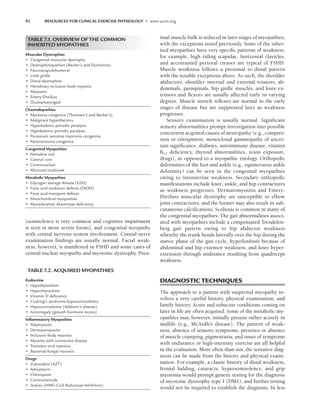
![CHAPTER 7 Muscular Dystrophy and Other Myopathies 93
classic cases, further testing is required, including serum
chemistries for creatine kinase (CK) activity, electrolytes,
and thyroid function as an initial screen. The finding of
rash, joint involvement, or other autoimmune features
would prompt further investigation for underlying con-
nective tissue disease as part of the initial investigation of
a potential inflammatory myopathy. In suspected meta-
bolic myopathy, forearm ischemic testing may be helpful
in showing absence of lactate rise with normal ammonia
rise (a classic glycogen storage disease pattern), or normal
lactate rise with absent ammonia rise (myoadenylate
deaminase deficiency). Evaluation of suspected free fatty
acid (FFA) oxidation defects, FFA transport abnormalities,
and mitochondrial cytopathy requires referral to tertiary
and quaternary centers that are skilled in the assessment
of these disorders (e.g., Institute for Exercise and Envi-
ronmental Medicine, Southwestern Medical Center, Uni-
versity of Texas, Dallas, Texas; Neuromuscular and Neu-
rometabolic Clinic, McMaster University Medical Center,
Hamilton, Ontario).
Most people with a suspected acquired myopathy
should have a number of routine investigations, includ-
ing a complete blood count, CK, creatinine, liver en-
zymes, thyroid function (thyroid-stimulating hormone
[TSH]), antinuclear antibody (ANA), and an erythrocyte
sedimentation rate (ESR). With any suspicion of respira-
tory involvement, both sitting and supine pulmonary
function studies and oximetry are of value. People with
suspected cardiac involvement or with possible conduc-
TABLE 7.3. MUSCULAR DYSTROPHIES/CONGENITAL MYOPATHIES/CHANNELOPATHIES
INHERITANCE PATTERN GENE PRODUCT SYMBOL
Duchenne muscular dystrophy XR Dystrophin DYS-DMD
Becker’s muscular dystrophy XR Dystrophin DYS-BMD
Limb girdle muscular dystrophy AR Dysferlin LG-MD2B
AR -sarcoglycan LG-MD2D
AR -sarcoglycan LG-MD2E
AR -sarcoglycan LG-MD2C
AR Calpain-3 LG-MD2A
Limb girdle muscular dystrophy (dominant) AD Caveolin-3 LG-MD1C
AD Laminin A/C LG-MD1B
Distal Muscular Dystrophy
• Miyoshi AR Dysferlin MM
• Miscellaneous (Nonaka, Udd, and Bethlem)
Hereditary inclusion body myopathy AR ?
IBM2
Oculopharyngeal muscular dystrophy AD Poly(A) binding OPMD
Protein
Congenital Myopathy/Dystrophy
• Myotubular myopathy XR Myotubularin MTMX
• Central core AD Ryanodine receptor CCD
• Nemaline myopathy AD -tropomyosin NEM1
AR Nebulin NEM2
• Emery-Dreifuss XR Emerin EMD1
AD Laminin A/C EMD2
Fascioscapulohumeral AD ? FSHD
Myotonic muscular dystrophy–1 AD Myotonin DM
Proximal myotonic myopathy AD Zinc finger protein 9 DM2
Congenital muscular dystrophy AR -2 laminin LAMA2
AR Integrin 7 ITG7
Congenital muscular dystrophy AR Fukutin FCMD
Channelopathies
Malignant hyperthermia AD Ryanodine receptor MHS1
Myotonia congenita AD Chloride channel CLC-1
AR Chloride channel CLC-1
Hyperkalemic periodic paralysis AD Sodium channel SCN4A
subunit
Hypokalemic periodic paralysis AD Dihydropyridine receptor CACNL1A3
TABLE 7.4. MYOPATHIES ASSOCIATED WITH
CARDIAC ABNORMALITIES
MYOPATHY CARDIAC DEFECT
Dystrophinopathies (Becker’s Cardiomyopathy
and Duchenne)
Myotonic dystrophy, types I II Cardiac conduction block
Emery-Dreifuss muscular dystrophy Cardiac conduction block
Nemaline rod myopathy Cardiomyopathy
Centronuclear myopathy Cardiomyopathy
LWBK191-4034G-C07_91-103.qxd 06/11/2008 10:03 AM Page 93](https://image.slidesharecdn.com/acsmsresourcesforclinicalexercisephysiology-230410064150-fb83bfa2/85/ACSM_s-Resources-for-Clinical-Exercise-Physiology-pdf-107-320.jpg)
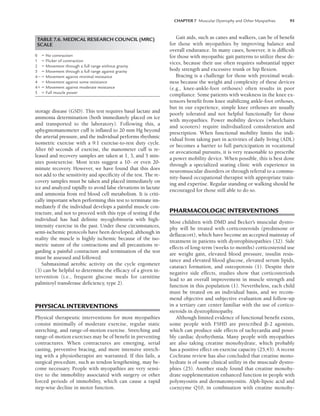
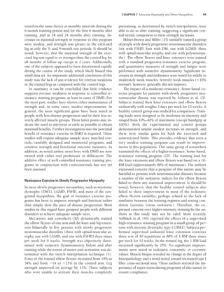
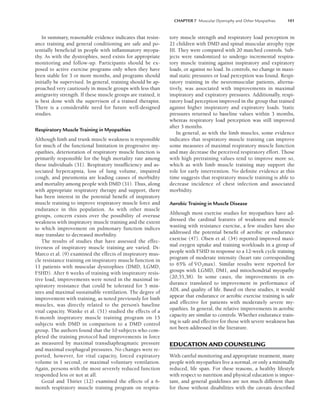
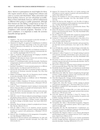

![104
Peripheral Neuropathy and
Neuropathic Pain
8
C
HA
P
TER
PATHOPHYSIOLOGY
Pain is a common experience, and the capacity to sense
pain plays a protective role in warning of current or po-
tential tissue damage. Response to tissue injury (as well
as painful stimuli) includes adaptive changes that pro-
mote healing and avoid further irritation to the injured
tissue.
Peripheral neuropathy is defined as deranged function
and structure of peripheral motor, sensory, and auto-
nomic neurons (outside the central nervous system
[CNS]), involving either the entire neuron or selected
levels (1). Pain and other sensory changes can be pro-
duced in a variety of ways and, in peripheral nerves,
motor or sensory fibers may be preferentially affected,
but in most neuropathies both are involved, leading to
various patterns of sensorimotor deficit (2). Pain is not
always associated with peripheral neuropathy. Painful
neuropathies are usually a result of damage to the axon,
in contrast to demyelinating neuropathies that tend, with
some exceptions, to cause pronounced motor or sensory
loss without pain (3).
The two basic mechanisms by which the experience of
pain can be evoked are somatic tissue injury (nociceptive)
or nerve injury (4). In the typical process of nociceptive
pain, stimulation of tissue nociceptors (found through-
out the musculoskeletal system) causes action potentials
to be propagated along nociceptive axons that ultimately
enter the limbic sectors of the cerebral cortex where the
pain is perceived. Nociceptive pain is typically described
by patients as deep, tender, dull, aching, and diffuse (4).
Pain induced by nociceptive pain mechanisms is the most
common variety seen in clinical practice (5).
In contrast to nociceptive pain syndromes that are typi-
cally related to musculoskeletal damage or injury, damage
to the peripheral or CNS produces a different type of pain.
This type of pain is known as neuropathic pain. Often neu-
ropathic pain is chronic and persistent, and patients are
more likely to use adjectives such as shooting, stabbing,
lancinating, burning, and searing to describe it and often
complain of pain worsening at night (6). Patients with
neuropathic pain may appear to have continuous or parox-
ysmal pain without any detectable relationship to stimulus
(7). The symptoms can be divided into those that are un-
provoked and those that are provoked by maneuvers such
as skin stimulation, pressure over affected nerves, changes
in temperature, or emotional factors (2).
One key diagnostic feature of neuropathic pain is the
presence of pain within an area of sensory deficit (8). Al-
lodynia is also a commonly seen hyperpathic state in neu-
ropathic pain, in which a normally innocuous stimulus
produces a sensation of pain whose quality is inappropri-
ate for the stimulus. An example of this is the patient who
cannot tolerate a blanket resting on an affected extremity.
Another feature suggestive of neuropathic pain is summa-
tion, which is the progressive worsening of pain evoked by
slow, repetitive stimulation with mildly noxious stimuli, for
example, pinprick (8).
Neuropathic pain can be classified on the basis of the
cause of the insult to the nervous system, the disease or
event that precipitated the pain syndrome, or the distri-
bution of pain. Certain medical conditions are associated
with neuropathic pain, and these commonly include dia-
betes, human immunodeficiency virus (HIV) infection or
acquired immunodeficiency syndrome (AIDS), multiple
sclerosis, cancer chemotherapy, malignancy, spinal sur-
gery, alcoholism, herpes zoster, and amputation (6). Can-
cer patients can develop neuropathy from tumor invasion
and also are at higher risk for neuropathic pain following
chemotherapy or radiation therapy.
Diabetes is associated with peripheral neuropathy and
radiculopathy (9,10). HIV is associated with a variety of
neuropathies and myelopathies. Multiple sclerosis is asso-
ciated with neuralgia and neuropathy (11). Failed spine
surgery is associated with radiculopathy. Amputation is
associated with neuroma and phantom limb pain (6).
Trauma can lead to the development of entrapment neu-
ropathies, as well as partial or complete nerve transection,
plexopathies, and painful scars. Entrapment neuropathies,
such as carpal tunnel syndrome, are usually characterized
in the early stages by paresthesia and pain (2).
CLASSIFICATION
A variety of disease processes can cause, or contribute to,
peripheral neuropathy and the possible pain associated
with it (12). A working knowledge of these is needed to
help identify or establish a specific cause or underlying
process and develop a prognosis and treatment plan
LWBK191-4034G-C08_104-112.qxd 06/11/2008 10:03 AM Page 104](https://image.slidesharecdn.com/acsmsresourcesforclinicalexercisephysiology-230410064150-fb83bfa2/85/ACSM_s-Resources-for-Clinical-Exercise-Physiology-pdf-118-320.jpg)
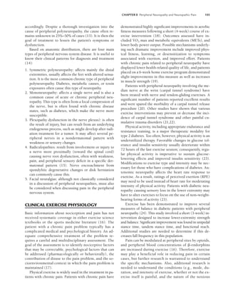
![106 RESOURCES FOR CLINICAL EXERCISE PHYSIOLOGY • www.acsm.org
stimulus) under which exercise-related analgesia is
produced (16).
PHARMACOLOGY
A variety of pharmacologic approaches to the manage-
ment of neuropathic pain are in use, although no current
consensus exists on optimal strategy. Pharmacologic
management is based on the response of the patient and
careful titration of dosage to achieve positive results
while minimizing side effects. Pharmacologic treatments
have generally been selected on the basis of evidence for
efficacy in randomized, placebo-controlled trials con-
ducted in disease-based groups of patients, notably in
postherpetic neuralgia and diabetic polyneuropathy (25).
These studies demonstrate efficacy of tricyclic antide-
pressants, standard and newer anticonvulsants, and opi-
oids. Evidence for efficacy is less for selective serotonin
reuptake inhibitors (SSRIs), antiarrhythmics (mexile-
tine), and capsaicin. Nonopioid analgesics (aspirin, acet-
aminophen, and other nonsteroidal anti-inflammatory
drugs [NSAIDs]) are often used by patients with neuro-
pathic pain, but they are typically more effective for mild
to moderate nociceptive pain. Emerging treatments in-
clude pharmocologic blockade of specific ion receptors,
ligand-gated channels, and nicotinic receptors, and in-
creasing spinal inhibition (26).
Many patients with mild to moderate pain are helped
by the use of aspirin, acetaminophen, and NSAIDs. Ac-
etaminophen is often used initially because of its lower
incidence of gastrointestinal toxicity when taken chroni-
cally. It has no anti-inflammatory effect and is not appro-
priate as a primary therapy for an inflammatory process.
A wide variety of NSAIDs are in use, and many patients
respond better to one over another, although this is un-
predictable in advance of starting on the drug. NSAID use
is associated with an increased incidence of gastrointesti-
nal toxicity and should be avoided in patients with peptic
ulcer disease or bleeding abnormalities (27). NSAIDs
should also be avoided in patients older than 70 years of
age and as unsupervised long-term treatment (28). Pa-
tients with neuropathic pain will typically be engaged in
low-level exercise programs, but it is important to ac-
knowledge a recent study that demonstrated that use of
NSAIDs was found to be a risk factor in exercise-associated
hyponatremia in high-level physical activity (29).
Many patients with neuropathic pain experience relief
with the use of antidepressants. Pain relief is often
achieved at doses that are subtherapeutic for the treat-
ment of depression. The analgesic effects of these agents
may result from their ability to block reuptake at presy-
naptic nerve endings of neurotransmitters, such as sero-
tonin and norepinephrine, which are involved in pain
and depression (30). Tricyclic antidepressants remain the
class most commonly used, and their use has been docu-
mented in a variety of pain-related conditions, including
postherpetic neuralgia, diabetic neuropathy, arthritis, low
back pain, myofascial pain, cancer pain, migraine and
tension-type headaches, central pain, and psychogenic
pain (30). Common side effects include sedation and dry
mouth. In light of recent reports of sudden death in chil-
dren being treated with desipramine (tricyclic antide-
pressant), three of which were associated with physical
exercise, the effects of this drug on exercise were evalu-
ated (31). Desipramine was found to have only minor ef-
fects on the cardiovascular response to exercise, and the
effects did not appear to be age related (31). The authors
stated that desipramine may increase the risk of exercise-
associated arrhythmias in some individuals.
When the previously described drugs are not sufficient,
the addition of an anticonvulsant drug to the antidepres-
sant may be useful (32). Anticonvulsants are generally
considered to be first-line agents for the treatment of neu-
ropathic pain with a predominantly paroxysmal or lanci-
nating quality, such as trigeminal neuralgia (30). The
mechanisms by which anticonvulsants relieve pain may
be related to stabilizing neuronal membranes (pheny-
toin), altering sodium channel activity (carbamazepine),
and modulating -aminobutyric acid activity (clon-
azepam and valproate) (30). Carbamazepine (anticonvul-
sant) produced a significant delay in pain increase in pa-
tients with peripheral neuropathy that was previously
mediated by spinal cord stimulation (33). In this study,
the pain-relieving spinal cord stimulation was inactivated
before introduction of medication. Anticonvulsant med-
ication demonstrated better delay in return of pain com-
pared with an opioid analgesic.
The role of opioid analgesics is controversial in the man-
agement of neuropathic pain. Most patients do not respond
to these drugs and should not receive them (32). Many pa-
tients need detoxification from opioids, sedative-hypnotics,
and muscle relaxants (32). Some evidence indicates that
some patients with neuropathic pain do function well while
taking low-dose, regularly scheduled opioids (32).
Neuropathic pain with a major cutaneous component
may respond well to topical therapy with the substance P
depletor, capsaicin, to reduce elevated prostaglandin
levels (32). Animal studies are demonstrating a rationale
for the selected use of topical capsaicin in the treatment
of pain (34).
Chronic pain and depression are often found to coex-
ist. The use of tricyclic antidepressants in higher thera-
peutic doses is indicated in these cases. SSRIs, such as
fluoxetine (Prozac), paroxetine (Paxil), and sertraline
(Zoloft), have generally replaced tricyclic antidepressants
for the treatment of depression (30). Although they are
typically well tolerated with fewer side effects, SSRIs have
not shown significant efficacy for treatment of pain.
Sometimes, patients with chronic pain and depression are
placed on bedtime dosing of tricyclics and concomitant
daytime dosing of SSRIs. Caution is required because
LWBK191-4034G-C08_104-112.qxd 07/11/2008 05:29 AM Page 106](https://image.slidesharecdn.com/acsmsresourcesforclinicalexercisephysiology-230410064150-fb83bfa2/85/ACSM_s-Resources-for-Clinical-Exercise-Physiology-pdf-120-320.jpg)
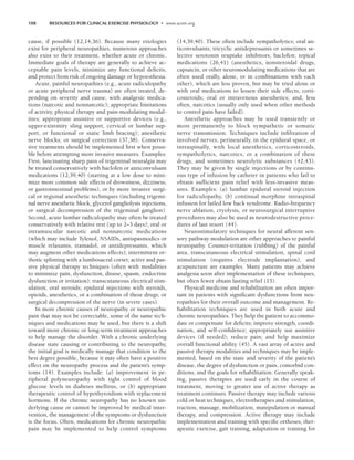
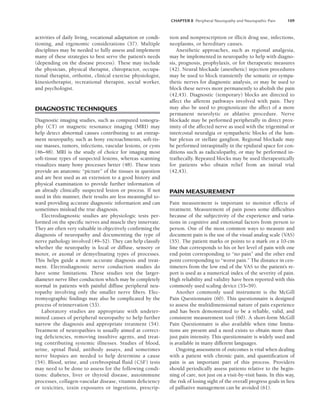
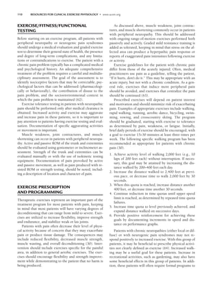
![114 RESOURCES FOR CLINICAL EXERCISE PHYSIOLOGY • www.acsm.org
usually equilibrium in pressure among tissue, blood, and
cerebrospinal fluid compartments. Should any one of
these compartments enlarge, the intracranial pressure will
increase and there is a risk of decreasing cerebral blood
flow, as well as tissue injury taking place as the swollen
brain attempts to squeeze across or through rigid tissues
or bony areas (herniation). The disruptive events at the
cellular level include the release of excitatory neurotrans-
mitters (so-called “excitotoxins”), free radicals, lipid per-
oxidases, and a variety of other proinflammatory media-
tors that can cause widespread, ongoing neuronal death
for a period of time after injury. Current acute treatment
for TBI is generally based on interventions directed to-
ward limiting these secondary consequences by control-
ling intracranial pressure, evacuating space-occupying
blood collections, or inhibiting the toxic cellular cascade
with treatments such as hypothermia (5,10). Despite
many promising animal laboratory findings, there are no
currently available, clinically effective, drug treatments
available to limit the secondary injury cascade.
SEVERITY OF INJURY
Traumatic brain injury severity is typically graded ac-
cording to the degree of compromise of neurologic func-
tion resulting from the injury. This grading can be done
in reference to the person’s degree of responsiveness and
ability to interact with the environment, or the duration
of the characteristic period of anterograde amnesia that
accompanies TBI. The most common way of grading re-
sponsiveness is the Glasgow Coma Scale (GCS). Initially
developed to allow accurate and reliable communication
of functional state (59), the GCS score ranges from 3 to
15 on three subscales based on eye-opening, best motor
response, and best verbal response. A GCS score of 8 or
less constitutes coma and represents severe brain injury, a
score of 9–12 constitutes moderate brain injury, and a
score of 13–15, mild brain injury. Some controversy re-
mains surrounding the accepted definition of mild TBI
(52), perhaps because disabilities associated with this
condition are often subtle and difficult to diagnose. It is
commonly accepted that injury to the brain may occur
without loss or significant alteration of consciousness.
About 80% of TBI falls into the mild category, with the re-
maining 20% representing moderate to severe injury.
Mild TBI frequently occurs in contact sports, such as ice
hockey, football, boxing, and soccer. These injuries have
been reported to alter the acute cardiovascular responses
(autonomic control) during exercise (5). Research has in-
dicated that even mild TBI can cause premature fatigue,
thereby affecting the person’s ability to perform routine
(43,73) activities of daily living (ADL).
The duration of posttraumatic amnesia (PTA) has also
been used to grade injury severity, with mild TBI defined
as PTA less than 1 hour and extremely severe TBI defined
as PTA lasting longer than 1 month (43). Both the initial
GCS score and the PTA duration are correlated (al-
though not perfectly) with functional outcome following
brain injury. In general, lower GCS scores and longer du-
rations of PTA are associated with poorer outcome and
increased disability. It should be noted that 10%–15% of
even mild TBI survivors have persisting symptoms and
impairments (52).
DIAGNOSTIC TECHNIQUES
AND NEUROIMAGING
A variety of techniques have been developed to assess
structure and function of the brain. Perhaps the most
widely available and useful neuroimaging technique is
computed tomography or CT scanning. CT is relatively
inexpensive and, because CT images clearly show bone,
acute blood collections, fluid-filled spaces, and brain tis-
sue, it is very useful in initial trauma evaluation and sur-
gical decision-making. Magnetic resonance imaging
(MRI) is a nonionizing radiation technique that has great
capacity to show structural details, but has two disadvan-
tages that limit its usefulness in the acute situation: it re-
quires longer scan acquisition times and more complex
technology. The greater spatial resolution and range of
image manipulation available with MRI does mean, how-
ever, that it is more effective than CT in situations where
structural injury may be more subtle, diffuse, or earlier in
evolution. Other techniques, such as CT angiography,
conventional angiography, magnetic resonance angiogra-
phy (MRA), or radionuclide scanning (single photo emis-
sion computed tomography [SPECT]) may be used in
some cases to evaluate the integrity of blood vessels and
cerebral perfusion (16). These imaging techniques are
limited, however, to assessing structure and give little in-
formation about function of the neural tissue. Functional
brain imaging may be done with positron emission to-
mography (PET) or functional MRI (fMRI), but these re-
main largely research techniques and are not presently in
widespread clinical use (39). The electrical activity of the
brain can also be used to assess brain function (71).
Recording of the minute electrical signals across the skull
(electroencephalography [EEG]), or recording stimulus-
locked brain electrical activity (evoked potentials [EP])
for different sensory systems (e.g., auditory evoked re-
sponses, visual-evoked responses, soma-to-sensory evoked
responses) can provide at least some crude assessment of
functional activity and the integrity of sensory pathways.
EEG also has a more specific application in the evaluation
of brain death.
Other diagnostic and evaluation techniques rely on be-
havioral responses to assess brain function. Some of these
techniques (e.g., the coma recovery scale-revised) (23) or
the Wessex head injury matrix (30) can be applied in the
case of the very low functioning patient. In the more alert
LWBK191-4034G-C09_113-130.qxd 06/11/2008 10:04 AM Page 114](https://image.slidesharecdn.com/acsmsresourcesforclinicalexercisephysiology-230410064150-fb83bfa2/85/ACSM_s-Resources-for-Clinical-Exercise-Physiology-pdf-128-320.jpg)
![116 RESOURCES FOR CLINICAL EXERCISE PHYSIOLOGY • www.acsm.org
drugs and sodium etidronate. Once ossification has oc-
curred, the only treatment is surgical resection of the
bony overgrowth.
BALANCE DISORDERS
Difficulty with balance is very common after TBI (18).
This may relate to motor problems, impaired postural con-
trol reflexes, or impaired central processing of balance and
stability cues, such as, proprioceptive, visual, and kines-
thetic feedback. Central or peripheral damage to the
vestibular apparatus may also occur and the patient should
be carefully evaluated for easily treatable causes of balance
dysfunction, such as benign positional vertigo (49).
MUSCULOSKELETAL INJURIES
Events such as motor vehicle accidents, falls, and artillery
blasts where the body is subjected to violence can result in
significant musculoskeletal injuries in addition to the
brain injury. Fractures, dislocations, tissue lacerations,
limb loss, and organ contusions are examples of the types
of injuries that can occur. Damage can occur to the me-
dian, ulnar, and radial nerves as well as the brachial plexus
when musculoskeletal damage involves the upper extrem-
ities and neck. The sciatic nerve or lumbosacral plexus
may also be damaged when injuries occur to the pelvis and
lower extremities. Damage to the spine can result in injury
to the spinal cord or the nerve roots innervating associated
organs and muscles. Damage to the central and peripheral
nervous systems can contribute to impairment in both the
motor and sensory systems and negatively affect involve-
ment in rehabilitation efforts. Poorly united fractures of
the pelvis and bones of the legs can result in leg length dis-
crepancies affecting gait and the sites of fractures may be
painful when stressed during activity.
NEUROCOGNITIVE FUNCTION, SENSORY
FUNCTION, SPEECH, AND COMMUNICATION
Behavioral psychiatric disturbance is common after TBI
(2). For example, some estimates have placed the inci-
dence of depression as high as 60% after brain injury (37)
and newly acquired psychiatric diagnoses, such as anxi-
ety and mood disorders, are common (2). Many individ-
uals experience personality changes, particularly those
with frontal lobe damage. Behavioral disturbance can also
take the form of agitated behavior, aggressive behavior, or
socially inappropriate behavior resulting from impair-
ments in judgment, temper control, planning and prob-
lem-solving, and self-awareness. Cognitive problems can
include deficits or delays in executive functioning, mem-
ory, attention, concentration, information processing,
and speech (e.g., word retrieval). Speech and communi-
cation disturbances can also occur because of damage to
the motor areas of the brain that control physical produc-
tion of speech. Sleep disturbances, chronic pain, and
headaches are commonly reported after TBI and can neg-
atively impact cognitive functioning. Individuals who be-
come neurocompromised as a result of TBI often experi-
ence changes in sensory systems that can range from
heightened sensitivity and difficulty filtering sensory
input, to sensory losses in vision, hearing, taste, and
smell (2). The importance of these behavioral, psychi-
atric, speech, and sensory consequences cannot be un-
derestimated because they may be the primary reason for
failure of successful community reintegration after brain
injury (41). In addition, many people with TBI have mul-
tiple symptoms, each of which can exacerbate the effects
of another. For example, someone with TBI from a bomb
blast may have chronic tinnitus, difficulty articulating,
poor concentration, depression, and difficulty self-
regulating. The individual gets frustrated because he or
she knows what to say during a conversation and under-
stands dialog, but his or her rate of speech is slow. As a
result, the person becomes frustrated, which causes trou-
ble with self-regulation and increases depression. Also,
the chronic tinnitus can lead to sleep problems, which in-
crease fatigue and have a negative impact on the ability to
concentrate, which can also increase depression.
Treatment may consist of psychotherapeutic interven-
tions, behavior modification techniques, speech therapy,
cognitive therapy, psychotropic pharmacotherapy, assis-
tive devices (e.g. hearing aids), or a combination of
methods. The use of psychopharmacologic agents is
somewhat controversial because some of the commonly
used drugs have been associated with side effects, such as
excessive sedation, cognitive impairment, or even the po-
tential for delaying or retarding functional recovery (24).
IATROGENIC AND
TREATMENT-RELATED ISSUES
A number of complications of severe TBI are related to non-
neurologic consequences. In cases of extended decreased
consciousness, some problems can simply be related to the
prolonged periods of bedrest and immobility (42). These
complications can include significant loss of lean muscle
mass (and associated strength loss) and overall body
weight, impaired skin integrity, joint contracture, periph-
eral neuropathy (intensive care unit [ICU] polyneuropathy
or focal pressure palsies), marked loss of cardiovascular
reflexes, and decreased general aerobic fitness. These con-
sequences may occur regardless of the extent and type of
neurologic injury, and may compound the functional im-
pairments resulting from the brain injury itself.
RISK OF SUBSEQUENT BRAIN INJURY AND
SECOND IMPACT SYNDROME
One concern with regard to return to activity after brain
injury is the risk associated with possible additional brain
LWBK191-4034G-C09_113-130.qxd 06/11/2008 10:04 AM Page 116](https://image.slidesharecdn.com/acsmsresourcesforclinicalexercisephysiology-230410064150-fb83bfa2/85/ACSM_s-Resources-for-Clinical-Exercise-Physiology-pdf-130-320.jpg)
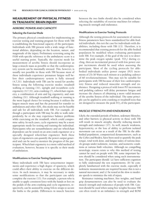
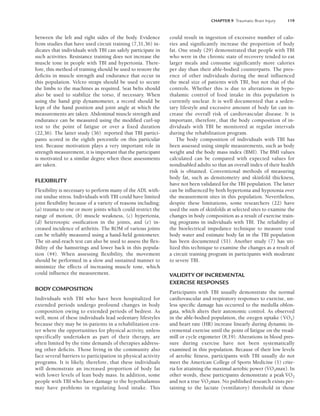
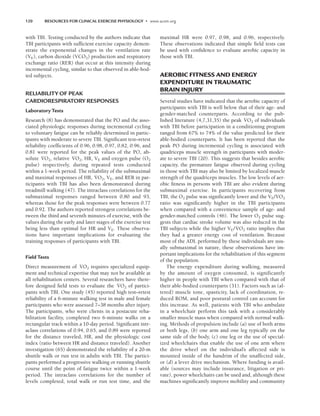
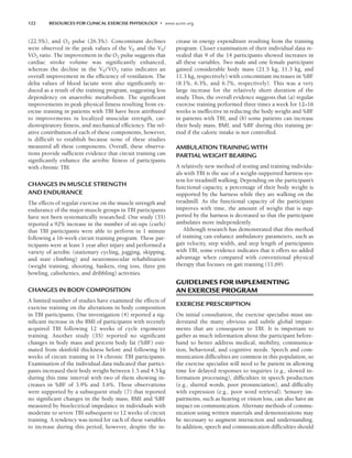

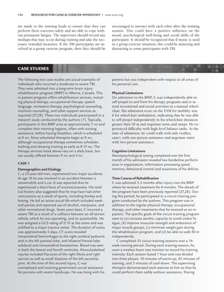
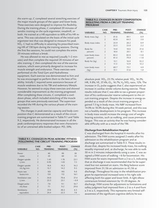
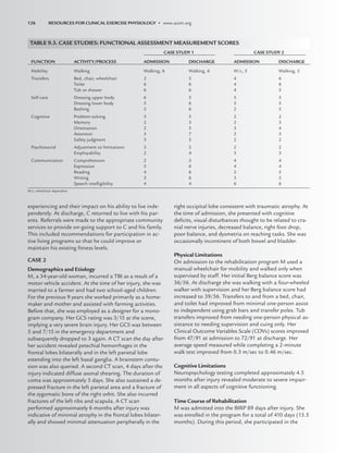
![128 RESOURCES FOR CLINICAL EXERCISE PHYSIOLOGY • www.acsm.org
management of severe traumatic brain injury. XIV. Hyperventila-
tion. J Neurotrauma 2007;24(Suppl 1):S87–S90.
11. Brown TH, Mount J, Rouland BL, Kautz KA, Barnes RM, Kim J.
Body weight-supported treadmill training versus conventional gait
training for people with chronic traumatic brain injury. J Head
Trauma Rehabil 2005;20:(5):402–415.
12. Butefisch CM. Neurobiological bases of rehabilitation. Neurol Sci
2006;27(Suppl 1):S18–S23.
13. Bhambhani Y, Coutts K, Gillespie M, et al. Inclusive Fitness and
Lifestyle Services of All Disabilities. Canadian Society for Exercise
Physiology; 2002.
14. Cantu RC. Posttraumatic retrograde and anterograde amnesia:
Pathophysiology and implications in grading and safe return to
play. Journal of Athletic Training 2001;36:244–248.
15. Centers for Disease Control and Prevention. Traumatic Brain Injury
in the United States: A Report to Congress. Atlanta, Georgia: Centers
for Disease Control and Prevention; 1999.
16. Coles JP
. Imaging after brain injury. Br J Anaesth 2997;99(1):49–60.
17. Convertino VA, Bloomfield SA, Greenleaf JE. An overview of the is-
sues: Physiological effects of bed rest and restricted physical activ-
ity. Med Sci Sports Exerc 1997;29(2):187–190.
18. Dault MC, Dugas C. Evaluation of a specific balance and coordina-
tion programme for individuals with a traumatic brain injury. Brain
Inj 2002;16(3):231–244.
19. Dawes H, Bateman A, Culpan J, Scott O, Wade DT, Roach N, Green-
wood R. The effect of increasing effort on movement economy dur-
ing incremental cycling exercise in individuals early after acquired
brain injury. Clin Rehabil 2003;17(5):528–534.
20. Dawes H, Scott OM, Roach NK, Wade DT. Exertional symptoms
and exercise capacity in individuals with brain injury. Disabil Reha-
bil 2006;28(20):1243–1250.
21. Dawes HN, Barker KL, Cockburn J, Roach N, Scott O, Wade D.
Borg’s rating of perceived exertion scales: Do the verbal anchors
mean the same for different clinical groups? Arch Phys Med Rehabil
2005;86(5):912–916.
22. Driver S, O’Connor J, Lox C, Rees K. Evaluation of an aquatics pro-
gramme on fitness parameters of individuals with a brain injury.
Brain Inj 2004;18(9):847–859.
23. Giacino JT, Kalmar K, Whyte J. The JFK Coma Recovery Scale-Re-
vised: Measurement characteristics and diagnostic utility. Arch Phys
Med Rehabil 2004;85(12):2020–2029.
24. Goldstein LB. Prescribing of potentially harmful drugs to patients
admitted to hospital after head injury. J Neurol Neurosurg Psychiatry
1995;58(6):753–755.
25. Gordon WA, Zafonte R, Cicerone K, et al. Traumatic brain injury reha-
bilitation: State of the science. Am J Med Rehabil 2006;85(4):343–382.
26. Gracies JM. Physical modalities other than stretch in spastic hyper-
tonia. Phys Med Rehabil Clin North Am 2001;12(4):769–792, vi.
27. Gray DS. Slow-to-recover severe traumatic brain injury: A review of
outcomes and rehabilitation effectiveness. Brain Inj 2000;14(11):
1003–1014.
28. Gray DS, Burnham RS. Preliminary outcome analysis of a long-term
rehabilitation program for severe acquired brain injury. Arch Phys
Med Rehabil 2000;81(11):1447–1456.
29. Henson MB, De Castro JM, Stringer AY, Johnson C. Food intake by
brain-injured humans who are in the chronic phase of recovery.
Brain Inj 1993;7(2):169–178.
30. Horn S, Watson M, Wilson BA, McLellan DL. The development of
new techniques in the assessment and monitoring of recovery from
severe head injury: A preliminary report and case history. Brain Inj
1992;6(4):321–325.
31. Hunter M, Tomberlin J, Kirkikis C, Kuna ST. Progressive exercise
testing in closed head-injured subjects: Comparison of exercise ap-
paratus in assessment of a physical conditioning program. Phys
Ther 1990;70(6):363–371.
32. Irdesel J, Aydiner SB, Akgoz S. Rehabilitation outcome after trau-
matic brain injury. Neurocirugia (Astur) 2007;18(1):5–15.
33. Iverson GL, Gaetz M, Lovell MR, Collins MW. Cumulative effects of
concussion in amateur athletes. Brain Inj 2004;18(5):433–443.
34. Jackson D, Turner-Stokes L, Culpan J, Bateman A, Scott O, Powell
J, Greenwood R. Can brain-injured patients participate in an aero-
bic exercise programme during early inpatient rehabilitation? Clin
Rehabil 2001;15(5):535–544.
35. Jankowski LW, Sullivan SJ. Aerobic and neuromuscular training: Ef-
fect on the capacity, efficiency, and fatigability of patients with trau-
matic brain injuries. Arch Phys Med Rehabil 1990;71(7):500–504.
36. Jankowski LW, Sullivan SJ. Aerobic and neuromuscular training: Ef-
fect on the capacity, efficiency, and fatigability of patients with trau-
matic brain injuries. Arch Phys Med Rehabil 1990;71(7):500–504.
37. Jorge RE, Robinson RG, Moser D, Tateno A, Crespo-Facorro B,
Arndt S. Major depression following traumatic brain injury. Arch
Gen Psychiatry 2004;61(1):42–50.
38. Kraus JF
, McArthur DL. Epidemiologic aspects of brain injury. Neu-
rol Clin 1996;14(2):435–450.
39. Laatsch L, Krisky C. Changes in fMRI activation following rehabil-
itation of reading and visual processing deficits in subjects with
traumatic brain injury. Brain Inj 2006;20(13–14):1367–1375.
40. LaRoche SM, Helmers SL. The new antiepileptic drugs: Clinical ap-
plications. JAMA 2004;291(5):615–620.
41. Lequerica AH, Rapport LJ, Loeher K, Axelrod BN, Vangel SJ, Jr,
Hanks RA. Agitation in acquired brain injury: Impact on acute
rehabilitation therapies. J Head Trauma Rehabil 2007;22(3):
177–183.
42. Lim HB, Smith M. Systemic complications after head injury: A clin-
ical review. Anaesthesia 2007;62(5):474–482.
43. McKinlay WW, Watkiss AJ. Cognitive and behavioral effects of
brain injury. In: Rosebthal M, Kreutzer J, Griffith E, Pentland B, eds.
Rehabilitation of the Adult and Child with Traumatic Brain Injury.
Philadelphia: FA Davis; 1999:74–86.
44. Mehrholz J, Major Y, Meissner D, Sandi-Gahun S, Koch R, Pohl M.
The influence of contractures and variation in measurement
stretching velocity on the reliability of the Modified Ashworth Scale
in patients with severe brain injury. Clin Rehabil 2005;19(1):63–72.
45. Mossberg KA. Reliability of a timed walk test in persons with ac-
quired brain injury. Am J Med Rehabil 2003;82(5):385–90; quiz
391–392.
46. Mossberg KA, Ayala D, Baker T, Heard J, Masel B. Aerobic capacity
after traumatic brain injury: Comparison with a nondisabled co-
hort. Arch Phys Med Rehabil 2007;88(3):315–320.
47. Mossberg KA, Greene BP
. Reliability of graded exercise testing after
traumatic brain injury: Submaximal and peak responses. Am J Med
Rehabil 2005;84(7):492–500.
48. Painter P
, Durstine JL, Rimmer J, Morgan D, Franklin B, Pitteti K.
Increasing physical activity in disabled populations. Medicine and
Science in Sports and Exercise 1998;30(5, Suppl):S86.
49. Parnes LS, Agrawal SK, Atlas J. Diagnosis and management of be-
nign paroxysmal positional vertigo (BPPV). CMAJ 2003;169(7):
681–693.
50. Putnam S, Fichtenberg N. Neuropsychological examination of the
patient with traumatic brain injury. In: Rosenthal M, Kreutzer J,
Griffith E, Pentland B, eds. Rehabilitation of the Adult and Child with
Traumatic Brain Injury. Philadelphia: FA Davis; 1999:147–166.
51. Raggueneau JL, Gambini D, Levante A, Riche F
, de Vernejoul P,
Echter E. [Monitoring of extra- and intra-cellular compartment
through total body impedance (author’s transl)]. Anesth Analg (Paris)
1979;36(9–10):439–443.
52. Ruff R. Two decades of advances in understanding of mild trau-
matic brain injury. J Head Trauma Rehabil 2005;20(1):5–18.
53. Salazar AM, Jabbari B, Vance SC, Grafman J, Amin D, Dillon JD.
Epilepsy after penetrating head injury. I. Clinical correlates: A re-
port of the Vietnam Head Injury Study. Neurology 1985;35(10):
1406–1414.
54. Satkunam LE. Rehabilitation medicine: 3. Management of adult
spasticity. CMAJ 2003;169(11):1173–1179.
LWBK191-4034G-C09_113-130.qxd 06/11/2008 10:04 AM Page 128](https://image.slidesharecdn.com/acsmsresourcesforclinicalexercisephysiology-230410064150-fb83bfa2/85/ACSM_s-Resources-for-Clinical-Exercise-Physiology-pdf-142-320.jpg)


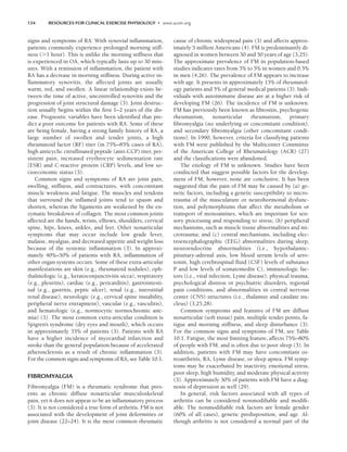
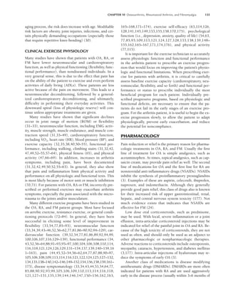
![136 RESOURCES FOR CLINICAL EXERCISE PHYSIOLOGY • www.acsm.org
disease onset) to prevent disability. Effectiveness of the
DMARDs for RA is determined by their ability to change
the course of RA by increasing physical function, decreas-
ing inflammatory synovitis, and slowing structural damage.
Examples of DMARDs are methotrexate, leflunomide,
sulfasalazine, hydroxychloroquine, and injectable gold.
Methotrexate, the most common DMARD, has been used
successfully in combination with other DMARDs and bio-
logics. As with the other arthritis medications, each
DMARD has its related toxic effects. For example, long-
term methotrexate use typically results in liver function
complications, whereas sulfasalazine can result in gas-
trointestinal and CNS toxicity. Biologics, which are also
considered DMARDs, specifically target the pathogenic
mediators of joint inflammation and damage (e.g., tumor
necrosis factor [TNF] antagonists, anti-IL-1 receptor an-
tagonists). Common biologic agents include the TNF in-
hibitors (i.e., etanercept, infliximab, and adalimumab),
which specifically inhibit the proinflammatory cytokine,
TNF-, and anti-IL-1 receptor antagonists (i.e.,
anakinra). For more severe RA, the biologics abatacept
and rituximab may be needed. Side effects of the TNF in-
hibitors may include an increased risk of lymphoma and
other malignancies, heart failure, and demyelinating dis-
orders (3). No evidence exists that suggests that any
DMARDs are effective or can modify OA.
Popular treatments for arthritis, especially OA, are
glucosamine, chondroitin sulfate, or the combination of
both. Their effectiveness for relieving pain is equivocal
(178). It appears that they are most effective for moderate
to severe knee pain (179). Currently, research is being
conducted to determine the use of vitamins C and D for
reducing the risk of progression of knee OA.
Specific to FM, medications are used to improve
restorative sleep and mood, and decrease pain to reduce
fatigue and decrease the symptomatology of FM. Some
of the pharmacologic agents used for sleep include
amitriptyline, cyclobenzaprine, zolpidem, and pramipex-
ole. Medications for pain include amitriptyline, tramadol,
duloxetine, and the anticonvulsants gabapentin and pre-
gabalin. Medications prescribed to improve mood in-
clude tricyclic antidepressants (amitriptyline and cy-
clobenzaprine), selective serotonin reuptake inhibitors
(SSRIs), such as fluoxetine and sertraline, and serotonin-
norepinephrine reuptake inhibitors (SNRIs) (venlafax-
ine and duloxetine) (3). Recently, pregabalin was the
first drug approved by the US Food and Drug Adminis-
tration (FDA) for treating FM after research showed that
it decreased pain and improved restorative sleep in pa-
tients with FM (180). As is typical with any medica-
tion, their effectiveness is not the same for all patients.
A meta-analysis of the efficacy of treatment outcomes
for FM indicates that nonpharmacologic treatments, es-
pecially exercise in combination with cognitive behav-
ioral therapy, give the best outcomes (181). However,
these can be supplemented by pharmacologic
treatments to reduce pain and sleep disturbances. See
Table 10.3 for the common pharmacologic treatments
for OA, RA, and FM.
Each pharmacologic agent for arthritis has side effects
and toxicity levels that can have an impact on different
physiologic systems. In general, there does not appear to be
TABLE 10.3. SOME COMMON PHARMACOLOGIC TREATMENTS FOR OSTEOARTHRITIS (OA),
RHEUMATOID ARTHRITIS (RA), AND FIBROMYALGIA (FM)
OA RA FM
Analgesics NSAIDs (see OA list) Pain
Acetaminophen (Tylenol) DMARDs Pregabalin (Lyrica)
Capsaicin (Capzasin-P) Methotrexate (Rheumatrex) Gabapentin (Neurontin)
NSAIDs Leflunomide (Arava) Duloxetine (Cymbalta)
Aspirin Sulfasalazine (Azulfidine) Tramadol (Ultram)
Celecoxib (Celebrex) Hydroxychloroquine (Plaquenil) Amitriptyline (Elavil)
Ibuprofen (Advil) Biologics Sleep
Naproxen (Aleve) Etanercept (Enbrel) Amitriptyline (Elavil)
Corticosteroids Infliximab (Remicade) Cyclobenzaprine (Flexeril)
Prednisone Adalimumab (Humira) Zolpidem (Ambien)
Viscosupplementation Abatacept (Orencia) Pramipexole (Mirapex)
Hyaluronan (Euflexxa) Rituximab (Rituxan) Pregabalin (Lyrica)
Hylan G-F 20 (Synvisc) Mood
SSRIs
Fluoxetine (Prozac)
Sertraline (Zoloft)
SNRIs
Venlafaxine (Effexor)
Duloxetine (Cymbalta)
Tricyclics
Amitriptyline (Elavil)
DMARDs, disease modifying antirheumatic drugs; NSAIDs, nonsteroidal anti-inflammatory drugs; SNRIs, serotonin-norepinephrine reuptake inhibitors; SSRIs, selective serotonin reuptake inhibitors.
LWBK191-4034G-C10_131-147.qxd 06/11/2008 10:05 AM Page 136](https://image.slidesharecdn.com/acsmsresourcesforclinicalexercisephysiology-230410064150-fb83bfa2/85/ACSM_s-Resources-for-Clinical-Exercise-Physiology-pdf-150-320.jpg)
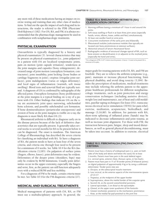
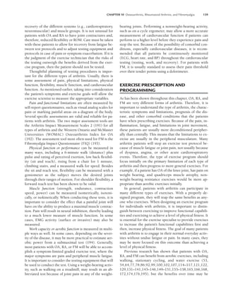
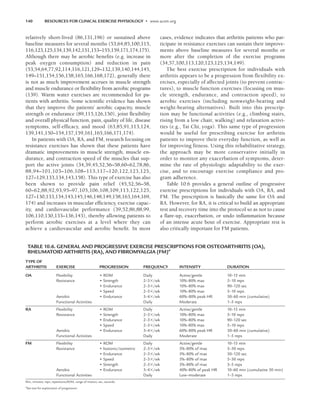
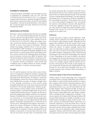
![142 RESOURCES FOR CLINICAL EXERCISE PHYSIOLOGY • www.acsm.org
in their programs than patients with OA and RA because
with FM, there is not as much concern for joint effusions.
It is recommended that for patients with OA and RA, con-
traction speed exercises should be added to the progres-
sion last, after the muscles around the affected joints have
begun to adapt and help support the joint. It is recom-
mended that this higher impact activity be performed 3
days per week and using lower resistances initially.
Within 6–12 weeks, the musculature should be trained to
a level where the patient starts to notice significant im-
provements in function and, in many cases, a reduction
in pain and fatigue levels.
CARDIOVASCULAR ACTIVITIES
Once the musculature has been trained sufficiently and
the muscles can be used more efficiently, it is recom-
mended that aerobic exercises and functional activities be
added to the exercise progression. Again, if the potential
exists to measure the patients’ maximal or peak aerobic
power, this will provide the tools to prescribe the best
aerobic program. An important consideration when pre-
scribing an aerobic training program for persons with
arthritis is to consider the types of aerobic activity they
are comfortable doing. For instance, many individuals
with OA and RA are overweight and will not do water ex-
ercises because they are uncomfortable in public wearing
a bathing suit. It is also important to consider if a non-
weight-bearing or weight-bearing aerobic exercise would
be more appropriate. Generally, whatever exercise the pa-
tient is willing to perform regularly will suffice. It is rec-
ommended that the exercise progression begin conserva-
tively by allowing the patient to adapt to aerobic activity
by using lower target heart rate ranges and then progress
up to the cardiovascular training ranges.
FUNCTIONAL ACTIVITIES
Functional activities can be defined as basic physical ac-
tivities that an individual does to participate in personal
daily leisure, recreational, family, and work activities. Ex-
amples of these activities are walking, climbing stairs, ris-
ing from a chair, and bending. As part of an overall exer-
cise program, it is important to include functional activities.
It has been shown that, although there is physiologic
adaptation with the traditional types of exercise, it does
not translate into better functioning unless individuals
can use their improved physiology to increase the effi-
ciency of their actual functional activities.
EDUCATION AND COUNSELING
Other than the medical, pharmacologic, and surgical
management of the different types of arthritis, patient
self-management is critical. It has been shown that arthri-
tis patients with higher self-efficacy report less pain and
impairment during physical activities (114,126,141,145,
149,153,155,158,170,198). Group programs, combining
strategies to improve fitness and flexibility with stress re-
duction techniques and support groups, seem to hold
promise as community or outpatient programs for indi-
viduals with arthritis (114,149,150,155,167,174,199).
Generally, any programs to promote physical activity and
weight reduction with which the patient will comply are
recommended. The Arthritis Foundation offers several
community-based programs for individuals with arthri-
tis. These include the land-based Arthritis Foundation
Exercise Program (formerly known as the People with
Arthritis Can Exercise [PACE] Program) (114) and the
warm water-based Arthritis Foundation Aquatic pro-
gram. Both programs focus on low-level flexibility,
strength, and endurance activities. Other Arthritis Foun-
dation programs include Tai Chi (Sun style) and the
Walk with Ease Program, which are designed to increase
quality of life and encourage regular physical activity. An-
other educational intervention is the Arthritis Founda-
tion Self-Help Program. It gives the patients information
on their disease, medications, and side effects, dealing
with their physicians, pain management, energy conser-
vation techniques, nutrition, and weight management,
relaxation techniques, and exercise and physical activity
(3,30). This program has been shown to reduce the costs
associated with arthritis care as well as reduce the pa-
tients’ perceptions of pain by 20% (200,201). Infre-
quently, patients are referred to physical or occupational
therapy, unless they experience an acute disease flare.
REFERENCES
1. Centers for Disease Control and Prevention (CDC). Prevalence of
disabilities and associated health conditions among adults: United
States, 1999. MMWR 2001;50:120–5.
2. Theis KA, Murphy L, Hootman JM, Helmick CG, Yelin E. Preva-
lence and correlates of arthritis-attributable work limitation in the
US population among persons ages 18–64:2002 National Health
Interview Survey data. Arthritis Care Res 2007;57(3):355–63.
3. Klippel JH, Stone JH, Crofford LJ, White PH, eds. Primer on the
Rheumatic Diseases. 13th ed. New York: Springer; 2008.
4. Lawrence RC, Felson DT, Helmick CG, et al. Estimates of the
prevalence of arthritis and other rheumatic conditions in the
United States. Part II. Arthritis Rheum 2008;58:26–35.
5. Peyron JG, Altman RD. The epidemiology of osteoarthritis. In:
Moskowitz RW, Howell DS, Goldberg VM, Mankin HJ, eds. Os-
teoarthritis: Diagnosis and Medical/Surgical Management. 2nd ed.
Philadelphia: WB Saunders; 1992:15–37.
6. Moskowitz RW. Clinical and laboratory findings in osteoarthritis.
In: Koopman WJ, editor. Arthritis and Allied Conditions: A Textbook
of Rheumatology. 13th ed. Baltimore: Williams Wilkins; 1997:
1985–2011.
7. Kellgren JH, Lawrence JS, Bier F
. Genetic factors in generalized
osteo-arthrosis. Ann Rheum Dis 1963;22:237–55.
8. Felson DT, Naimark A, Anderson JJ, Kazis L, Castelli W, Meenan
RF
. The prevalence of knee osteoarthritis in the elderly: The Fram-
ingham Osteoarthritis Study. Arthritis Rheum 1987;30:914–8.
9. Felson DT. Epidemiology of the rheumatic diseases. In: Koopman
WJ, editor. Arthritis and Allied Conditions: A Textbook of Rheuma-
tology. 13th ed. Baltimore: Williams Wilkins; 1997:3–34.
LWBK191-4034G-C10_131-147.qxd 06/11/2008 10:05 AM Page 142](https://image.slidesharecdn.com/acsmsresourcesforclinicalexercisephysiology-230410064150-fb83bfa2/85/ACSM_s-Resources-for-Clinical-Exercise-Physiology-pdf-156-320.jpg)


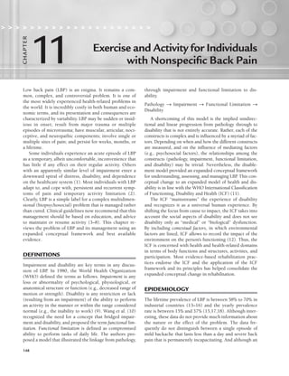
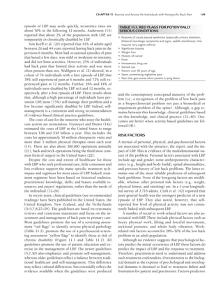
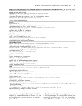
![CHAPTER 11 Exercise and Activity for Individuals with Nonspecific Back Pain 153
substances (e.g., prostaglandins, leukotrienes, bradykinin,
histamine, sustance P calcitonin gene-related peptide)
either sensitize or directly stimulate receptors involved
in nociception, including “silent nociceptors.” Silent no-
ciceptors are normally unresponsive to mechanical stim-
uli, but following tissue injury, they respond to mechan-
ical stimuli (even very weak mechanical stimuli). Silent
nociceptor sensitization accounts for the increased sen-
sitivity (hyperalgesia) to noxious and non-noxious stim-
uli in the directly injured tissues and in adjacent tissues.
Hence accurate location of specifically injured tissue is
difficult and error prone. Although the area of injury can
be identified with confidence, this confidence is mis-
placed in regard to the accurate location of a specifically
injured tissue.
Persistent or intense noxious input also leads to sensi-
tization of cells in the central nervous system. Nocicep-
tive neurons in the dorsal horn are classified as nocicep-
tive specific (NS) and wide dynamic range (WDR)
neurons, based on their response characteristics. As their
name suggests, nociceptive neurones respond to noxious
input, whereas WDR neurons respond to both noxious
and non-noxious input. Persistent noxious input to WDR
neurons leads to a progressive increase in their response,
processes known as “wind-up” and central sensitization.
Wind-up is largely mediated through N-methyl-D-aspar-
tate (NMDA) glutamate receptors, whereas central sensi-
tization is largely mediated through excitatory amino
acids, and substance P and can last for hours (54).
Central sensitization also leads to enhanced responses
of spinothalmamic tract neurons. In essence, peripheral
nociceptive input results in a cascade of activity through-
out the neural system. The threshold for nociceptive sig-
nals is decreased and, once generated, nociceptive signals
can persist even after the initiating noxious stimulus is
removed (56). This aberrant nociceptive sensitivity and
enhanced nociceptive signaling make it difficult to ifen-
tify accurately the injured tissue based on location of pain
or on specific provocative movements that may be used
as part of a clinical assessment.
Finally, the neural system is plastic. Persistent noci-
ceptive input leads to neuroplastic changes throughout
the neural system and these changes help account for
persistent or chronic pain that is no longer associated
with ongoing tissue injury. Thus, chronic pain is not simply
acute pain that lasts a long time. Indeed, chronic pain is a
pathologic condition in and of itself (similar to epilepsy).
Acute pain should be managed appropriately in an effort
to inhibit neuroplastic changes that contribute to persist-
ent and disabling chronic pain.
Psychological Factors and Pain
Clearly, the nociceptive signal that arrives at the cortex is
distinctly different from that generated in the periphery.
Whether the signal is enhanced or inhibited depends on
the relative strength of the opposing neuromodulatory
processes. Whether the signal is interpreted as painful or
not, however, it is influenced by a myriad of psychologi-
cal and social factors. The individual’s expectancy, mood,
attention, sense of control, current activity, past experi-
ence, and the social and environmental context in which
the signal is received influence the interpretation of that
signal. Indeed, the meaning that the individual attaches
to the pain will influence his or her emotional response
and subsequent behavior and that behavior outlasts the
sensory signal and contributes to disability. (See the ex-
cellent review by Vlaeyen and Linton, on this topic [57]).
The beliefs that individuals has about their pain will
influence what they do about it. Some individuals will
consider the pain a minor inconvenience and attempt to
ignore it, whereas others will worry about its meaning
and even think the pain is catastropic and immediately
seek professional help. If pain is aggravated by activity,
they may avoid activities that are painful and even those
that they anticipate will be painful. Although this action
may be appropriate in the short term for acute pain it is
not appropriate for the long term and indeed will aggra-
vate the problem.
An important point to consider in this context is that
pain, mood, stress, and exercise or activity all have neu-
robiochemical bases. The interactions among these con-
structs are complex and not well understood, either at
the molecular or the person level. It is intriguing to con-
sider whether such central mechanisms account for the
reported nonspecific effects of specific exercise regimens
(58,59).
In summary, LBP is a multidimensional experience. It
has sensory, emotional, cognitive, and behavioral compo-
nents. The relative magnitude of each component helps
determine how an individual’s problem should be man-
aged (i.e., modify sensory input, address misunderstand-
ings about the meaning of the pain and the relationship
to injury, and address anxieties about pain and activity).
The simple adage “let pain be the guide” belies the com-
plexity of the construct.
For a significant acute back injury (where pain and in-
jury are related) it is reasonable to reduce activity for a
day or two, treat the pain (e.g., with analgesic medication
a physical modality, such as ice, or both) and be guided
by pain intensity and duration as normal activity is re-
sumed (1–2 days). The period of inactivity should be lim-
ited by time not pain, however. An early return to normal
activities should be encouraged and expected. Any advice
to rest must be accompanied by advice on activity resump-
tion.
For chronic or recurrent LBP
, this approach is inap-
propriate and even harmful. Pain is not indicative of on-
going tissue injury and it is likely to persist, therefore it
cannot be used to guide the amount of activity. Practi-
tioners must be sensitive to the degree of pain and to its
effect on an individual’s psychological and physical state.
LWBK191-4034G-C11_148-161.qxd 06/11/2008 10:05 AM Page 153](https://image.slidesharecdn.com/acsmsresourcesforclinicalexercisephysiology-230410064150-fb83bfa2/85/ACSM_s-Resources-for-Clinical-Exercise-Physiology-pdf-167-320.jpg)
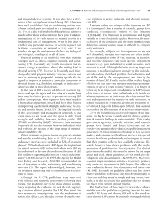
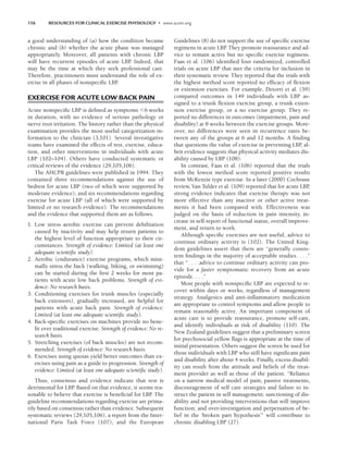
![CHAPTER 11 Exercise and Activity for Individuals with Nonspecific Back Pain 157
EXERCISE FOR CHRONIC LOW
BACK PAIN
Chronic nonspecific LBP is characterized by the persist-
ence of symptoms beyond 12 weeks. However, with the
exception of sharing a minimal time of symptom dura-
tion, individuals with chronic LBP are characterized by
heterogeneity. Most individuals with chronic and persist-
ent symptoms of LBP do not seek healthcare, they have
little disability, continue to work, and are generally not
distressed about their back pain. Those who do seek pro-
fessional care vary widely in terms of the total duration of
symptoms, symptom severity, disability level, distress,
and overall physical condition resulting from those
symptoms. Clearly, the approach to management has to
be individualized, holistic, and rational, with considera-
tion given to biological, psychological, and sociological
factors.
Clinical guidelines now address the assessment and
management of chronic LBP (7). Also, in the United
Kingdom, the Clinical Standards Advisory Group (4) has
suggested that as LBP becomes chronic, psychological
factors become more important and purely physical man-
agement should be avoided in favor of active exercise and
multidisciplinary rehabilitation based on a biopsychoso-
cial model. These recommendations are essentially con-
cordant with the European Guidelines for the Manage-
ment of Chronic Nonspecific Low Back Pain (7):
education, cognitive behavioral therapy, supervised activ-
ity, and active multidisciplinary biopsychosocial treat-
ment. Although the Guidelines suggest that a short
course of mobilization can be considered, physical
modalities (i.e., heat, cold, laser, transcutaneous electri-
cal nerve stimulation [TENS], massage, corsets) are not
recommended. The 2000 report from the International
Paris Task Force on Back Pain (107) recommends the
prescription of physical, therapeutic, or recreational ex-
ercise in cases of chronic nonspecific back pain.
A number of randomized, controlled trials and sys-
tematic reviews have addressed exercise and activity in
individuals with chronic nonspecific LBP
. It seems clear
that exercise and activity are beneficial for individuals
with LBP because they can reduce the perception of pain
and enhance the sense of well-being. It is less clear
whether the type, intensity, frequency, or duration of ex-
ercise or activity is important. The Paris Task Force rec-
ommends that exercise programs should combine
strength training, stretching and fitness, whereas other
guidelines are less prescriptive. But perhaps the most ef-
fective exercise or activity regimen is that which is done.
In individuals with LBP
, barriers to, and motivators of,
physical activity are similar to those in the general popu-
lation (e.g., lack of time, inclement weather, and family
commitments) (111). However, it is interesting that back
problems are identified as both barriers to, and motiva-
tors of, activity. In a qualitative study Keen et al. (111) in-
terviewed 27 individuals who were participants in a ran-
domized, controlled trial of a progressive exercise pro-
gram. They reported that some individuals believed that
being more physically active helped ease their back pain
and made them feel better. They were worried about
stopping exercise for fear that their back pain would re-
turn. Others did not exercise on a regular basis but re-
sumed exercise when reminded to by their backache. Still
others avoided physical activity for fear of an aggravation
of their LBP
. Although all subjects identified the avoid-
ance of some physical activity (e.g., lifting and garden-
ing), not all were fearful or anxious about such activity. It
appears that those individuals in the latter category, re-
ported that their confidence was restored over time
through (a) reassurance and advice from health profes-
sionals, (b) modifying the way an activity was done (e.g.,
less vigorous), and (c) a progressive exercise program
(111). It appears that a change in behavior led to a
change in belief about the ability to be active.
Participation in exercise or activity is essential if bene-
fits are to accrue, whether those benefits are physical or
attitudinal. Friedrich et al. (112) conducted a double-
blind, randomized study and evaluated the effect of a mo-
tivation program on exercise compliance (adherence)
and disability. A total of 93 patients with LBP were ran-
domly assigned to either a standard exercise program
(n 49) group or a combined exercise and motivation
group (n 44). The exercise program consisted of an in-
dividual submaximal gradually increased training ses-
sion. Each patient was prescribed 10 sessions that each
lasted about 25 minutes. The specific exercises were
aimed at “improving spinal mobility, as well as trunk and
lower limb muscle length, force, endurance, and coordi-
nation, thereby restoring normal function.” Flexibility
exercises for the trunk and lower limbs preceded
strengthening exercises for the trunk. The motivation
program consisted of five sessions that included counsel-
ing, information about LBP and exercise, reinforcement,
forming a treatment contract between patient and thera-
pist, and keeping an exercise diary.
The combined exercise and motivation group in-
creased the rate of attendance and reduced disability and
pain in the short term (4 and 12 months). However, there
was no difference in exercise adherence in the long term.
Long-term adherence to exercise is an acknowledged
problem in the general population and is no different in
those with LBP
. In the case of LBP
, when exercise benefits
may not be immediate or even apparent, and when recur-
rence of LBP is inevitable anyway, it is hardly surprising
that adherence to exercise is problematic. For some indi-
viduals, encouraging and facilitating them to have a more
active lifestyle may be more beneficial for them than pre-
scribing a specific exercise regimen that they do not do.
That no evidence supports the notion that specific regi-
mens are differentially more effective for nonspecific
back pain supports this position. Some evidence,
LWBK191-4034G-C11_148-161.qxd 06/11/2008 10:05 AM Page 157](https://image.slidesharecdn.com/acsmsresourcesforclinicalexercisephysiology-230410064150-fb83bfa2/85/ACSM_s-Resources-for-Clinical-Exercise-Physiology-pdf-171-320.jpg)
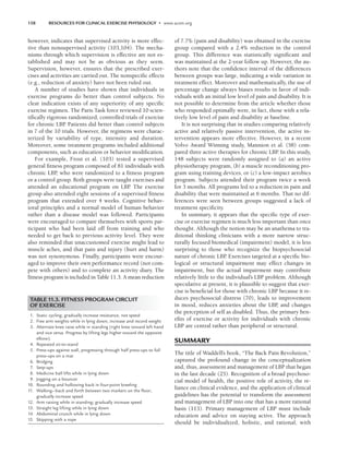
![164 RESOURCES FOR CLINICAL EXERCISE PHYSIOLOGY • www.acsm.org
is associated with low BMD in the hip but not the spine
(118). The findings were similar for diet colas but were
weaker when the colas were decaffeinated.
Lower body weight is directly correlated to lower bone
density (3,98) and is a determinant of bone density in
adults (34). Smoking is associated with low BMD (91),
an increased risk of fracture (54), decreased levels of vi-
tamin D (121), and decreased calcium absorption (91). It
is thought that smoking also interferes with estrogen me-
tabolism (114). The gonadal hormones, particularly es-
trogen, are essential for maintaining bone mass. Estrogen
directly affects bone turnover by binding to estrogen re-
ceptors on the osteoblasts. Estrogen also enhances cal-
cium absorption from the intestines (16). Several med-
ications, in particular the glucocorticoids, have adverse
effects on the skeleton. These include increased urinary
excretion and decreased intestinal absorption of calcium,
reduced levels of gonadal hormones, inhibition of os-
teoblast function, and increased bone resorption (16).
Another risk factor for osteoporosis is decreased activ-
ity level. It is well known that increased activity level and
exercise will aid in maintaining or improving BMD. In a
recent study it was determined that among many lifestyle
risk factors, high school sports participation appeared to
have the greatest influence on BMD levels in the femoral
neck (3). This supports the need for physical activity, es-
pecially during adolescence, for accruing and improving
bone mass.
Several of these risk factors are interrelated. For exam-
ple, peak bone mass is largely determined by genetic fac-
tors; however, failure to reach one’s genetic potential is
often the result of inadequate calcium intake and exercise
(45). Also, risk of osteoporosis increases with advancing
age, particularly after menopause in women, with early
menopause, either natural or surgical, causing an even
greater risk. Postmenopausal bone loss is largely the re-
sult of estrogen deficiency, but pharmacologic therapy
can dramatically attenuate postmenopausal bone loss.
CLINICAL EXERCISE PHYSIOLOGY
Bone density does not respond to acute exercise bouts
and cardiovascular responses to acute exercise have not
been well studied in osteoporotic populations. Lombardi
et al. (69) examined physical capacity during exercise in
women with osteoporosis, with and without vertebral
fracture, and compared their responses with those of
women without osteoporosis. Very few differences were
found in measures of physical capacity during walking
exercise at 3 or 4 mph (V
.
O2, metabolic equivalents
[METs], heart rate) although energy expenditure was
positively associated with the degree of kyphosis (69).
The primary purposes of acute exercise testing are typi-
cally to aid in the diagnoses of coronary artery disease
(CAD) and to determine appropriate levels of exercise
training. Osteoporosis can sometimes mask the presence
of CAD if it prevents an individual from achieving the ad-
equate heart rate and blood pressure necessary for accu-
rate diagnoses. In addition, severe thoracic kyphosis can
impair respiration and limit the test (24). Nonetheless,
no specific recommendations from the American College
of Sports Medicine (ACSM) would suggest that osteo-
porosis is an absolute contraindication to exercise testing
(1). If an exercise stress test is to be used in patients with
osteoporosis, one utilizing a bicycle protocol would prob-
ably be the best choice, because that would involve the
least trauma and impact on the bones. Caution, however,
must still be taken when utilizing a bike protocol. An up-
right posture should be maintained by the patient at all
times because spinal flexion is contraindicated in people
with osteoporosis. Treadmill protocols can be utilized if
need be, but a walking protocol should be used and care
should be taken to ensure the patient does not trip or fall.
Bone mass responses to chronic exercise in the osteo-
porotic population and in postmenopausal women have
been well studied (11,26,43,64,78,89,120). The primary
purpose of prescribing exercise in these populations
would be to increase both BMD and overall fitness and
balance to aid in fall prevention (62). In this regard, most
studies have shown positive results. A number of studies
in postmenopausal women have shown that exercise can
increase BMD or prevent further bone loss when com-
pared with nonexercising controls (11,26,64,78).
Although bone mass responds positively to chronic
exercise in adults, the use of exercise as a treatment for
older adults with low bone mass is limited (61) because
increases in BMD with exercise are generally small (2% or
less). A better utilization of exercise may lie in prevention
of osteoporosis by improving peak bone mass. Recent re-
views of the literature highlight the importance of exer-
cise in establishing optimal levels of bone mineral during
the growing years when bone may respond better to
chronic exercise (4,6). Further, the timing of exercise in-
tervention in childhood has been demonstrated to affect
bone mineral status in adulthood. Female tennis and
squash players who started their playing careers before or
at menarche were found to have a two- to threefold
greater dominant arm bone mineral content than those
who had started playing more than 15 years after menar-
che (55). So, the chronic responses of bone to exercise
would appear to differ, depending at what point in life the
exercise is initiated.
PHARMACOLOGY
Most women diagnosed with osteoporosis, and post-
menopausal women in general, likely will be taking some
form of calcium and vitamin D supplements. Other com-
mon drugs available that may be used for treatment of os-
teoporosis include estrogen alone or in combination with
progesterone, bisphosphonates, calcitonin, and selective
estrogen receptor modulators (SERM). Other less
LWBK191-4034G-C12_162-174.qxd 06/11/2008 10:06 AM Page 164](https://image.slidesharecdn.com/acsmsresourcesforclinicalexercisephysiology-230410064150-fb83bfa2/85/ACSM_s-Resources-for-Clinical-Exercise-Physiology-pdf-178-320.jpg)
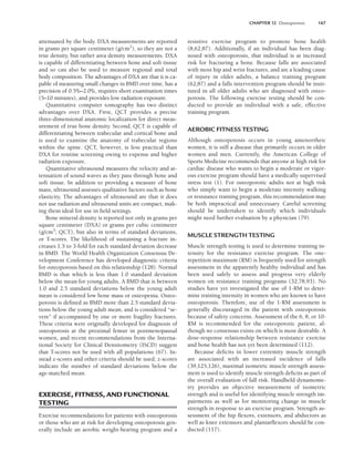
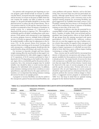
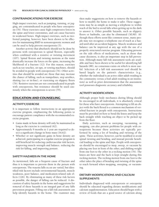
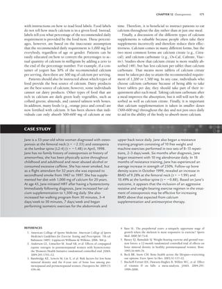
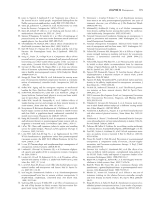
![175
Clinical exercise practitioners may work with individuals
who experience acute, subacute, or chronic spinal pain.
Because the spine is critical in posture, movement, and
protection, pain and loss of function hinders an individ-
ual’s ability to perform routine activities. As with any
complex structure, the potential for problems is great. Al-
though “vertebral disorders” can occur in any part of the
spine (cervical, thoracic, lumbar, sacral, and coccygeal),
most vertebral disorders are lumbar. Lumbar disorders
result in the highest medical, financial, legal, and psy-
chosocial costs to society. Consequently, lumbar disor-
ders have attracted international medical, scientific, eco-
nomic, and popular attention. This chapter is not
necessarily intended to help readers diagnose or treat
back pain. Individuals experiencing new or worsening
low back pain should be referred back to the appropriate
clinician for diagnosis and treatment. This chapter, there-
fore, focuses on lumbar disorders, and specifically, on
what is termed nonspecific, mechanical low back pain.
EPIDEMIOLOGY AND IMPACT
Low back pain is defined as “pain, muscle tension, or stiff-
ness localized below the costal margin and above the infe-
rior gluteal folds, with or without leg pain (sciatica)” (1,
p. 134). Low back pain afflicts nearly everyone at some
stage in life. Reported prevalence rates vary according to
study methodology used, but a 60%–80% individual life-
time prevalence of low back pain is often reported (2–4).
The prevailing notion is that episodes of low back pain are
short-lived (1 month). Evidence suggests, however, that
42%–75% of people still have symptoms after 1 year (5),
although, these individuals are not necessarily seeking or
receiving treatment. A disturbing statistic is that the re-
currence rate after an episode of low back pain is esti-
mated at 60% within the first year (6) and the lifetime re-
currence rate may be as high as 85% (7). Consequently,
low back pain is one of the most prevalent and costly
health problems in industrialized countries. In 1990, di-
rect medical costs for low back pain exceeded $24 billion.
When disability costs are included, the total annual cost
for low back disorders is estimated at $50 billion (8,9).
Low back pain is among the leading reasons people seek
healthcare and, coupled with neck problems, is the sec-
ond leading cause of disability in the United States (10).
Despite that, by definition, nonspecific low back pain
does not involve perceptible structural changes, it can
cause limitations in function, activities, and participation
as defined within the “biopsychosocial” framework of the
World Health Organization’s (WHO) International Clas-
sification of Functioning, Disability and Health (ICF)
(11). The ICF model can be used to visualize the impact
of low back pain on both populations and individuals
(12). An example of the potential impact of nonspecific
low back pain on an individual using the integrated ICF
model is shown in Figure 13.1.
PATHOPHYSIOLOGY
Pain is considered “an unpleasant sensory and emotional
experience associated with actual or potential tissue
damage or described in terms of such damage” (13). No-
ciceptive nerve endings (pain fibers) are activated with
mechanical, thermal, or chemical stimuli. Mechanical
pain is caused by deformation of tissues and varies with
physical activity. That is, certain movements or positions
make mechanical pain worse and other movements or
positions relieve mechanical pain. Specific low back pain
has an identifiable cause, such as fractures, cancer, or in-
fection. Approximately 90% of back pain has no identifi-
able cause and is termed nonspecific (1).
In general, pain can be somatic (arising from stimula-
tion of nerve endings in a musculoskeletal site [joint, mus-
cle, ligament, bone]), visceral (arising from a body organ),
neurogenic (arising from irritation of the axons or cell bod-
ies of peripheral nerves, spinal nerves, or nerve roots), or
psychogenic. Pain resulting from irritation of spinal nerves
or roots is more specifically called radicular pain. Further-
more, pain can be referred to, and perceived in, an area re-
mote from the source of the pain. Pain referred elsewhere
from the viscera is called visceral referred pain, and pain
from somatic sources is described as somatic referred pain
(14). An example of a visceral referred pain is the arm pain
sometimes associated with a myocardial infarction. An ex-
ample of somatic referred pain is diffuse pain in the but-
tock or leg associated with low back pain.
Vertebral Disorders
13
CHA
P
TER
LWBK191-4034G-C13_175-196.qxd 06/11/2008 11:10 AM Page 175](https://image.slidesharecdn.com/acsmsresourcesforclinicalexercisephysiology-230410064150-fb83bfa2/85/ACSM_s-Resources-for-Clinical-Exercise-Physiology-pdf-189-320.jpg)
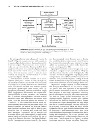
![CHAPTER 13 Vertebral Disorders 177
formation, and loss of joint cartilage, characterize stage 3
(the stage of stabilization). These changes can contribute
to central and spinal stenosis. This degenerative cascade
model helps explain the age-related incidences of spinal
disorders. Discogenic sources of pain are more common
in the fourth and fifth decades (stages of dysfunction and
instability), and stenosis is more common in the sixth
and seventh decades (stage of stabilization).
Thus, a pathoanatomic diagnosis is often not possible.
Indeed, the symptoms are frequently caused by movement
disorders rather than structural, morphologic disorders.
Because back pain is difficult to diagnose, various classifi-
cation systems have evolved. Waddell (20) suggested a
three-category triage system consisting of “simple back-
ache,” “nerve root pain,” and “serious spinal pathology.”
Simple (i.e., nonspecific, low back pain) includes a variety
of disorders and is the most common type of back pain.
Nerve root pain could be attributable to disorders such as
disc prolapse or spinal stenosis and is present less than 5%
of the time. Serious spinal pathology (and red flags) in-
cludes diseases such as tumors, infections, and inflamma-
tory disorders, such as ankylosing spondylitis, and repre-
sents less than 1% of the persons presenting with low back
pain. The value of further subclassifying low back pain
will be discussed later in this chapter.
RISK FACTORS
Because low back pain is a multifactorial disorder with a
number of potential causes, determining risk factors is
difficult. Hence, a multitude of risk factors linked to non-
specific low back pain has been identified. None of these
factors is considered causal and the strength of the evi-
dence supporting each factor is variable, depending on
the sources of the data. Overall risk factors can be divided
into risk factors for primary occurrences and risk factors
associated with chronicity. Risk factors can be further
subcategorized generally into individual, psychosocial,
health behavior, and occupational risk factors. Table 13.1
lists commonly identified risk factors associated with low
back pain (1,5,21–23).
CLINICAL PICTURE
Typically, individuals with vertebral disorders present
with one or more of the following physical complaints:
back pain, leg pain, stiffness, muscle tension, neurologic
symptoms, and spinal deformity. Pain is usually the pri-
mary complaint.
According to Waddell (20) persons with “simple back-
ache” are usually healthy individuals, between 20 and 55
years of age, who present with pain in the lumbosacral re-
gion, buttocks, or thighs. Pain varies in intensity and may
be produced, aggravated, or relieved with general or spe-
cific spinal movements, activities, positions, and time.
Morning stiffness or pain is common, and pain may
worsen over the course of the day. A lateral spinal shift
can be present, where the spine is pulled to one side, and
the lordotic lumbar curve can be lost. Persons presenting
with specific spinal conditions, such as “nerve root pain,”
complain of unilateral leg pain that is worse than their
back pain, pain that radiates below the knee, numbness
or altered sensation in the same distribution, nerve root
signs (e.g., a positive straight leg raise [SLR] test), and
motor, sensory, or reflex changes in one nerve root. Frank
nerve root compression signs are reflex changes, muscle
weakness, muscle atrophy, and sensory loss over a de-
fined area. Spinal stenosis is a condition where the spinal
canal or nerve root formina becomes narrowed (usually
because of degenerative changes) and compresses the
spinal cord or nerve roots. This compression, depending
on location, can cause low back pain, leg pain in one or
both legs, and weakness and numbness of the legs. These
symptoms are characteristically aggravated by walking
and relieved by sitting, lying and bending forward. Red
TABLE 13.1. POTENTIAL RISK FACTORS
ASSOCIATED WITH THE OCCURRENCE OF
NONSPECIFIC LOW BACK PAIN AND WITH
CHRONICITY (1,12,23,74)
FACTORS OCCURRENCE CHRONICITY
Individual Heredity/genetica
Educational level
Agea
Unemployment
Prior back pain historya
High levels of
Sex pain/disability
Anatomic/biomechanical Socioeconomic
variations status
General health status
High birth weight (males)
Psychosocial Stress Distress
Depressiona
Depression
Pain behavior Fear-avoidance
behavior
Anxiety Somatization
Health Behaviors Smoking Obesity/Body Mass
Obesity/Body Mass Indexa
Indexa
Strength of trunk
musculature
Physical fitness level
Occupational Heavy physical work Unavailability of light
Lifting, bending and duty
twisting Daily repetitive lifting
Monotonous tasks Job dissatisfaction
Whole-body vibration
Static work postures
Job dissatisfaction
Night shifts
Control at work
Other Healthcare provider
attitudes
a
Predictors with stronger evidence.
LWBK191-4034G-C13_175-196.qxd 06/11/2008 11:10 AM Page 177](https://image.slidesharecdn.com/acsmsresourcesforclinicalexercisephysiology-230410064150-fb83bfa2/85/ACSM_s-Resources-for-Clinical-Exercise-Physiology-pdf-191-320.jpg)
![CHAPTER 13 Vertebral Disorders 179
the reported intensity of pain reflect the amount of per-
ceived disability (28). Pain intensity does not necessarily
signal that tissue damage is occurring (i.e., pain can
occur in structurally normal tissues). Simply put, hurt
does not always mean harm. Stretching one’s hamstrings
can be uncomfortable, but is not typically harmful. Pa-
tients and clinicians need to make this distinction to help
prevent chronic pain behavior and permanent disability
by keeping patients generally active and functioning dur-
ing their recovery or rehabilitation.
DIAGNOSTIC TECHNIQUES
Accurate diagnosis is, of course, the cornerstone of effec-
tive intervention. But, as previously pointed out, in the
case of low back pain, diagnosis rarely identifies the
pathologic source of the pain (29,30). In addition, evi-
dence suggests that using specific anatomic diagnoses
does not improve outcomes for most patients (31). Wad-
dell’s (20) triage system gives a broad clinical picture of
low back pain and offers a screening system that can be
used to guide in further diagnostic testing. A similar
three-category initial triage was also recommended in a
recent clinical guideline jointly developed by the Ameri-
can College of Physicians/American Pain Society Low
Back Pain Guidelines Panel (31). In general the cate-
gories are “nonspecific low back pain,” “back pain poten-
tially associated with radiculopathy or spinal stenosis,”
or “back pain potentially associated with another specific
spinal cause.” The key points in diagnosing low back
pain are determining whether the pain is indeed coming
from the spine, ruling out specific disorders and poten-
tially serious disorders, and acquiring information neces-
sary to develop a care plan. Diagnostic procedures always
include a clinical examination and evaluation (31) and
may include movement system, diagnostic imaging, electro-
diagnostic, and laboratory testing when further informa-
tion is needed. Each of these diagnostic techniques is dis-
cussed briefly in the following paragraphs.
CLINICAL EXAMINATION AND EVALUATION
The primary diagnostic procedure in the case of low back
pain is the clinical examination and evaluation. The clin-
ical examination consists of two parts, the subjective ex-
amination, or history, and the objective examination, or
physical. Based on the results of the examination, the cli-
nician evaluates (or assesses) the findings of the exami-
nation and either makes a diagnosis or determines the
need for further testing. Typical components of a clinical
examination are outlined in Table 13.3.
HISTORY
The history, or subjective examination, consists of gath-
ering information from patient reports in five major
TABLE 13.3. OUTLINE OF TYPICAL COMPONENTS
OF A CLINICAL EXAMINATION OF A PATIENT
WITH LOW BACK PAINa
CLINICAL EXAMINATION
History
(Subjective Examination)
Present Condition
Mechanism and date of onset or duration
Location of symptoms
Nature, quality and intensity of pain (assessed using a pain scale)
Behavior of symptoms (better and worse)
Types of limitations/disabilities (including use of a standardized,
condition-specific outcome measure [e.g., Oswestry])
Previous Incidents
Including prior treatment and outcomes
Medical and Surgical History
Including medications
Personal Information
Demographic
Occupational
Social
Living environment
Patient Goals
Using the Patient Specific Functional Scale
Physical Examinationa
(Objective Examination)
PART 1: SYSTEMS REVIEW/MEDICAL SCREENING
Systems Review and Observations
General appearance
Communication ability, affect and behavior
Gross symmetry, structure, and skin integrity
Locomotion, balance, and transitional movements
Heart and respiratory rate and blood pressure
Height, weight, body mass index (BMI)
Other
PART 2: REGIONAL TESTS AND MEASURES TO VERIFY LUMBAR
SPINE INVOLVEMENT AND GENERAL NATURE OF INVOLVEMENT
Posture/Alignment
Movement Examination: Active, Passive, and Resistive Lumbar
Movements
Quantity (range) of movement
Quality and pattern of movement
Effect of movement on symptoms
Neurologic Screening Lower Quarter
Cutaneous sensation in dermatomes
Myotome (groups of muscles supplied by a single nerve root) testing
Deep tendon reflexes
Upper motor neuron screen (e.g., pathologica reflexes)
Neurodynamic tests (e.g., straight leg raising, dural mobility tests)
Vascular Screening of Lower Quadrant
Pulses, for example
Peripheral and Sacroiliac Joint Screening
Special Tests
As indicated, based on symptoms (e.g., prone instability test, active
straight raise leg test)
Palpation
Temperature
Tenderness
Tissue condition: tension, texture, thickness, and so forth
Intervertebral Joint Movement Testing
Movement and end feel
Functional Assessment Screening
Task performance ability
a
Content and extent of the physical examination varies with the practitioner’s discipline, back-
ground, and experience, the results of the subjective examination, and the purpose of the ex-
amination.
LWBK191-4034G-C13_175-196.qxd 06/11/2008 11:10 AM Page 179](https://image.slidesharecdn.com/acsmsresourcesforclinicalexercisephysiology-230410064150-fb83bfa2/85/ACSM_s-Resources-for-Clinical-Exercise-Physiology-pdf-193-320.jpg)
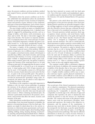
![CHAPTER 13 Vertebral Disorders 181
many diagnostic labels suggesting essentially the same
thing (e.g., sprain or strain, degenerative disc or joint dis-
ease) is insufficient for a physical therapist, for example,
to initiate intervention. A physical therapist needs de-
tailed information about the quality and quantity of ac-
tive and passive movements and the effect of movements
and positions on pain and function as well as the results
of several selected special tests. Assessment of the move-
ments and postures affecting the problem is used in con-
junction with other information to direct treatment.
Potential components of a physical examination are
outlined in Table 13.3. The examination consists of two
parts. The first part is the systems review (also called,
medical screening or scan examination), which is used
primarily to confirm or refute any red flags suggested by
the history. The second part of the examination is to de-
termine the presence of signs of nerve irritation, decide
whether the pain actually involves the lumbar spine, and
obtain further clarifying information to make a differen-
tial diagnosis and develop a care plan. The general intent
of the physical examination is to reproduce the compara-
ble (or asterisk) sign through selected tests, measures,
and patient responses, and to determine the nature and
extent of impairments, function, and disability (activity
limitations and participation restrictions).
Evaluation
Based on evaluation (interpretation) of the results of the
history and physical examinations, the clinician can usu-
ally decide on one of two courses of action:
• Diagnose the disorder (see section entitled, The Med-
ical Clinical Diagnosis), make a prognosis, design
and implement the care plan, or
• Tentatively diagnose the disorder and determine the
need for further testing to confirm or refute this work-
ing diagnosis. Trial treatment, based on the tentative
diagnosis, may or may not be initiated at this time.
If further diagnostic procedures are indicated, this
testing might consist of more in-depth movement system
testing, diagnostic imaging, electrodiagnostic testing, or
laboratory testing to clarify the diagnosis. The tests de-
pend on the discipline and background of the practi-
tioner and on the needs of the patient.
MOVEMENT SYSTEM TESTING
Movement is a physiologic system with several contribut-
ing components. Movement system impairments are the-
orized to lead to pain, functional limitations, disability,
and to pathology (36). Thorough movement system test-
ing is performed when the systems review scan examina-
tion does not yield an adequate specific medical diagno-
sis or additional information on movement system
impairments is needed to make a clinical diagnosis and
develop a care plan. Impairments are defined as alter-
ations in anatomic, physiologic, or psychologic struc-
tures or functions (27). For the purposes of this discus-
sion, movement dysfunctions are as follows: reduced mo-
tion, excessive motion, aberrant motion, uncoordinated
movement, or atypical recruitment patterns or timing.
Movement dysfunctions can occur because of impair-
ments in elements of the movement system at the base
(musculoskeletal) level (e.g., extensibility, mobility,
strength, endurance), modulator (nervous system) level
(e.g., muscle recruitment, feedback, feed forward), bio-
mechanical level (e.g., statics, dynamics), support level
(e.g., cardiac, pulmonary, metabolic), or a combination
(36). The movement system evaluation is used to deter-
mine which elements or which combination of these ele-
ments is causing or perpetuating the pain and dysfunc-
tion. Examples follow.
The routine clinical examination may have included
screening of passive physiologic intervertebral mobility
as listed in Table 13.3. If indicated by the screen, more in-
depth and specialized testing of articular (joint) mobility
(termed a biomechanical examination by Meadows [37])
may be needed. When restricted physiologic interverte-
bral movements are found, specific passive arthrokine-
matic (movement at the level of the joint surface) inter-
vertebral movement tests are used to detect articular
dysfunctions potentially amenable to manipulation or
mobilization. When excessive or aberrant movement is
detected with physiologic intervertebral movement test-
ing, segmental stability is examined to test for articular
integrity using one or more specific stability tests, such as
a torsion test or the prone instability test. Clinical insta-
bility (38) is potentially amenable to stabilization exer-
cises, which are discussed later in this chapter. Clinical
instability can involve uncoordinated movement, aber-
rant movement, or movement with atypical recruitment
patterns or timing (dynamic instability).
Movement system impairments occurring primarily at
the musculoskeletal and neuromuscular levels (e.g., prob-
lems with muscle strength, muscle extensibility, muscle
length-tension properties, endurance, mobility, alignment,
stability, coordination, or muscle recruitment patterns) can
also cause movement-related disorders. More in-depth
movement system testing is likely to include examination
and evaluation of the impairments hypothesized to relate
to the patient’s functional limitations and disability. This
testing might consist of traditional testing of muscle force
or torque capacity, such as with a dynamometer, and mo-
bility testing using goniometry, and so on (see section on
Exercise, Fitness, and Functional Testing). Or, regional, inte-
grated, standardized movement impairment assessment
protocols, such as that developed by Sahrmann (39), can
be used to identify interrelationships among systems that
affect movement quality and to detect the offending “di-
rectional susceptibility to movement (DSM)”. Specific
movement system disorders are corrected with individual-
ized exercise and retraining programs.
LWBK191-4034G-C13_175-196.qxd 06/11/2008 11:10 AM Page 181](https://image.slidesharecdn.com/acsmsresourcesforclinicalexercisephysiology-230410064150-fb83bfa2/85/ACSM_s-Resources-for-Clinical-Exercise-Physiology-pdf-195-320.jpg)
![182 RESOURCES FOR CLINICAL EXERCISE PHYSIOLOGY • www.acsm.org
DIAGNOSTIC IMAGING
Diagnostic imaging may be indicated in certain circum-
stances (e.g., in the presence of progressive neurologic
deficit) and in recommended time frames. Imaging stud-
ies are not routinely, initially recommended in patients
with nonspecific low back pain (31). Because anatomic
anomalies and changes associated with aging will be
present on diagnostic images, the patient’s clinical find-
ings must be correlated with radiologic findings. Typical
imaging used to assist in diagnosis and treatment plan-
ning might include one or more of the following: plain
film radiography, myelography, computerized tomogra-
phy, bone scans, discography, fluoroscopy, magnetic reso-
nance imaging, and rehabilitation ultrasound imaging.
Each of these procedures is briefly discussed.
Plain film radiography (x-ray) is a primary means of
diagnostic imaging for musculoskeletal disorders. The
main purpose of plain film radiography is to rule out frac-
tures, infection, serious disease, and structural abnormal-
ities. Disc space narrowing of the lumbar vertebra ap-
pears more strongly associated with back pain than other
radiographic findings, such as degenerative disc disease
and facet joint arthritis (40). Radiographs poorly differ-
entiate most soft-tissue structures.
Computed tomography (CT) scans produce cross-
sectional images taken at specific levels and axial projec-
tions. Based on the amount of radiation absorbed by differ-
ent structures from multiple angles, 2- or 3-dimensional
images are reconstructed. CT is particularly useful for vi-
sualizing bone and has good resolution of soft tissue
structures, such as the paravertebral muscles. CT scans
can detect disc protrusions, spinal stenosis, joint disease,
tumors, epidural scarring after surgery, and fractures.
Bone scans (radionuclide imaging) consist of intra-
venous injections of radioactive tracers (isotopes) to lo-
calize specific areas of high level turnover of bone. These
areas of high turnover are then detected, usually with ra-
diographs, as “hot spots.” Bone scans are used to detect
bone loss, active bone disease, fracture, infection, arthri-
tis, and tumors. Bone scans are sensitive to bone abnor-
malities, but do not identify the specific abnormality.
Discography is an invasive and less common tech-
nique that involves injecting radiopaque dye into the nu-
cleus pulposus of an intervertebral disc under radi-
ographic guidance. Discography is used to determine
internal disc derangement, especially when magnetic res-
onance imaging and myelography findings are normal.
Discography is also used to determine whether the injec-
tion reproduces the patient’s symptoms.
Myelography is an invasive imaging procedure used to
visualize soft tissues within the spine. A radiopaque dye is
injected into the epidural space and allowed to flow to dif-
ferent levels of the spinal cord. A plain film or CT scan is
then taken. The procedure is used to detect such disorders
as disc herniation, spinal stenosis, osteophytes, tumor,
and nerve root entrapment. Magnetic resonance imaging
(MRI) has largely replaced myelography, which can have a
number of side effects. Myelography, however, is used as a
surgical screen when the MRI or CT is equivocal.
Fluoroscopy is an infrequently performed technique
used to show motion in joints using x-ray imaging. Fluo-
roscopy is frequently used, however, to direct the needle
in injection therapy.
Magnetic resonance imaging (MRI) is a noninvasive,
multiplanar imaging technique that uses exposure to
magnetic fields to image bone and soft tissue. MRI has
the advantage of no exposure to x-ray. Delineation of soft
tissues is greater with MRI than with CT. MRI is the pre-
ferred technique for imaging disc disease. MRI can be
used to detect tumors, disc pathology, infection, and soft
tissue lesions. MRI can also be used with contrasts, for
example, to enhance imaging of intrathecal nerve roots.
Emerging developments in MRI, including imaging in
weight-bearing positions, such as standing, and use of
cinematographic MRI, may yield even more sophisticated
diagnostic capabilities for low back disorders, especially
for identifying movement disorders.
Rehabilitation ultrasound imaging (RUSI), or real-
time or musculoskeletal, ultrasound imaging, is a radia-
tion-free imaging modality that uses sound-wave tech-
nology to reflect off soft tissues. RUSI can be used to
examine and measure selected structure and form prop-
erties of muscles. In patients with low back pain, RUSI
has emerged as a tool useful for determining altered mus-
cular control and function, particularly of the deep stabi-
lizing musculature. RUSI can also be used as biofeedback
in helping patients activate the stabilizing muscles.
ELECTRODIAGNOSTIC TESTING
Electrodiagnostic testing (e.g., electromyography [EMG]
and nerve conduction velocity [NCV] studies) is some-
times indicated to localize dysfunction along lower
motor neurons. Specifically, disease processes can be lo-
calized to the level of the anterior horn cell, nerve root,
plexus, or peripheral nerve, neuromuscular junction, or
muscle. Kinesiologic EMG can be used to determine the
function and coordination of muscles during activities.
LABORATORY TESTING
Sometimes laboratory-screening tests, such as erythro-
cyte sedimentation rate (ESR), blood count, or urinalysis,
are requested when there are clinical red flags suggesting
disease or infection. Otherwise, use of laboratory tests in
the case of low back pain is not common.
THE MEDICAL AND CLINICAL DIAGNOSIS
A diagnosis, even one made on interpretation of the most
reliable and valid tests and measures, is largely provisional
LWBK191-4034G-C13_175-196.qxd 06/11/2008 11:10 AM Page 182](https://image.slidesharecdn.com/acsmsresourcesforclinicalexercisephysiology-230410064150-fb83bfa2/85/ACSM_s-Resources-for-Clinical-Exercise-Physiology-pdf-196-320.jpg)
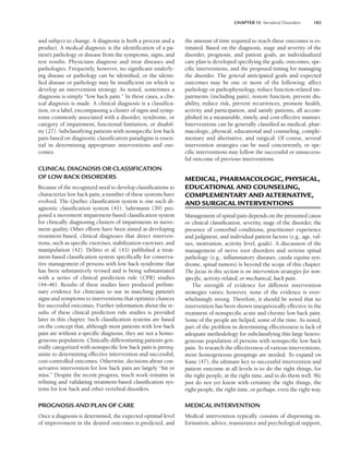
![184 RESOURCES FOR CLINICAL EXERCISE PHYSIOLOGY • www.acsm.org
making referrals, and managing pharmacologic interven-
tion. Dispensing information, advice, reassurance, and
psychological support are discussed in the subsection on
educational and counseling interventions. Recognizing
the need for appropriate referrals is essential for each
practitioner dealing with persons with low back pain.
Pharmacologic intervention is discussed in the next sub-
section.
PHARMACOLOGIC INTERVENTION
Pharmacologic intervention is common for reducing the
symptoms of low back pain and for maintaining function.
Drugs may be nonprescription or prescription, oral, topi-
cal, or injected. Medication types used in the treatment of
low back pain are anti-inflammatories (nonsteroidal anti-
inflammatory drugs [NSAIDS], and steroids), muscle re-
laxants, analgesics (opioid and nonopioid), antidepres-
sants, anticonvulsants, and anesthetics. Patients need to
understand, however, that medications do not cure low
back pain, but medications may be useful in assisting
them in tolerating activity.
Commonly used nonprescription medications are
acetaminophen (Tylenol), aspirin, Aleve, and ibuprofen
(Motrin and Advil). Acetaminophen is a simple analgesic
with strong evidence for effectiveness in relieving the
symptoms of low back pain and promoting activity and
participation (12). The other medications mentioned are
anti-inflammatory analgesics (NSAIDS).
Widely prescribed for acute and chronic low back pain
(48), NSAIDs purportedly reduce swelling and inflamma-
tion and promote healing. Examples of prescription
NSAIDs are Dolobid, Naprosyn, Relafen, Ansaid,
Voltaren, and Celebrex (Cox-2 inhibitor). NSAIDs have a
number of potentially serious adverse drug reactions
(ADRs), especially gastrointestinal tract irritation and
renal effects. Cox-2 inhibitors have been associated with
risk of stroke and myocardial infarction. Both prescrip-
tion and nonprescription NSAIDs must be used with dis-
cretion. Even use of over-the-counter NSAIDs is not
recommended for more than 10 days. NSAIDs are some-
times augmented with acetaminophen and a muscle re-
laxant (49) (e.g., Flexeril, Soma, Valium) to relieve mus-
cle spasms in acute low back pain. Drowsiness is a
common side effect of muscle relaxants. Oral steroids
(e.g., Prednisone, Medrol) are strong anti-inflammatory
medications and are occasionally used short term (1–2
weeks) for more severe inflammation. Ultram (tramadol)
is a narcotic-like analgesic used to treat moderate to se-
vere pain. A short course of narcotic analgesics (e.g., Dar-
vocet, Tylenol with Codeine, Vicodin, OxyContin) is oc-
casionally prescribed for more severe pain, but less likely
with nonspecific low back pain. Narcotic analgesics are
addictive and usually avoided. Antiseizure medications,
such as Neurontin, and antidepressants are sometimes
used to help control symptoms of chronic low back pain.
In addition, injection therapy is sometimes used for
symptom control. Typical injections consist of myofas-
cial trigger point, intra-articular (facet), or epidural in-
jections and nerve blocks. Myofascial trigger points can
be injected with an anesthetic to relieve pain and spasm.
Intra-articular injections, epidural injections, and nerve
blocks consist of local injections of a mixture that typi-
cally includes steroids and anesthetics into a specific
area under x-ray (fluoroscopy) guidance. These injec-
tions are used both diagnostically and as therapy. That is,
if the symptoms are relieved, the injection was treat-
ment. If the symptoms were not affected, then presum-
ably the injection sites were not the sources of pain.
Intra-articular injections are made directly into the of-
fending lumbar joints relieve pain and inflammation.
Epidural injections consist of injecting into the epidural
space close to the affected area. Epidural injections are
used for patients with nerve root irritation, or compro-
mise, and presumably decrease inflammation of nerve
roots and relieve pain. A lumbar sympathetic nerve
block involves injecting around the sympathetic nerves
to “block” neuropathic pain. Blocks can also be selective
nerve root blocks for specific spinal roots. In severe
cases, injections are used to permanently destroy nerves
to relieve pain. The efficacy of injection therapy is in-
conclusive (13).
PHYSICAL INTERVENTION
Physical intervention consists of a broad range of treat-
ments that might be categorized as thermal modalities,
electrotherapeutic modalities, mechanical modalities, or-
thotics, protective and supportive devices, manual ther-
apy, and exercise. In general, the evidence supporting use
of various physical interventions is inconclusive or found
to be useful only during specific stages (i.e., acute or
chronic) and for patients who fit well into current treat-
ment-based classifications. The Philadelphia Panel Evi-
dence-Based Clinical Guideline on Selected Rehabilita-
tion Interventions for Low Back Pain (50) provided the
status of the information on the effectiveness of various
management strategies for use with adults with low back
pain, but did not include evidence for the effectiveness of
manipulation in their review.
Thermal modalities are physical agents that use heat,
cold, sound or light energy to decrease pain, increase tis-
sue extensibility, reduce soft inflammation, decrease
swelling, remodel scar tissue, and so forth. Ultrasound
(which has both thermal and mechanical effects), hy-
drotherapy, hot packs, and cold packs are examples of
thermal modalities used in treatment of low back pain.
Typically, thermal agents, if used, are used in the acute
stages of low back pain or as adjunctive interventions to
other physical interventions, such as using heat com-
bined with stretching. Generally, the effectiveness of ther-
mal therapy has not been substantiated.
LWBK191-4034G-C13_175-196.qxd 06/11/2008 11:10 AM Page 184](https://image.slidesharecdn.com/acsmsresourcesforclinicalexercisephysiology-230410064150-fb83bfa2/85/ACSM_s-Resources-for-Clinical-Exercise-Physiology-pdf-198-320.jpg)
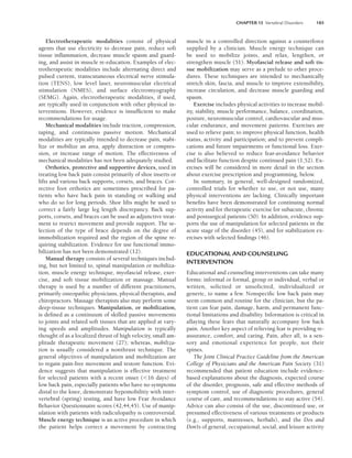
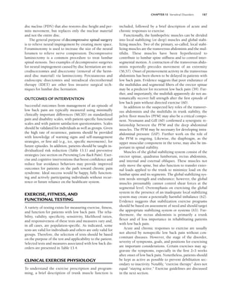
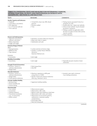
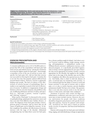
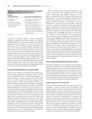
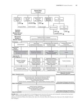
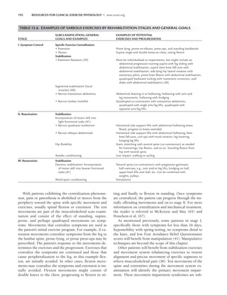
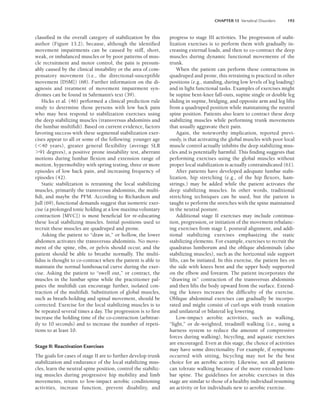
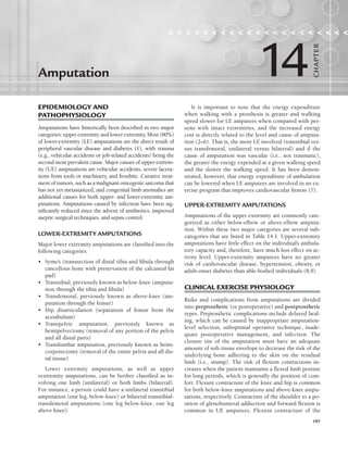
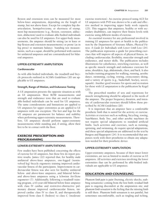


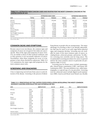
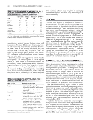
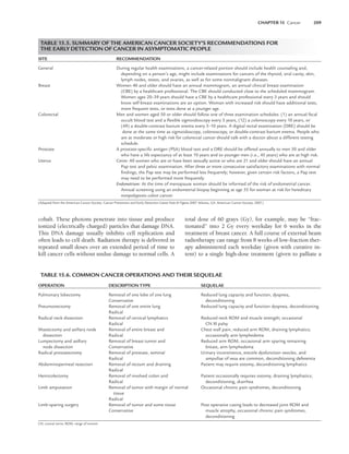
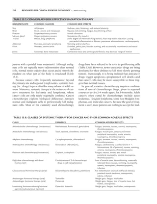
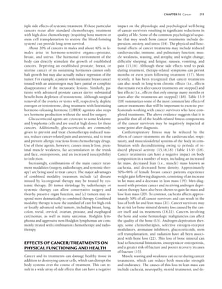
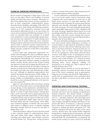
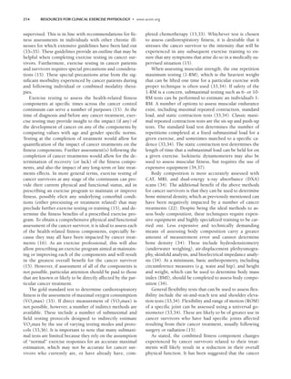
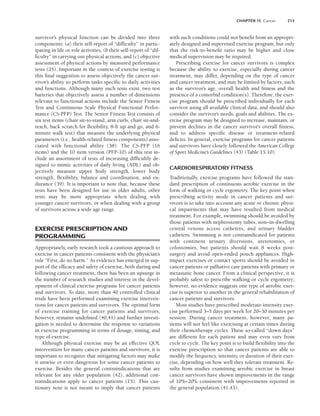
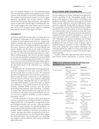
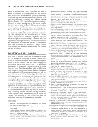
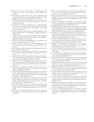
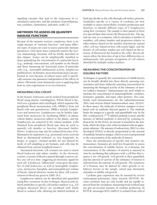
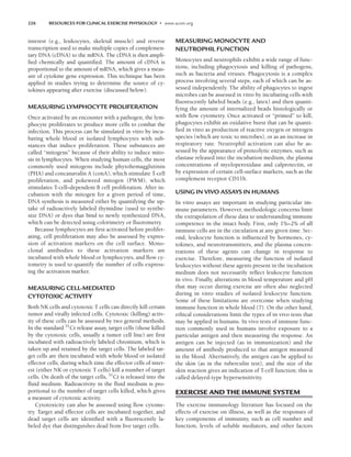
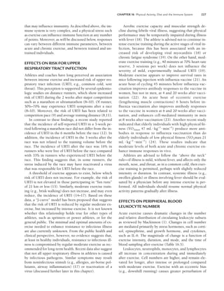
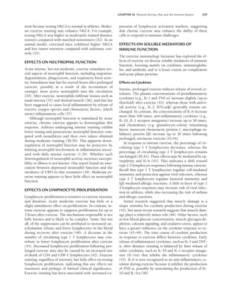
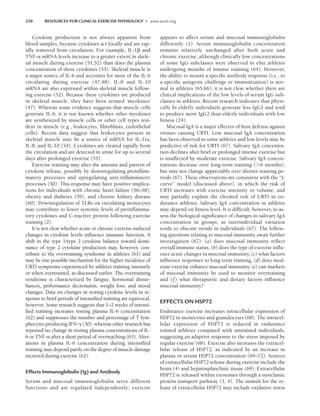
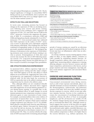
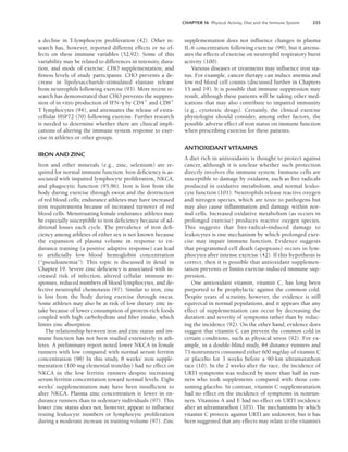
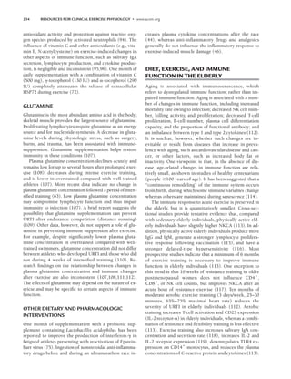
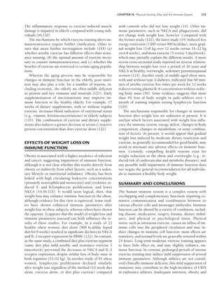
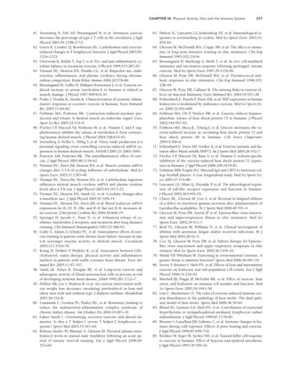
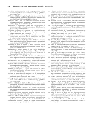
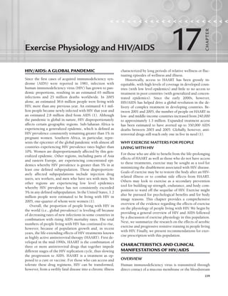
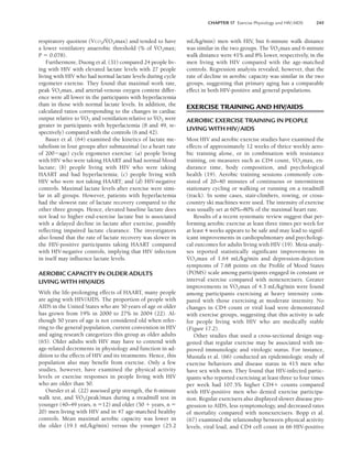
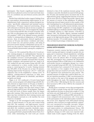
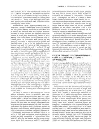
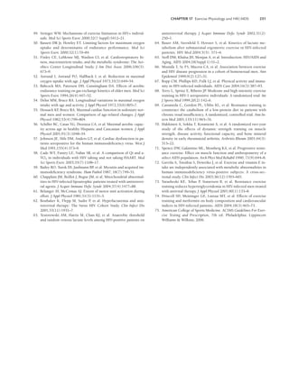
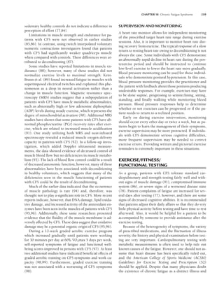
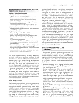
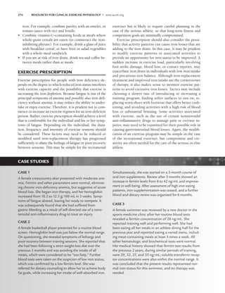
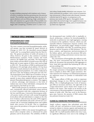
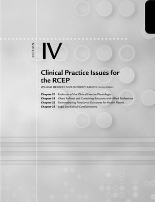

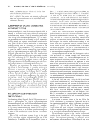
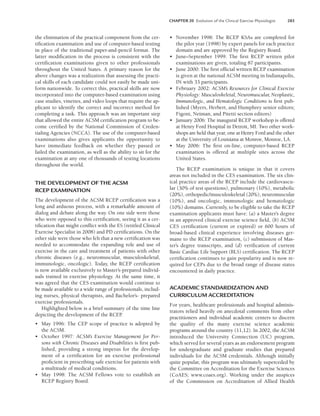
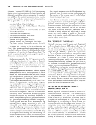
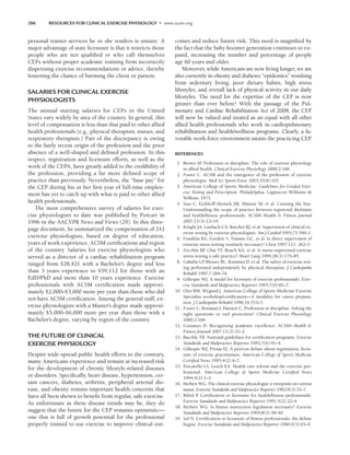

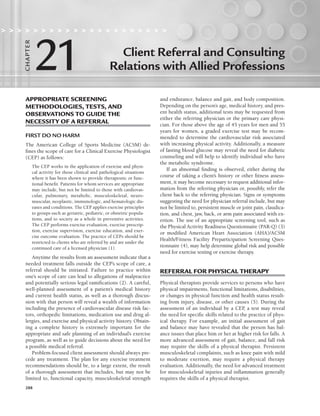
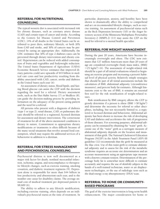

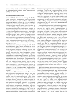
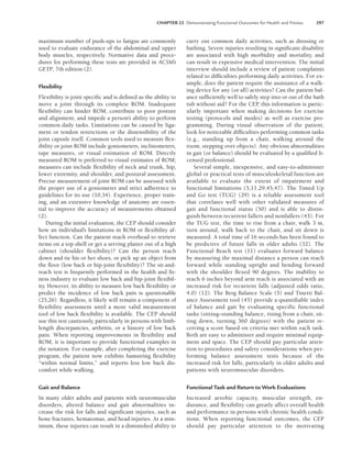
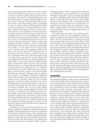

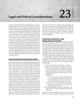
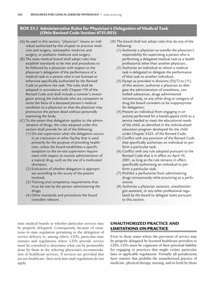
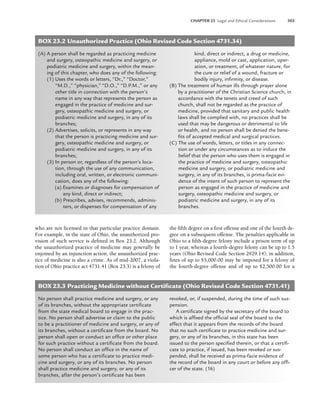
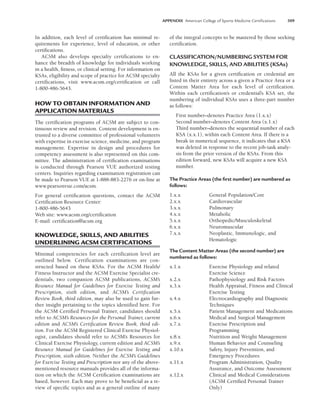
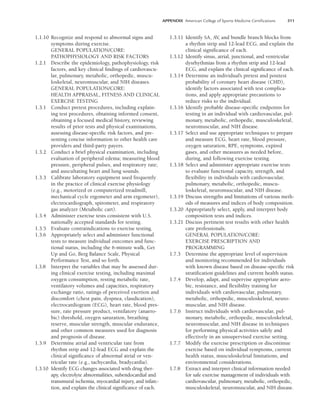
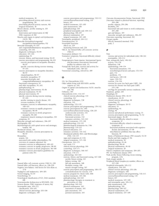
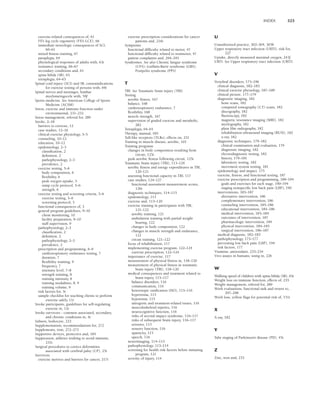


![PROFESSIONAL ISSUES [INCLUDING ETHICS] IN PHYSIOTHERAPY SRS](https://cdn.slidesharecdn.com/ss_thumbnails/professionalissuesandethicssrs-200404153912-thumbnail.jpg?width=640&height=640&fit=bounds)


