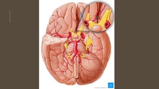
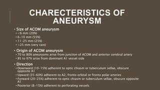
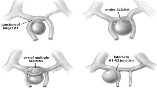
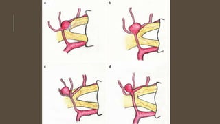
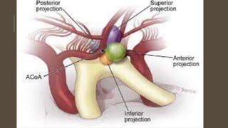
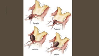
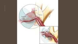
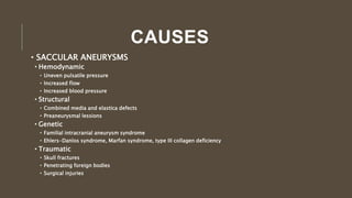
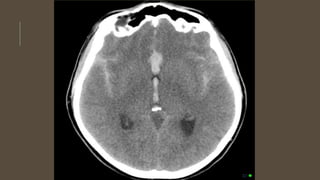
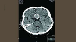
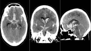
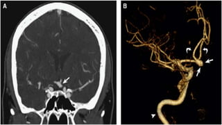
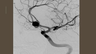
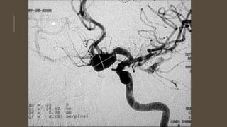
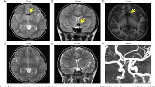
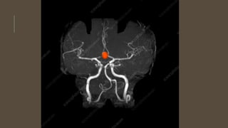
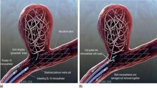
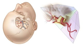
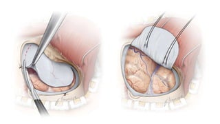
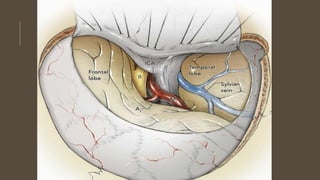
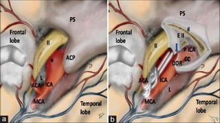
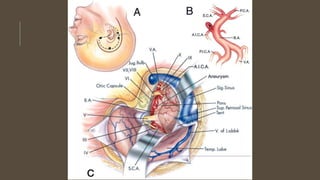
ACOM ANEURYSM BY:
DR. ANAS AHMED
OUTLINES
• INTRODUCTION
• ANATOMY
• CAUSES
• CLINICAL
• INVESTIGATIONS
• MANAGEMENT
INTRODUCTION
• Anterior communicating artery is the most common site for
aneurysms
• These aneurysms are clinically silent until they rupture
• They can produce symptoms due to subarachnoid hemorrhage or
due to mass effect
• There are 2 types of aneurysms
• Saccular
• Fusiform
ANATOMY
• Anterior communicating artery communicate two anterior cerebral
arteries
• A1 segment of the Anterior cerebral artery runs from internal carotid
artery to anterior communicating arteries
• The junction of A1 and anterior communicating artery lies over optic
chiasm in 70% cases while over optic nerve in 30% of cases
• Rarely A1 segment is replaced by artery arising from carotid at level
of anterior clinoid and passes under ipsilateral optic nerve perceived
as misplaced A1 segment and this anomaly is strongly associated
with aneurysm
• Cholinergic nuclei are located in paraolfactory area and around the
inferior third ventricle which function in memory and arousal and are
• Anterior communicating artery gives perforating branches to lamina
terminalis, anterior hypothalamus, septal nucleus, medial
paraolfactory nuclei, subcallosal cingulum and genu of corpus
callosum
• Two larger branches are subcallosal and medial callosal arteries
CHARECTERISTICS OF
ANEURYSM
• Size of ACOM aneurysm
• <6 mm (20%)
• 6-10 mm (55%)
• 11-25 mm (25%)
• >25 mm (very rare)
• Origin of ACOM aneurysm
• 70 to 80% aneurysms arise from junction of ACOM and anterior cerebral artery
• 85 to 97% arise from dominant A1 vessel side
• Direction
• Downward (10-15%) adherent to optic chiasm or tuberculum sellae, obscure
opposite A1
• Upward (35-60%) adherent to A2, fronto orbital or fronto polar arteries
• Forward (20-25%) adherent to optic chiasm or tuberculum sellae, obscure opposite
A1
• Posterior (8-15%) adherent to perforating vessels
CAUSES
• SACCULAR ANEURYSMS
• Hemodynamic
• Uneven pulsatile pressure
• Increased flow
• Increased blood pressure
• Structural
• Combined media and elastica defects
• Preaneurysmal lessions
• Genetic
• Familial intracranial aneurysm syndrome
• Ehlers-Danlos syndrome, Marfan syndrome, type III collagen deficiency
• Traumatic
• Skull fractures
• Penetrating foreign bodies
• Surgical injuries
• Infectious
• Bacterial
• Fungal
• Neoplastic
• Metastatic
• Primary Neoplasms
• Aneurysms associated with neoplasms
• Radiation
• Others
• Granulomatous angitis
• Systemic Lupus erythematosus
• Moyamoya disease
• Sickle cell anemia
• FUSIFORM ANEURYSMS
• Atherosclerosis
• Structural
• Long areas of loss of elastica and media
• Diffuse fibromuscular dysplasia
• Genetic
• Marfan’s syndrome, pseudoxanthoma elasticum
• Infectious
• Syphilis
• Hemodynamic
• Coaractation of aorta
• Radiations
• Others
• Giant cell artritis
CLINICAL
• Ruptured Aneurysm
• Meningism
• Coma
• Nausea and vomiting
• Headache
• Neurological deficit
• Dysphasia
• Homonymous hemianopia
• Third nerve palsy
• Unruptured aneurysm
• Headache
• Visual loss
• Hormonal imbalance
• Personality changes
• Thrombo-embolism
• Seizures
• Focal neurological deficits
INVESTIGATIONS
• CT scan
• CT angiogram
• MRI and MRA
• DSA
MANAGEMENT
• GENERAL MANAGEMENT
• Hemodilution
• Hypervolumia
• Hypertention
• Hydrocephalus
• Seizures
• Compression stockings
• Calcium channel blockers
MANAGEMENT
• SPECIFIC MANAGEMENT
• Clipping
• Pterional approach
• Bifrontal approach
• Coilling
MANAGEMENT
• Advantages of early surgery
• Eliminates chances of rehemorrhage
• Removes clots, decreasing chances of vasospasm
• Allows hypertensive and endovascular treatment of vasospasm
• Prevents complication s of bed rest
• Soft clot easier to dissect
• Shorten hospital stay
•Advantages of delayed surgery
• Improved hemodynamic status of brain
• Brain slack
• Proven excellent surgical results
COMPLICATIONS
• Aneurysmal Rupture
• Damage to temporal lobe
• Nerve injury
• Artery injury
• CSF leak
• Hemtoma
• Memory loss
• Personality changes
• Visual loss
• Endocrine disturbance
ACOM ANEURYSM Presentation slide share..

ACOM ANEURYSM Presentation slide share..

  • 1.
  • 2.
    OUTLINES • INTRODUCTION • ANATOMY •CAUSES • CLINICAL • INVESTIGATIONS • MANAGEMENT
  • 3.
    INTRODUCTION • Anterior communicatingartery is the most common site for aneurysms • These aneurysms are clinically silent until they rupture • They can produce symptoms due to subarachnoid hemorrhage or due to mass effect • There are 2 types of aneurysms • Saccular • Fusiform
  • 4.
    ANATOMY • Anterior communicatingartery communicate two anterior cerebral arteries • A1 segment of the Anterior cerebral artery runs from internal carotid artery to anterior communicating arteries • The junction of A1 and anterior communicating artery lies over optic chiasm in 70% cases while over optic nerve in 30% of cases • Rarely A1 segment is replaced by artery arising from carotid at level of anterior clinoid and passes under ipsilateral optic nerve perceived as misplaced A1 segment and this anomaly is strongly associated with aneurysm • Cholinergic nuclei are located in paraolfactory area and around the inferior third ventricle which function in memory and arousal and are
  • 5.
    • Anterior communicatingartery gives perforating branches to lamina terminalis, anterior hypothalamus, septal nucleus, medial paraolfactory nuclei, subcallosal cingulum and genu of corpus callosum • Two larger branches are subcallosal and medial callosal arteries
  • 10.
    CHARECTERISTICS OF ANEURYSM • Sizeof ACOM aneurysm • <6 mm (20%) • 6-10 mm (55%) • 11-25 mm (25%) • >25 mm (very rare) • Origin of ACOM aneurysm • 70 to 80% aneurysms arise from junction of ACOM and anterior cerebral artery • 85 to 97% arise from dominant A1 vessel side • Direction • Downward (10-15%) adherent to optic chiasm or tuberculum sellae, obscure opposite A1 • Upward (35-60%) adherent to A2, fronto orbital or fronto polar arteries • Forward (20-25%) adherent to optic chiasm or tuberculum sellae, obscure opposite A1 • Posterior (8-15%) adherent to perforating vessels
  • 16.
    CAUSES • SACCULAR ANEURYSMS •Hemodynamic • Uneven pulsatile pressure • Increased flow • Increased blood pressure • Structural • Combined media and elastica defects • Preaneurysmal lessions • Genetic • Familial intracranial aneurysm syndrome • Ehlers-Danlos syndrome, Marfan syndrome, type III collagen deficiency • Traumatic • Skull fractures • Penetrating foreign bodies • Surgical injuries
  • 17.
    • Infectious • Bacterial •Fungal • Neoplastic • Metastatic • Primary Neoplasms • Aneurysms associated with neoplasms • Radiation • Others • Granulomatous angitis • Systemic Lupus erythematosus • Moyamoya disease • Sickle cell anemia
  • 18.
    • FUSIFORM ANEURYSMS •Atherosclerosis • Structural • Long areas of loss of elastica and media • Diffuse fibromuscular dysplasia • Genetic • Marfan’s syndrome, pseudoxanthoma elasticum • Infectious • Syphilis • Hemodynamic • Coaractation of aorta • Radiations • Others • Giant cell artritis
  • 19.
    CLINICAL • Ruptured Aneurysm •Meningism • Coma • Nausea and vomiting • Headache • Neurological deficit • Dysphasia • Homonymous hemianopia • Third nerve palsy
  • 20.
    • Unruptured aneurysm •Headache • Visual loss • Hormonal imbalance • Personality changes • Thrombo-embolism • Seizures • Focal neurological deficits
  • 21.
    INVESTIGATIONS • CT scan •CT angiogram • MRI and MRA • DSA
  • 31.
    MANAGEMENT • GENERAL MANAGEMENT •Hemodilution • Hypervolumia • Hypertention • Hydrocephalus • Seizures • Compression stockings • Calcium channel blockers
  • 32.
    MANAGEMENT • SPECIFIC MANAGEMENT •Clipping • Pterional approach • Bifrontal approach • Coilling
  • 34.
    MANAGEMENT • Advantages ofearly surgery • Eliminates chances of rehemorrhage • Removes clots, decreasing chances of vasospasm • Allows hypertensive and endovascular treatment of vasospasm • Prevents complication s of bed rest • Soft clot easier to dissect • Shorten hospital stay •Advantages of delayed surgery • Improved hemodynamic status of brain • Brain slack • Proven excellent surgical results
  • 49.
    COMPLICATIONS • Aneurysmal Rupture •Damage to temporal lobe • Nerve injury • Artery injury • CSF leak • Hemtoma • Memory loss • Personality changes • Visual loss • Endocrine disturbance