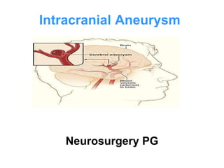
Intracranial Aneurysm
Neurosurgery PG
•Cerebral aneurysms are pathologic focal dilatations of the
cerebrovasculature that are prone to rupture.
• Saccular, berry, or congenital aneurysms constitute 90%
and located at branch points.
• Dolichoectatic, fusiform, or arteriosclerotic aneurysms
are elongated outpouchings of proximal arteries that
account for 7%
• Infectious or mycotic aneurysms are situated
peripherally and comprise 0.5% of all cerebral
aneurysms.
• Other peripheral lesions include neoplastic aneurysms,
rare sequelae of embolized tumor fragments, and
traumatic aneurysms.
• Trauma can also cause dissecting aneurysms
Intracranial Aneurysm
• Artery junction points may become
weak, causing ballooning of the
blood vessel wall that can form a
small sac or aneurysm.
• The integrity of the internal elastic lamina is
compromised, with associated elastic defects
in the adjacent layers of the tunica media and
adventitia.
• Muscular defects of the tunica media and
minimal support of adjacent brain
parenchyma augment the pathologic potential
of chronic hemodynamic stress on the arterial
wall.
• Focal turbulence and discontinuity of the
normal architecture at vessel bifurcations may
account for the propensity of saccular
aneurysm formation at these locations.
Pathophysiology
Pathophysiology
Distal aneurysms may be smaller compared with proximal sites,
yet the risk of rupture may be dissimilar due to the relatively
thinner parent artery wall thickness.
Abnormalities of the internal elastic lamina may be congenital or
degenerative. Multiple conditions have been associated with
cerebral aneurysms; they include the following:
Autosomal dominant inherited polycystic kidney disease
Fibromuscular dysplasia
Arteriovenous malformations
Osler-Weber-Rendu syndrome
Coarctation of the aorta
Other vascular anomalies
Moyamoya syndrome
Marfan syndrome
Ehlers-Danlos syndrome, type IV
Other collagen type III disorders
Pseudoxanthoma elasticum
Alpha1-antitrypsin deficiency
Systemic lupus erythematosus
Pathophysiology
Sickle cell anemia
Bacterial endocarditis
Fungal infections
Neurofibromatosis type 1
Tuberous sclerosis
Dolichoectatic aneurysms of proximal vessels most likely have
an arteriosclerotic etiology. These tortuous, elongated dilatations
devoid of a true aneurysmal neck.
Infectious aneurysms typically are situated in distal branches of
the middle cerebral artery (MCA; 75-80% of cases), reflecting the
embolic origin of these lesions.
Traumatic aneurysms may be located in peripheral cortical
branches secondary to contact with the falcine edge or skull
fractures associated with penetrating or closed head injury.
Incidence
• Headache: This is characterized by the
acute onset of severe pain, which patients
often describe as "the worst headache of
my life.“
• Facial pain: Aneurysms may produce facial
pain.
• Manifestations of meningeal irritation:
Neck pain or stiffness
• Alterations in consciousness: The
sudden elevation of ICP associated with
aneurysmal rupture may lead to a severe
decline in cerebral perfusion pressure,
causing syncope (50% of cases). Confusion
or mild impairment in alertness also may be
noted.
• Seizures: are present in 25% of
aneurysmal SAH cases, with most events
occurring within 24 hours of onset.
• Autonomic
disturbanc
es:
Subarachnoid
accumulation of products of blood degradation
may elicit fever. Nausea or vomiting, sweating,
chills, and cardiac arrhythmias also may be
present.
• Visual symptoms: Blurring of
vision, diplopia, or visual field defects may be
present.
Investigation
CT brain Aneurysmal SAH may be detected in 90-95% of cases.
MRI brain Fluid-attenuated inversion recovery (FLAIR)
sequences are very sensitive for SAH, although the comparison of CT
scan and MRI in detection of SAH is controversial.
CT angiography Conventional angiography is the
definitive procedure for the detection and characterization of cerebral
aneurysms. Aneurysm location, size, and morphology may be
evaluated in the acute or chronic setting with this modality.
Digital subtraction angiography with biplanar magnification views
provides details that may be helpful in identifying an acutely ruptured
aneurysm.
Transcranial Doppler TCD facilitates the diagnosis of
vasospasm and serial monitoring of cerebral blood flow at the bedside.
TCD has exhibited close correlation with angiography in the setting of
vasospasm, typically manifesting 3-21 days following aneurysmal SAH.
Aim Allow the brain to
recover from initial
insult ( bleeding)
Prevent or
treat other
complication
Vasospasm
Management
Surgical
Clipping Coiling
Medical
Medication
Management
Fischer’s grading of subarachnoid haemorrhage
Grade 1 No detectable blood on CT scan
Grade 2 Diffuse thin SAH
Vertical layers < 1 mm thickness
Grade 3 Localised clot and/or thick SAH vertical layer > 1 mm
thickness
Grade 4 Intraventricular or intracerebral clot with diffuse or no
subarachnoid haemorrhage
Management
Nimotop
• Calcium channel blockers
(Nimodipine:
, Verapamil (Isoptin)
• Osmotic diuretic (Mannitol 20%)
• Antiepileptics (Phenytoin)
• Antihypertensives (Nitroprusside)
• If surgery is delayed or
contraindicated (antifibrinolytic
agents)
• Analgesics (acetaminophen)
• Laxatives to prevent straining to avoid
BP
Medical Management
Surgical Management
• Clipping: A neurosurgeon can operate on
the brain by cutting open the skull,
identifying the damaged blood vessel and
putting a clip across the aneurysm.
• This prevents blood from entering the
aneurysm and causing further growth or
blood leakage.
• Coiling: A neurosurgeon or interventional
radiologist can thread a tube through the
arteries, as with an angiogram, identify the
aneurysm, and fill it with coils of platinum
wire or with latex.
• This prevents further blood from entering
the aneurysm and resolves the problem.
Aneurysms.pptx

Aneurysms.pptx

  • 1.
  • 2.
    •Cerebral aneurysms arepathologic focal dilatations of the cerebrovasculature that are prone to rupture. • Saccular, berry, or congenital aneurysms constitute 90% and located at branch points. • Dolichoectatic, fusiform, or arteriosclerotic aneurysms are elongated outpouchings of proximal arteries that account for 7% • Infectious or mycotic aneurysms are situated peripherally and comprise 0.5% of all cerebral aneurysms. • Other peripheral lesions include neoplastic aneurysms, rare sequelae of embolized tumor fragments, and traumatic aneurysms. • Trauma can also cause dissecting aneurysms Intracranial Aneurysm
  • 3.
  • 4.
    • Artery junctionpoints may become weak, causing ballooning of the blood vessel wall that can form a small sac or aneurysm.
  • 10.
    • The integrityof the internal elastic lamina is compromised, with associated elastic defects in the adjacent layers of the tunica media and adventitia. • Muscular defects of the tunica media and minimal support of adjacent brain parenchyma augment the pathologic potential of chronic hemodynamic stress on the arterial wall. • Focal turbulence and discontinuity of the normal architecture at vessel bifurcations may account for the propensity of saccular aneurysm formation at these locations. Pathophysiology
  • 11.
    Pathophysiology Distal aneurysms maybe smaller compared with proximal sites, yet the risk of rupture may be dissimilar due to the relatively thinner parent artery wall thickness. Abnormalities of the internal elastic lamina may be congenital or degenerative. Multiple conditions have been associated with cerebral aneurysms; they include the following: Autosomal dominant inherited polycystic kidney disease Fibromuscular dysplasia Arteriovenous malformations Osler-Weber-Rendu syndrome Coarctation of the aorta Other vascular anomalies Moyamoya syndrome Marfan syndrome Ehlers-Danlos syndrome, type IV Other collagen type III disorders Pseudoxanthoma elasticum Alpha1-antitrypsin deficiency Systemic lupus erythematosus
  • 12.
    Pathophysiology Sickle cell anemia Bacterialendocarditis Fungal infections Neurofibromatosis type 1 Tuberous sclerosis Dolichoectatic aneurysms of proximal vessels most likely have an arteriosclerotic etiology. These tortuous, elongated dilatations devoid of a true aneurysmal neck. Infectious aneurysms typically are situated in distal branches of the middle cerebral artery (MCA; 75-80% of cases), reflecting the embolic origin of these lesions. Traumatic aneurysms may be located in peripheral cortical branches secondary to contact with the falcine edge or skull fractures associated with penetrating or closed head injury.
  • 13.
  • 14.
    • Headache: Thisis characterized by the acute onset of severe pain, which patients often describe as "the worst headache of my life.“ • Facial pain: Aneurysms may produce facial pain. • Manifestations of meningeal irritation: Neck pain or stiffness
  • 15.
    • Alterations inconsciousness: The sudden elevation of ICP associated with aneurysmal rupture may lead to a severe decline in cerebral perfusion pressure, causing syncope (50% of cases). Confusion or mild impairment in alertness also may be noted. • Seizures: are present in 25% of aneurysmal SAH cases, with most events occurring within 24 hours of onset.
  • 16.
    • Autonomic disturbanc es: Subarachnoid accumulation ofproducts of blood degradation may elicit fever. Nausea or vomiting, sweating, chills, and cardiac arrhythmias also may be present. • Visual symptoms: Blurring of vision, diplopia, or visual field defects may be present.
  • 17.
    Investigation CT brain AneurysmalSAH may be detected in 90-95% of cases. MRI brain Fluid-attenuated inversion recovery (FLAIR) sequences are very sensitive for SAH, although the comparison of CT scan and MRI in detection of SAH is controversial. CT angiography Conventional angiography is the definitive procedure for the detection and characterization of cerebral aneurysms. Aneurysm location, size, and morphology may be evaluated in the acute or chronic setting with this modality. Digital subtraction angiography with biplanar magnification views provides details that may be helpful in identifying an acutely ruptured aneurysm. Transcranial Doppler TCD facilitates the diagnosis of vasospasm and serial monitoring of cerebral blood flow at the bedside. TCD has exhibited close correlation with angiography in the setting of vasospasm, typically manifesting 3-21 days following aneurysmal SAH.
  • 18.
    Aim Allow thebrain to recover from initial insult ( bleeding) Prevent or treat other complication Vasospasm
  • 19.
  • 20.
  • 21.
    Fischer’s grading ofsubarachnoid haemorrhage Grade 1 No detectable blood on CT scan Grade 2 Diffuse thin SAH Vertical layers < 1 mm thickness Grade 3 Localised clot and/or thick SAH vertical layer > 1 mm thickness Grade 4 Intraventricular or intracerebral clot with diffuse or no subarachnoid haemorrhage Management
  • 22.
    Nimotop • Calcium channelblockers (Nimodipine: , Verapamil (Isoptin) • Osmotic diuretic (Mannitol 20%) • Antiepileptics (Phenytoin) • Antihypertensives (Nitroprusside) • If surgery is delayed or contraindicated (antifibrinolytic agents) • Analgesics (acetaminophen) • Laxatives to prevent straining to avoid BP Medical Management
  • 24.
  • 25.
    • Clipping: Aneurosurgeon can operate on the brain by cutting open the skull, identifying the damaged blood vessel and putting a clip across the aneurysm. • This prevents blood from entering the aneurysm and causing further growth or blood leakage.
  • 26.
    • Coiling: Aneurosurgeon or interventional radiologist can thread a tube through the arteries, as with an angiogram, identify the aneurysm, and fill it with coils of platinum wire or with latex. • This prevents further blood from entering the aneurysm and resolves the problem.