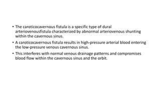
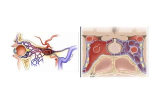
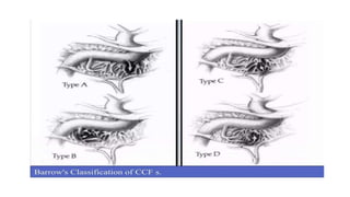
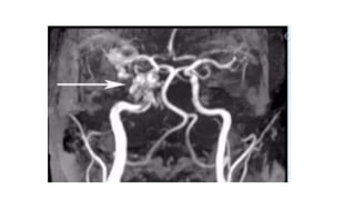
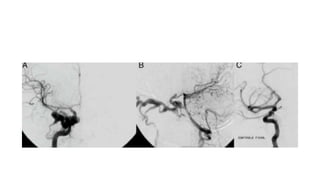
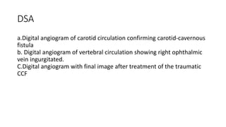
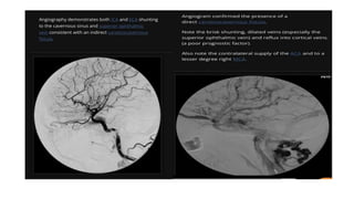
Caroticocavernous fistula
• The caroticocavernous fistula is a specific type of dural
arteriovenousfistula characterized by abnormal arteriovenous shunting
within the cavernous sinus.
• A caroticocavernous fistula results in high-pressure arterial blood entering
the low-pressure venous cavernous sinus.
• This interferes with normal venous drainage patterns and compromises
blood flow within the cavernous sinus and the orbit.
Epidemiology
Caroticocavernous fistulas represent approximately 12% of all dural
arteriovenous fistulas.
Direct CCFs are often secondary to trauma: head trauma: Youngs:
Presentation: acute/rapid.
indirect CCFs : Post menopause: insidious.
Classification
direct indirect
CLASSIFICATION
Type A: direct connection between the intracavernous ICA and CS
Type B: dural shunt between intracavernous branches of the ICA
and CS
Type C: dural shunts between meningeal branches of the ECA and
CS
Type D: B + C
OTHER CLASSIFICATION
• Traumatic or spontaneous fistulas.
• Flow:
-Direct high flow
-Indirect low flow fistula
Pathophysiology
Direct: type A: ICA>CS Indirect: Br of ICA/ECS> CS; types B, C, D
The most frequent among indirect is type C, with meningeal branches of
the ECA forming the fistula
Direct
-Trauma
-Ruptured intracavernous
carotid aneurysms
-Collagen deficiency
syndromes
-arterial dissection
-Fibromuscular dysplasia
-Direct surgical trauma
Indirect
Cause often unknown:
-Pregnancy
-Sinusitis
-Trauma
-Surgical procedures
-Cavernous sinus thrombosis
They are postulated to occur
secondary to cavernous sinus
thrombosis with revascularisation
Clinical presentation
• Their symptoms range from benign to extremely severe
ophthalmologic or neurologic complications.
• Clinical presentation is consequence of the elevated intracavernous
pressure.
• In direct, high-flow CCF s, symptoms appear suddenly.
• Symptoms caused by CCFs are related to their size, duration, location,
adequacy and route of venous drainage, and presence of arterial and
venous collaterals
CLINICAL PRESENTATION
• Pulsatile exophthalmos(75%)
• Chemosis and subconjunctival
• haemorrhage
• Proptosis
• Progressive visual loss: 25-32%
• Raised intracranial pressure
• Cranial nerve (III, IV, Vc, VI) palsies
The Dandy's triad: pulsatile exophthalmos, bruit and chemosis
• Moreover, other factors like dominant pattern of venous drainage the
size and location of CCF and the presence of collateral vessels (arterial
or venous) are important in this setting.
• Diplopia, pain, cephalic bruit, ophtalmoplegia, visual loss (Ophth.
vein)
• Intracranial hemorrhage : sphenoparietal sinus and deep middle
cerebral vein)
External haemorrhage: Otorrhagia, epistaxis (Pterygoid plexus)
Radiographic features
• CT:
-Proptosis Enlarged superior ophthalmic veins
-Extraocular muscles may be enlarged
-Orbital oedema
-May show SAH/ICH from a ruptured cortical vein
• Angiography (DSA)
-Rapid shunting from ICA to CS
-Enlarged draining veins
-Retrograde flow from CS, most commonly into the ophthalmic veins
• Ultrasound
Arterialised ophthalmic veins may be seen on Doppler study
Magnetic Resonance Angiogram
(MRA) image demonstrating an
enlarged superior ophthalmic vein
MA demonstrating a right
carotid cavernous fistula
DSA
a.Digital angiogram of carotid circulation confirming carotid-cavernous
fistula
b. Digital angiogram of vertebral circulation showing right ophthalmic
vein ingurgitated.
C.Digital angiogram with final image after treatment of the traumatic
CCF
Treatment and prognosis
• The natural history of CF is highly varied, ranging from spontaneous
closure to rapidly progressive symptoms.
• Poor treatment outcome indicators include feeding vessel aneurysms
(indirect CF) and retrograde filling of cortical veins (increased risk of
haemorrhage).
• Direct fistulas have a relatively high spontaneous rate of haemorrhage
• subarachnoid, intracerebral or external haemorrhage (epistaxis, or
otorrhagia).
Subconjunctival hemorrhage is also common but does not carry the
same poor prognosis
GOAL OF TREATMENT
• Direct CF: Occlude the tear between ICAand CS, preserving the
patency of ICA
• Indirect CCF : Interrupt fistulous communications/reduce CS pressure
Treatment modalities
• Conservative management
(manual compression therapy
and medical therapy)
• Surgical management
• Stereotactic radiosurgery
• Endovascular management
Direct fistula
Transarterial treatment (preferred approach
for direct CCF)
Detachable balloon occlusion
Transarterial coil and material embolization
Covered stent graft placement
(endovascular reconstruction of the parent
artery)
Parent artery occlusion
Transvenous treatment
Transvenous detachable coil embolization
Liquid embolizing agents (n-BCA, Onyx)
Indirect fistula
Transvenous treatment (preferred approach
for indirect CCF)
Transvenous detachable coil embolization
Liquid embolizing agents (n-BCA, Onyx)
Transarterial treatment
Transarterial coil and material embolization
Carotid compression therapy
• Contralateral hand: 10sec: 4-6/hr: Reduces AV shunting + Increase
outlet
venous pressures > Thrombosis.
• Most useful in the treatment of indirect fistulas resulting in
spontaneous closure in up to 30% of cases.
Surgery
• Options:
-Ligation of the CC
-Surgical trapping of the fistula, and
-Surgical transvenous packing.
• Both direct and indirect CCFs:
• Disadv: Cranial nerve deficits and residual fistulous communications.
• Indications for surgical repair include
1. Compromised proximal arterial access that prevents endovascular
repair or causes it to fail.
2. Salvage:failed endovascular treatments.
PARENT ARTERY OCCLUSION
• Arterial sacrifice may be required as a life-saving emergency
treatment
• Indication: Difficult case:
Extensive traumatic vessel wall damage
Active hemorrhage
Transarterial balloon embolisation
• TOC: Symptomatic direct CCF.
If not possible, detachable coils may be use.
• Both arterial and venous access (including superior ophthalmic vein)
• Indirect fistulas typically require a combined transarterial (closure of
feeders) and transvenous (closure of cavernous sinus) approach.
• Indirect types are more difficult to treat and have a higher rate of
spontaneous closure
Balloon Occlusion
• This procedure requires that the CS must be large enough to put the
balloon for embolization and the size of fistula must be smaller than the
inflated balloon, but large enough to allow a deflated balloon.
• The balloon has the advantage of being able to be flow-directed through
the fistula and CS, and must be inflated to a volume larger than the fistula
orifice to prevent its retrograde migration into ICA.
• Angiography is repeated to ensure closure of the fistula and patency of
the ICA
Transarterial embolization
• Mainstay of treatment in high-flow direct CCF's.
• It's an alternative when residual AV shunt remains in dural CCF.
• Embolization can be made with detachable platinum coils and liquid
embolic agents (n-buty| cyanoacrylate, ethylene-vinyl alcohol
copolymer);
• Coils are preferred because of their reliable and controlled deployment
into CS.
• Complications of this procedure includes thromboembolus and ICA
dissection
Covered stent graft placement
• Recent Advance: poly flurotetraethylene-covered stents
Traumatic arterial damage
• immediate obliteration of a direct CF, while preserving ICA patency
• Disadv:
Longitudinal flexibility: difficult navigation: tortuosity of the
intracranial vasculature.
• Vasospasms: Intra-arterial nimodipine and papaverine infusion
Endoleak, coverage of vital perforators, dissection and rupture
Transvenous embolization
• Is the current method of choice in treatment of indirect CCF's.
• The goal of this technique is to catheterize the abnormal CS
superselectively and occlude the fistula without re-routing venous
drainage to cortical structures.
Several routes: Most: inferior petrosal sinus (IPS)
RADIOSURGERY
• Indirect CCFs.
Gamma knife radiosurgery can be used either alone or as an adjunct
therapy before/after endovascular intervention.
Preliminary data : safe and effective alternative treatment
Drawback: 22-mo average lag
Carotico-cavernous fistula Presentation.

Carotico-cavernous fistula Presentation.

  • 1.
  • 4.
    • The caroticocavernousfistula is a specific type of dural arteriovenousfistula characterized by abnormal arteriovenous shunting within the cavernous sinus. • A caroticocavernous fistula results in high-pressure arterial blood entering the low-pressure venous cavernous sinus. • This interferes with normal venous drainage patterns and compromises blood flow within the cavernous sinus and the orbit.
  • 6.
    Epidemiology Caroticocavernous fistulas representapproximately 12% of all dural arteriovenous fistulas. Direct CCFs are often secondary to trauma: head trauma: Youngs: Presentation: acute/rapid. indirect CCFs : Post menopause: insidious.
  • 7.
  • 8.
    CLASSIFICATION Type A: directconnection between the intracavernous ICA and CS Type B: dural shunt between intracavernous branches of the ICA and CS Type C: dural shunts between meningeal branches of the ECA and CS Type D: B + C
  • 10.
    OTHER CLASSIFICATION • Traumaticor spontaneous fistulas. • Flow: -Direct high flow -Indirect low flow fistula
  • 11.
    Pathophysiology Direct: type A:ICA>CS Indirect: Br of ICA/ECS> CS; types B, C, D The most frequent among indirect is type C, with meningeal branches of the ECA forming the fistula Direct -Trauma -Ruptured intracavernous carotid aneurysms -Collagen deficiency syndromes -arterial dissection -Fibromuscular dysplasia -Direct surgical trauma Indirect Cause often unknown: -Pregnancy -Sinusitis -Trauma -Surgical procedures -Cavernous sinus thrombosis They are postulated to occur secondary to cavernous sinus thrombosis with revascularisation
  • 12.
    Clinical presentation • Theirsymptoms range from benign to extremely severe ophthalmologic or neurologic complications. • Clinical presentation is consequence of the elevated intracavernous pressure. • In direct, high-flow CCF s, symptoms appear suddenly. • Symptoms caused by CCFs are related to their size, duration, location, adequacy and route of venous drainage, and presence of arterial and venous collaterals
  • 13.
    CLINICAL PRESENTATION • Pulsatileexophthalmos(75%) • Chemosis and subconjunctival • haemorrhage • Proptosis • Progressive visual loss: 25-32% • Raised intracranial pressure • Cranial nerve (III, IV, Vc, VI) palsies The Dandy's triad: pulsatile exophthalmos, bruit and chemosis
  • 15.
    • Moreover, otherfactors like dominant pattern of venous drainage the size and location of CCF and the presence of collateral vessels (arterial or venous) are important in this setting. • Diplopia, pain, cephalic bruit, ophtalmoplegia, visual loss (Ophth. vein) • Intracranial hemorrhage : sphenoparietal sinus and deep middle cerebral vein) External haemorrhage: Otorrhagia, epistaxis (Pterygoid plexus)
  • 16.
    Radiographic features • CT: -ProptosisEnlarged superior ophthalmic veins -Extraocular muscles may be enlarged -Orbital oedema -May show SAH/ICH from a ruptured cortical vein • Angiography (DSA) -Rapid shunting from ICA to CS -Enlarged draining veins -Retrograde flow from CS, most commonly into the ophthalmic veins • Ultrasound Arterialised ophthalmic veins may be seen on Doppler study
  • 17.
    Magnetic Resonance Angiogram (MRA)image demonstrating an enlarged superior ophthalmic vein MA demonstrating a right carotid cavernous fistula
  • 20.
    DSA a.Digital angiogram ofcarotid circulation confirming carotid-cavernous fistula b. Digital angiogram of vertebral circulation showing right ophthalmic vein ingurgitated. C.Digital angiogram with final image after treatment of the traumatic CCF
  • 22.
    Treatment and prognosis •The natural history of CF is highly varied, ranging from spontaneous closure to rapidly progressive symptoms. • Poor treatment outcome indicators include feeding vessel aneurysms (indirect CF) and retrograde filling of cortical veins (increased risk of haemorrhage). • Direct fistulas have a relatively high spontaneous rate of haemorrhage • subarachnoid, intracerebral or external haemorrhage (epistaxis, or otorrhagia). Subconjunctival hemorrhage is also common but does not carry the same poor prognosis
  • 23.
    GOAL OF TREATMENT •Direct CF: Occlude the tear between ICAand CS, preserving the patency of ICA • Indirect CCF : Interrupt fistulous communications/reduce CS pressure
  • 24.
    Treatment modalities • Conservativemanagement (manual compression therapy and medical therapy) • Surgical management • Stereotactic radiosurgery • Endovascular management Direct fistula Transarterial treatment (preferred approach for direct CCF) Detachable balloon occlusion Transarterial coil and material embolization Covered stent graft placement (endovascular reconstruction of the parent artery) Parent artery occlusion Transvenous treatment Transvenous detachable coil embolization Liquid embolizing agents (n-BCA, Onyx) Indirect fistula Transvenous treatment (preferred approach for indirect CCF) Transvenous detachable coil embolization Liquid embolizing agents (n-BCA, Onyx) Transarterial treatment Transarterial coil and material embolization
  • 25.
    Carotid compression therapy •Contralateral hand: 10sec: 4-6/hr: Reduces AV shunting + Increase outlet venous pressures > Thrombosis. • Most useful in the treatment of indirect fistulas resulting in spontaneous closure in up to 30% of cases.
  • 26.
    Surgery • Options: -Ligation ofthe CC -Surgical trapping of the fistula, and -Surgical transvenous packing. • Both direct and indirect CCFs: • Disadv: Cranial nerve deficits and residual fistulous communications. • Indications for surgical repair include 1. Compromised proximal arterial access that prevents endovascular repair or causes it to fail. 2. Salvage:failed endovascular treatments.
  • 27.
    PARENT ARTERY OCCLUSION •Arterial sacrifice may be required as a life-saving emergency treatment • Indication: Difficult case: Extensive traumatic vessel wall damage Active hemorrhage
  • 28.
    Transarterial balloon embolisation •TOC: Symptomatic direct CCF. If not possible, detachable coils may be use. • Both arterial and venous access (including superior ophthalmic vein) • Indirect fistulas typically require a combined transarterial (closure of feeders) and transvenous (closure of cavernous sinus) approach. • Indirect types are more difficult to treat and have a higher rate of spontaneous closure
  • 29.
    Balloon Occlusion • Thisprocedure requires that the CS must be large enough to put the balloon for embolization and the size of fistula must be smaller than the inflated balloon, but large enough to allow a deflated balloon. • The balloon has the advantage of being able to be flow-directed through the fistula and CS, and must be inflated to a volume larger than the fistula orifice to prevent its retrograde migration into ICA. • Angiography is repeated to ensure closure of the fistula and patency of the ICA
  • 30.
    Transarterial embolization • Mainstayof treatment in high-flow direct CCF's. • It's an alternative when residual AV shunt remains in dural CCF. • Embolization can be made with detachable platinum coils and liquid embolic agents (n-buty| cyanoacrylate, ethylene-vinyl alcohol copolymer); • Coils are preferred because of their reliable and controlled deployment into CS. • Complications of this procedure includes thromboembolus and ICA dissection
  • 31.
    Covered stent graftplacement • Recent Advance: poly flurotetraethylene-covered stents Traumatic arterial damage • immediate obliteration of a direct CF, while preserving ICA patency • Disadv: Longitudinal flexibility: difficult navigation: tortuosity of the intracranial vasculature. • Vasospasms: Intra-arterial nimodipine and papaverine infusion Endoleak, coverage of vital perforators, dissection and rupture
  • 32.
    Transvenous embolization • Isthe current method of choice in treatment of indirect CCF's. • The goal of this technique is to catheterize the abnormal CS superselectively and occlude the fistula without re-routing venous drainage to cortical structures. Several routes: Most: inferior petrosal sinus (IPS)
  • 33.
    RADIOSURGERY • Indirect CCFs. Gammaknife radiosurgery can be used either alone or as an adjunct therapy before/after endovascular intervention. Preliminary data : safe and effective alternative treatment Drawback: 22-mo average lag