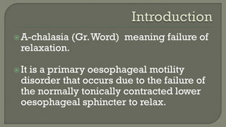
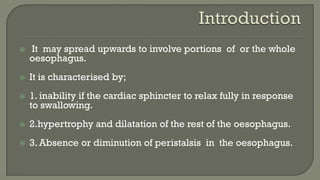
ARANSI RILWAN A.
 Introduction
 Epidemiology
 Aetiology
 Pathogenesis
 Clinical Features
 Investigation
 Treatment
 Complications
 Differential diagnosis
A-chalasia (Gr.Word) meaning failure of
relaxation.
It is a primary oesophageal motility
disorder that occurs due to the failure of
the normally tonically contracted lower
oesophageal sphincter to relax.
 It may spread upwards to involve portions of or the whole
oesophagus.
 It is characterised by;
 1. inability if the cardiac sphincter to relax fully in response
to swallowing.
 2.hypertrophy and dilatation of the rest of the oesophagus.
 3. Absence or diminution of peristalsis in the oesophagus.
Achalasia may occur at any age, however,
incidence peaks in individuals in the 3rd
and 5th decade of life.

No sex predilection.
 Idiopahic
 Proposed causes include;
 1.Neuronal degeneration
 2.viral infection
 3.Chagas disease
 4.gastroesophageal junction obstruction
 5.genetic inheritance
 6.Autoimune disease.
Insults to the oesophagus perhaps a viral
infection or some other external factors
results in myenteric plexus inflammation.
Inflammation leads to autoimmune
response in a susceptible population who
may be genetically predisposed.
 Subsequent chronic inflammation leads to
destruction of the inhibitory nitrigenic myenteric
neurons resulting in inability of the L.O .S to
relax in response to swallowing.
 The muscle, especially the circular muscle, of
the rest of the oesophagus overworks to propel
food through the L. O. S and thus undergoes
hypertrophy.
 The disease process spreads upwards,
propulsive peristalsis ceases
 Food and saliva now accumulate in the distal
oesophagus and only trickle through the
L.O.S when the hydrostatic pressure is
high enough to overcome the intracardiac
pressure.
Intermittent dysphagia.
Retrostemal pain which may radiate to
the neck and interscapular and subcostal
regions.
Halithosis
 Regurgitation of food.
 Some may present with pulmonary
symptoms such as dyspnoea, pneumonitis
and chronic cough and purulent
expectoration indicative of lung abscess
due to aspiration of accumulated food.
 Weight loss.
Aim:
Confirm diagnosis
Complications
Optimize for treatment.
 Gold standard for diagnosis is oesophageal
manometry, however, these investigations
can be done in suspected cases;
 Chest radiograph:
retrocardiac dilation of the esophagus
retrocardiac air-fluid level
minimal or abscent gastric bubble
signs of aspiration.
 Barium Swallow
o Dilated, tortuous, oesophagus that smoothly
tapers down at the OGJ giving the "Bird's
beak" appearance.
o absence of gas in the fundus of the
stomach.
o weak, irregular, uncoordinated or absent
peristalsis on fluoroscopy.
Oesophagoscopy:
o The oesophagus contains food debris
and is dilated.
o Possibility of associated tumour (seen in
5-10%)
 Oesophageal manometry may show a high
resting pressure in the cardiac sphincter.
 Normal Manometric Findings
 LOS pressure 15-25mmHg with normal relaxation
on swallowing.
 Mean amplitude distal oesophageal peristaltic
w.ave is 30-100mmHg
 Resting/ excercise ECG: to rule out cardiac
cause of pain.
 FBC.
 Serum electrolyte, urea and creatinine.
 Urinalysis.
 Aim is to reduce the pressure of the L.O.S. so as to allow
food to pass into the stomach unimpeded.
 Achieved by
 i) pharmacological manipulation
 ii) dilatation or stretching and disruption of the circu1ar
muscle of the L.O.S to render it incompetent and
 iii) surgical division of the circu1ar muscle of the L.O.S
(cardiomyotomy).
 Calcium channel blockers (e.g. nifedipine) and
nitrates (e.g. glyceryl trinitrate), which relax the
smooth muscle of the L.O.S have been used.
 Taken 10-30minutes before meals .
 These are reserved primarily for patients who
refuse or are not good candidates for more
effective and invasive forms of therapy.
 Botulinum toxin injection:
 Botulinum toxin (Botox) is a potent inhibitor
of the release of acetylcholine from nerve
endings.
 It is injected endoscopically into the L.O.S
and poisons the excitatory (acetylcholline
releasing) neurons that increase the L.O.S
tone.
 Cardiomyotomy
Surgical intervention is indicated after
failure of repeated dilatation,
in mega-oesophagus,
when associated carcinoma is suspected
or as first line treatment.
 1. Shock.
 2. Perforation of the oesophagus
 3. Bleeding.
 4. Mediastinitis.
 5. Pneumonia.
 6. Septicaemia.
 7. Oesophageal Stricture.
 8. Gastric outlet obstruction.
 9. Malignant change may occur in a strictured
oesophagus of more than 16 years duration.
Carcinoma of lower end of oesophagus
Stricture of lower end of oesophagus
Hiatus hernia
Scleroderma
Principles and Practice of Surgical
Practice( Badoe)
The American Journal of
Gastroenterology(2005)

Achalasia

  • 1.
  • 2.
     Introduction  Epidemiology Aetiology  Pathogenesis  Clinical Features  Investigation  Treatment  Complications  Differential diagnosis
  • 3.
    A-chalasia (Gr.Word) meaningfailure of relaxation. It is a primary oesophageal motility disorder that occurs due to the failure of the normally tonically contracted lower oesophageal sphincter to relax.
  • 4.
     It mayspread upwards to involve portions of or the whole oesophagus.  It is characterised by;  1. inability if the cardiac sphincter to relax fully in response to swallowing.  2.hypertrophy and dilatation of the rest of the oesophagus.  3. Absence or diminution of peristalsis in the oesophagus.
  • 5.
    Achalasia may occurat any age, however, incidence peaks in individuals in the 3rd and 5th decade of life.  No sex predilection.
  • 6.
     Idiopahic  Proposedcauses include;  1.Neuronal degeneration  2.viral infection  3.Chagas disease  4.gastroesophageal junction obstruction  5.genetic inheritance  6.Autoimune disease.
  • 7.
    Insults to theoesophagus perhaps a viral infection or some other external factors results in myenteric plexus inflammation. Inflammation leads to autoimmune response in a susceptible population who may be genetically predisposed.
  • 8.
     Subsequent chronicinflammation leads to destruction of the inhibitory nitrigenic myenteric neurons resulting in inability of the L.O .S to relax in response to swallowing.  The muscle, especially the circular muscle, of the rest of the oesophagus overworks to propel food through the L. O. S and thus undergoes hypertrophy.
  • 9.
     The diseaseprocess spreads upwards, propulsive peristalsis ceases  Food and saliva now accumulate in the distal oesophagus and only trickle through the L.O.S when the hydrostatic pressure is high enough to overcome the intracardiac pressure.
  • 10.
    Intermittent dysphagia. Retrostemal painwhich may radiate to the neck and interscapular and subcostal regions. Halithosis
  • 11.
     Regurgitation offood.  Some may present with pulmonary symptoms such as dyspnoea, pneumonitis and chronic cough and purulent expectoration indicative of lung abscess due to aspiration of accumulated food.  Weight loss.
  • 12.
  • 13.
     Gold standardfor diagnosis is oesophageal manometry, however, these investigations can be done in suspected cases;  Chest radiograph: retrocardiac dilation of the esophagus retrocardiac air-fluid level minimal or abscent gastric bubble signs of aspiration.
  • 14.
     Barium Swallow oDilated, tortuous, oesophagus that smoothly tapers down at the OGJ giving the "Bird's beak" appearance. o absence of gas in the fundus of the stomach. o weak, irregular, uncoordinated or absent peristalsis on fluoroscopy.
  • 17.
    Oesophagoscopy: o The oesophaguscontains food debris and is dilated. o Possibility of associated tumour (seen in 5-10%)
  • 18.
     Oesophageal manometrymay show a high resting pressure in the cardiac sphincter.  Normal Manometric Findings  LOS pressure 15-25mmHg with normal relaxation on swallowing.  Mean amplitude distal oesophageal peristaltic w.ave is 30-100mmHg
  • 20.
     Resting/ excerciseECG: to rule out cardiac cause of pain.  FBC.  Serum electrolyte, urea and creatinine.  Urinalysis.
  • 21.
     Aim isto reduce the pressure of the L.O.S. so as to allow food to pass into the stomach unimpeded.  Achieved by  i) pharmacological manipulation  ii) dilatation or stretching and disruption of the circu1ar muscle of the L.O.S to render it incompetent and  iii) surgical division of the circu1ar muscle of the L.O.S (cardiomyotomy).
  • 22.
     Calcium channelblockers (e.g. nifedipine) and nitrates (e.g. glyceryl trinitrate), which relax the smooth muscle of the L.O.S have been used.  Taken 10-30minutes before meals .  These are reserved primarily for patients who refuse or are not good candidates for more effective and invasive forms of therapy.
  • 23.
     Botulinum toxininjection:  Botulinum toxin (Botox) is a potent inhibitor of the release of acetylcholine from nerve endings.  It is injected endoscopically into the L.O.S and poisons the excitatory (acetylcholline releasing) neurons that increase the L.O.S tone.
  • 24.
     Cardiomyotomy Surgical interventionis indicated after failure of repeated dilatation, in mega-oesophagus, when associated carcinoma is suspected or as first line treatment.
  • 25.
     1. Shock. 2. Perforation of the oesophagus  3. Bleeding.  4. Mediastinitis.  5. Pneumonia.  6. Septicaemia.  7. Oesophageal Stricture.  8. Gastric outlet obstruction.  9. Malignant change may occur in a strictured oesophagus of more than 16 years duration.
  • 26.
    Carcinoma of lowerend of oesophagus Stricture of lower end of oesophagus Hiatus hernia Scleroderma
  • 29.
    Principles and Practiceof Surgical Practice( Badoe) The American Journal of Gastroenterology(2005)