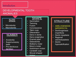
(1) The document discusses amelogenesis imperfecta (AI), which refers to hereditary defects in enamel formation. It describes several classifications of AI based on clinical, radiographic, and histological findings. (2) The main types are hypoplastic (reduced enamel), hypocalcified (soft enamel), and hypomaturation (improperly formed enamel). Clinical features, inheritance patterns, and prevalence are provided for each type. (3) The case report describes a 26-year-old female patient diagnosed with hypoplastic AI based on yellow-brown tooth discoloration and decreased vertical dimension. She was treated with periodontal therapy and full-mouth fixed bridges.
This document discusses genetic conditions that affect dental enamel formation called amelogenesis imperfecta. It describes four main types of enamel defects: hypoplastic (thin enamel), hypomaturation (soft enamel), hypocalcified (soft, friable enamel), and hypomatured/hypoplastic. The types are caused by defects in genes encoding enamel matrix proteins and result in discolored, sensitive teeth prone to disintegration. Amelogenesis imperfecta can be passed through autosomal dominant, recessive or sex-linked inheritance patterns. Diagnosis is based on family history, clinical observations and radiographs. Non-genetic conditions like dental fluorosis can also affect enamel formation.
Amelogenesis imperfecta is a hereditary condition that affects the development of dental enamel. There are three main types - hypoplastic, hypocalcified, and hypomaturation. The enamel is either not fully formed, not properly mineralized, or does not mature correctly. Treatment depends on the specific type but can include preventative care with sealants, bonding, crowns, veneers, and in severe cases, full coverage restorations. Managing the condition over many years using various techniques can lead to excellent functional and aesthetic outcomes.
Amelogenesis imperfecta is a hereditary disorder affecting enamel formation. There are three main types - hypoplastic (thin enamel), hypomaturation (improperly mineralized enamel), and hypocalcified (soft enamel). It is caused by defects during the three stages of enamel development: matrix formation, mineralization, and maturation. The condition has various inheritance patterns and clinical presentations depending on the genetic subtype. Treatment aims to restore aesthetics, sensitivity, and function and depends on the severity of enamel defects in each case.
Dentinogenesis imperfecta is a hereditary condition that affects the formation of dentin in both primary and permanent teeth. It is classified into two main types - dentinogenesis imperfecta type 1 and type 2. Type 1 is caused by mutations in the DSPP gene and affects only the teeth. Type 2 may be caused by mutations in two tightly linked genes and is characterized by multiple pulp exposures and shell-like teeth. Treatment aims to prevent wear of enamel and dentin through full coverage restorations.
This document discusses pulp calcification and pulp stones. It notes that pulp stones are a physiological manifestation that may increase in number or size due to local or systemic pathology. The etiological factors involved in their formation are not fully understood. As people age, the pulp space decreases in size and the blood vessels, nerves, and cells in the pulp also decrease. Pulp stones can form due to factors like age, circulatory disturbances, orthodontic tooth movement, and genetic predisposition. They are typically composed of calcium and phosphorus. Pulp stones may block access to canal orifices or engage instruments, but can usually be removed during root canal treatment with magnification, access, and proper instruments.
Matrices are used in operative dentistry to support and give form to dental restorations during placement and hardening. The document discusses the importance of matrices, their functions and characteristics of a good matrix. It describes different types of matrices including metallic matrices like Tofflemire, automatrix and sectional matrices like Palodent. Techniques for proper matrix selection, adaptation and wedge placement are also covered.
Kennedy’s Classification in Cast Partial DentureAamir Godil
This document discusses Kennedy's classification system for partially edentulous arches and Applegate's rules for applying the Kennedy classification. It provides details on Kennedy's four basic classes for partial edentulism and Applegate's eight rules to govern the application of Kennedy's classification. Examples are given to demonstrate how to use Kennedy's classification and Applegate's rules to classify different clinical scenarios of partial edentulism.
pre natal &; post-natal growth of maxilla & palate mahesh kumar
This document discusses the prenatal and postnatal development of the maxilla and palate.
During prenatal development, the maxilla forms from the maxillary prominences. The palate develops from the maxillary processes and palatal shelves. The palatal shelves initially grow vertically but then reorient horizontally and fuse in the midline.
Postnatally, the maxilla grows through processes like displacement, growth at sutures, and surface remodeling. Displacement includes primary displacement from growth of structures like the maxillary tuberosity, and secondary displacement from growth of structures it is attached to like the cranial base. Growth occurs at sutures connecting the maxilla. Surface remodeling increases the size, shape
This document provides an overview of periodontal instruments, including their classification, parts, materials used, and specific uses. It describes various assessment instruments like mouth mirrors and probes, as well as therapeutic instruments such as scalers, curettes, files, chisels, and surgical tools. The key instruments discussed in detail include mirrors, probes, explorers, sickle scalers, and curettes. It explains the design and uses of each instrument in assessing and treating periodontal disease.
The document discusses various types of major connectors used in removable partial dentures. It defines a major connector as the part that joins components on one side of the dental arch to the other. The main types described are the lingual bar, linguoplate, double lingual bar, labial bar, and swing lock design. Each has specific indications, advantages, and disadvantages. For example, the lingual bar is most commonly used but care must be taken with design to avoid weakness, while the linguoplate is indicated when space is limited between the gingiva and floor of the mouth. Factors such as tooth positions, soft tissue contours, and oral hygiene influence the choice of major connector.
The document discusses different types of articulators used in dentistry based on various classification systems. It describes Bonwill's theory of condylar guidance which defines a triangle formed by the condylar contact points and incisal edge. It also outlines Sharry's four class classification of articulators based on their adjustability and ability to accept registrations. Key articulator types mentioned are non-adjustable, semi-adjustable, and fully-adjustable.
The document discusses balanced occlusion in prosthodontics. It defines balanced occlusion as simultaneous contact of opposing teeth in centric relation position, with smooth bilateral gliding to eccentric positions. It describes Hanau's quint, which are the five factors that determine balanced occlusion: condylar guidance, incisal guidance, occlusal plane, compensating curves, and cusp inclination. It also discusses selection of posterior teeth based on ridge morphology, and arrangements for different molar and arch relationships. Examples are provided for managing resorbed ridges and flabby tissues. The goal is to understand principles of occlusion to provide patients with balanced occlusion.
This document discusses occlusion and its development from birth through adulthood. It begins by defining static and dynamic occlusion. It then discusses ideal, normal, and physiologic occlusion. It describes the periods of occlusal development from pre-dental through deciduous, mixed, and permanent dentition. It provides details on eruption sequences, spacing, and transitional periods. It also discusses occlusal curvatures like the Curve of Spee and Wilson. In summary, it provides a comprehensive overview of occlusion, its classifications, development through life stages, and related anatomical concepts.
The document summarizes the histopathology of dental caries in enamel and dentine. It describes the four zones seen in enamel caries: the translucent zone, dark zone, body of the lesion, and surface zone. It then discusses the five zones of dentine caries: the zone of sclerosis, zone of demineralization, zone of bacterial invasion, zone of destruction, and reactionary dentine. The zones represent areas of increasing demineralization and bacterial involvement as the caries progresses from enamel to dentine.
Orthodontic Case History and ExaminationAhmed Gamil
The document discusses essential and supplemental diagnostic aids used in orthodontic diagnosis. Essential aids include case history, clinical examination, study models, and certain radiographs. Supplemental aids provide additional information and include specialized radiographs, electromyography, and biopsy. A thorough case history covers chief complaint, medical/dental history, and family history. Clinical examination assesses facial symmetry, profile, jaw relationships, and soft tissues. Essential radiographs aid in case evaluation and treatment planning.
This document discusses different types of compensating curves used in dental prosthetics to establish balanced articulation during jaw movements. It describes the anteroposterior and mediolateral compensating curves that begin with the first replacement tooth and continue through the second molar. It also outlines the curve of spee from the mandibular canine through the condyle, the curve of Wilson to arrange the lower molars, and the curve of Monson connecting the curve of spee and Wilson to all cusps on a 4 inch radius.
Amelogenesis imperfecta is a hereditary condition affecting the development of enamel of the teeth. It results from mutations in genes responsible for enamel formation and maturation. There are three main types - hypoplastic AI (deficient enamel amount), hypomaturation AI (defective enamel structure), and hypocalcified AI (soft, easily worn enamel). Clinical features vary depending on type and include discolored, chipped teeth and increased sensitivity. It can be associated with other dental anomalies and syndromes like tricho-dento-osseous syndrome. The condition is caused by mutations in various enamel matrix protein genes, proteinase genes and other genes involved in enamel development.
The periodontal ligament is a connective tissue that connects the tooth to the alveolar bone. It contains collagen fibers, fibroblasts, cementoblasts, osteoblasts and other cells. The principal collagen fibers of the periodontal ligament originate on the cementum and insert into the alveolar bone in different orientations to provide structural support to the tooth and resist various forces. The periodontal ligament is essential for functions such as tooth eruption and maintains the space between the tooth and bone.
Anatomy and clinical significance of denture bearing areasOgundiran Temidayo
A presentation on the anatomy and clinical significance of the denture bearing areas by Ogundiran Temidayo who is a dental student at OBAFEMI AWOLOWO UNIVERSITY ILE-IFE
This document provides information on dental hand instruments. It discusses the history and development of instruments, materials used, classification systems, instrument parts and nomenclature, design principles like balance and contrangling, and applications of different instrument types like chisels, excavators, and plastic instruments. It also covers sharpening, sterilization, and references related to dental hand instruments.
Jaw relations refer to the spatial relationship between the maxilla and mandible. There are several types of jaw relations including orientation, vertical, and horizontal relations. The vertical jaw relation is the distance between two selected points on the maxilla and mandible. It is important to accurately record the vertical jaw relation to establish proper esthetics, phonetics, and function. There are various methods for determining the vertical jaw relation including physiologic methods and using interocclusal records or prior dentures. Facebows are used to transfer the maxillomandibular spatial relationship to articulators.
Andrew identified 6 keys to normal occlusion based on a study of non-orthodontic models. The 6 keys are:
1. Proper molar relationship between the upper first molar and lower second molar.
2. Positive crown angulation for all teeth.
3. Negative crown inclination for most teeth, except upper front teeth.
4. Teeth should be free from undesirable rotations.
5. Tight contact points between all teeth.
6. An occlusal plane that is relatively flat, rather than a deep or reverse curve of Spee.
When these 6 keys are present, it results in optimal intercuspation and occlusion. Deviations
This document discusses bone destruction patterns caused by periodontal disease. It identifies the main causes of bone destruction as the extension of gingival inflammation, trauma from occlusion, and systemic disorders. It describes several patterns of bone loss seen in periodontal disease, including horizontal, vertical, osseous craters, bulbous contours, reversed architecture, ledges, and furcation involvement. The rate and episodic nature of bone destruction in periodontal disease is also covered.
An Adams clasp is a component used to retain a custom-made medical device in the mouth. The clasp functions by engaging the mesiobuccal and distobuccal undercuts of a tooth, typically the maxillary first molar and is used to retain a wide range of devices prescribed in a variety of medical and dental specialties
The document discusses various theories of craniofacial growth:
1. Remodelling theory states growth occurs through bone remodeling.
2. Genetic theory views growth as genetically programmed but influenced by environmental factors.
3. Sutural theory sees sutures as primary growth centers controlled by heredity and environment.
4. Cartilaginous theory views cartilage, not sutures, as primary growth centers, with cartilage transplants demonstrating growth potential.
5. Functional matrix theory proposes bone growth is primarily influenced by soft tissue function through adaptation.
That's a high-level three sentence summary of the key points made in the document about different theories of craniofac
This document discusses acid etching of dental surfaces. It describes how acid etching was first proposed in 1955 to increase bond strength between composite resin and enamel. Acid etching removes enamel and creates an irregular porous surface that allows resin to penetrate through micromechanical interlocking, improving bond strength. Factors like acid type/concentration, etching duration, and fluoride levels can affect bonding. While acid etching is effective, some alternatives under investigation include crystal growth solutions, air abrasion, and lasers, but they have not achieved bond strengths as high as acid etching.
Clinical consideration in tooth development, eruption and sheddingShashibhal Maurya
This document discusses various clinical considerations related to tooth development, eruption, and shedding. It begins with an introduction on tooth composition and classifications of developmental defects. It then examines specific defects that can occur during different stages of tooth development, such as anodontia, supernumerary teeth, gemination and fusion during the initiation stage. Enamel hypoplasia, dens invaginatus, taurodontism and amelogenesis imperfecta are among the defects discussed during the apposition stage. Syndromes associated with certain defects and systemic diseases that can cause tooth abnormalities are also reviewed.
This document provides an overview of amelogenesis imperfecta (AI), a hereditary condition affecting the enamel of teeth. It discusses the classification, pathogenesis, clinical features, diagnosis and treatment of AI. AI results from genetic mutations that disrupt enamel formation and can be autosomal dominant, recessive or X-linked. Clinically, AI presents with hypoplastic, hypomineralised or hypocalcified enamel. Treatment involves restoring aesthetic and functional deficits through methods like crowns, composites or prosthetics.
This document summarizes amelogenesis imperfecta (AI), which encompasses conditions that affect the structure and appearance of dental enamel. It describes the normal development of enamel and classifications of AI types based on the developmental stage disturbed (formative, calcification, maturation). The types include hypoplastic, hypocalcified, and hypomaturation AI. Genetic causes are discussed, including genes involved and inheritance patterns. Clinical features of each type are provided along with radiographic characteristics.
This document provides an overview of intrinsic and extrinsic tooth discoloration. It begins with an introduction discussing the importance of correctly diagnosing the cause of discoloration for treatment planning. It then covers color perception and how surrounding conditions can affect perceived tooth color.
The document goes on to classify discoloration based on location (intrinsic vs extrinsic vs internalized) and etiology (pre-eruptive vs post-eruptive causes). Intrinsic discoloration results from changes to dental hard tissues during development due to certain metabolic disorders or local injuries. Extrinsic discoloration occurs on tooth surfaces from external factors like metals, foods/drinks, or smoking. Internalized stains were previously extrinsic
Dentin dysplasia is a rare genetic disorder that affects dentin formation, resulting in abnormal dentin and pulpal morphology. There are two types: type 1 affects the roots and causes them to be short and conical, while type 2 affects the crowns and causes bulbous or rounded crowns. Symptoms include premature loss of teeth due to extreme mobility from short roots or wearing away of rounded crowns. Radiographs show obliterated pulp chambers in primary teeth and crescent-shaped remnants in permanent teeth for type 1, while type 2 shows "thistle tube"-like enlarged pulp chambers in permanent tooth crowns. There is no treatment for the disease other than dental monitoring and extraction of mobile teeth.
This document summarizes structural anomalies of teeth, focusing on enamel hypoplasia. It describes the causes of enamel hypoplasia including nutritional deficiencies, infections, trauma, and fluoride ingestion. The key types of enamel hypoplasia are discussed - hypoplastic, hypocalcified, and hypomaturation. Clinical features and classifications of amelogenesis imperfecta are provided for each type. Histopathological features and treatments are also briefly mentioned.
The document discusses clinical considerations in tooth development. It outlines four key signaling pathways - BMP, FGF, SHH, and Wnt - that are critical for proper tooth development. Disruption of these pathways can lead to developmental defects of enamel, dentin, and cementum. The document also reviews genetic and molecular factors involved in stages of tooth development including bud formation, crown patterning, root formation, and mineralization.
This document discusses various types of environmental alterations that can affect tooth enamel formation and structure, including different forms of amelogenesis imperfecta where enamel is pitted, irregular, thin or translucent. It also mentions Turner's tooth which results from a periapical infection of a deciduous tooth. Dentinogenesis imperfecta is described as a severe form resulting in a shell tooth. Finally, syphilitic hypoplasia is discussed as a rare congenital condition from syphilis that causes distinctive alterations to anterior and posterior teeth.
Amelogenesis Imperfecta is a condition affecting the development of dental enamel. It has several classifications depending on the specific enamel defect present, including hypoplastic (inadequate enamel), hypocalcified (no mineralization), and hypomaturation (failure of enamel to mature). The condition can be inherited in autosomal dominant, recessive, x-linked, or sporadic patterns. Diagnosis is based on family history, clinical examination showing discolored, sensitive teeth prone to disintegration, and radiographic findings of abnormal enamel appearance. It may occur alone or be associated with other systemic abnormalities in syndromes.
This document discusses various developmental disturbances that can affect teeth, including disturbances in number, size, shape, and structure. It describes conditions like hypodontia, supernumerary teeth, microdontia, macrodontia, gemination, fusion, dens invaginatus, taurodontism, and enamel pearls. It provides details on the classification, etiology, clinical features, and radiographic presentation of these different developmental disturbances that occur due to abnormalities during tooth development.
A rare condition affecting the primary and permanent by dr ibrahimDr Ibrahim
Dentin dysplasia is a rare genetic condition that affects tooth development. There are two types:
Type I affects both dentitions and causes teeth to have normal crowns but abnormal, stunted roots. The pulp chambers and root canals are often completely obliterated. Teeth frequently exfoliate prematurely.
Type II primarily affects the primary dentition, causing teeth to appear translucent. However, the permanent dentition appears normal. It is characterized by an enlarged pulp chamber and abnormal pulp canal morphology.
The document describes a case study of a 7-year-old girl diagnosed with Type I dentin dysplasia based on clinical and radiographic features including rootless teeth and obliterated
Dental anomalies and its prosthetic implications.pptxKanmaniAthi
This document discusses various dental anomalies including differences in tooth size, number, morphology, and location. It describes developmental anomalies such as microdontia, macrodontia, hypodontia, anodontia, and hyperdontia. Characteristics and treatments for conditions like ectodermal dysplasia, amelogenesis imperfecta, and dentinogenesis imperfecta are provided. The conclusion emphasizes that dental anomalies can cause clinical problems and their treatment requires knowledge of classification criteria to properly diagnose and manage each condition.
Developmental disturbances of tooth structureHagir Taha
This document discusses dental enamel and dentin development and defects. It provides details on:
- The normal development of enamel and causes of enamel defects including environmental factors and hereditary conditions like amelogenesis imperfecta.
- Different types of dentin defects caused by conditions like dentin dysplasia and dentinogenesis imperfecta which are inherited and involve genes responsible for dentin formation.
- Characteristics and classifications of various enamel and dentin defects like enamel hypoplasia, fluorosis, regional odontodysplasia and ghost teeth. Radiographic and microscopic features are also described.
This document discusses several dental anomalies including amelogenesis imperfecta, molar incisal hypomineralisation, dentinogenesis imperfecta, dentinal dysplasia, taurodontism, and dens invaginatus vs evaginatus. It provides details on the characteristics, clinical features, classifications, diagnostic criteria, management challenges, and treatment approaches for each condition. The document is intended as an introduction and overview for dental professionals on managing these developmental anomalies.
Etiology, clinical picture and treatment of pathology of hard tissues of teet...HamzahAbuhashish
Etiology, clinical picture and treatment of pathology of hard tissues of teeth in orthopedic dentistry
Indications and contraindications for inlay
Features of the preparation of cavities for inlay
Inlay manufacturing technique
Inlay cementing
Comparative characteristics of indirect composite and ceramic restorations
Dentinogenesis imperfecta is a genetic disorder that causes teeth to be discolored and translucent, as well as weaker and prone to breakage. It is caused by abnormal dentin formation and can affect primary and permanent teeth. The condition is inherited in an autosomal dominant pattern and affects about 1 in 6,000-8,000 people. While the teeth may appear more susceptible to cavities, they are not actually more prone to dental caries. Treatment focuses on bonding to strengthen and whiten teeth, as other cosmetic procedures could cause further damage.
Dentinogenesis imperfecta is a genetic disorder that causes teeth to be discolored and translucent, as well as weaker and prone to breakage. It is caused by abnormal dentin formation and is inherited in an autosomal dominant pattern. The condition affects an estimated 1 in 6,000 to 8,000 people and can involve both primary and permanent teeth. Treatment focuses on bonding to strengthen enamel and make teeth appear whiter, as common cosmetic procedures are inappropriate due to the weakened state of the teeth.
This document summarizes several developmental alterations of teeth structures. It begins by describing Amelogenesis Imperfecta, which is a group of hereditary conditions that affect the quality and quantity of dental enamel. It classifies AI based on the stage of development affected and lists associated clinical features. It then discusses Dentinogenesis Imperfecta, Dentin Dysplasia, Regional Odontodysplasia, and Dentin Hypocalcification - each of which involve hereditary defects in dentin formation. For each condition, it provides details on classification, clinical presentation, radiographic findings, diagnosis, and management.
This document discusses several conditions related to abnormalities in dentin formation, including dentinogenesis imperfecta and dentin dysplasia. It describes the genetic basis, clinical and radiographic features, classifications, and histopathological characteristics of these inherited disorders. The key features include opalescent or discolored teeth, bulbous crowns, thin dentin, enlarged pulp chambers, shortened roots, and premature tooth loss. Classification systems include those proposed by Shields and Witkop. Treatment may involve extraction and dental prosthetics due to poor cosmetic outcomes and functional complications.
Similar to Amelogenesis imperfecta, hypoplastic type - Dr Sanjana Ravindra (20)
Review on the applications of ultrasonography in dentistry - Dr Sanjana RavindraDr. Sanjana Ravindra
Ultrasonography has various applications in the dentomaxillofacial region for evaluating soft tissue masses, salivary gland and duct calculi, vascular structures, and assisting with biopsies. It can assess conditions like TMJ disorders, intraosseous lesions, lymph node metastasis, and soft tissue lesions. Ultrasonography is a useful imaging technique as it uses non-ionizing sound waves to generate real-time images without radiation exposure. However, its ability to image deep structures and penetrate bone is limited compared to other modalities.
Basic principles of magnetic resonance imaging for beginner - Dr Sanjana Ravi...Dr. Sanjana Ravindra
This document provides an overview of the basic principles of magnetic resonance imaging (MRI) for beginner oral and maxillofacial radiologists. It discusses the discovery of MRI and its use as a noninvasive tool. The basic physics of MRI are described, including proton spin and precession in magnetic fields. The document outlines the principles of MRI, including indications, advantages, limitations and basic sequences. It provides details on image formation, including the nuclear magnetic resonance phenomenon and T1- and T2-weighted images. In summary, the document serves as an introduction to MRI for oral radiologists by describing the fundamental concepts and physics that underlie MRI.
This document provides an overview of panoramic radiography. It begins with an introduction and then discusses the history, terminology, principles, concepts, equipment, positioning requirements, and indications of panoramic radiography. The key points covered include that panoramic radiography produces a single tomographic image of the facial structures using a rotating x-ray beam and that the image layer is a curved zone where structures appear in focus on the final image. Advantages are that it shows a wide area in a single image while disadvantages include some distortion and structures being out of focus above and below the image layer.
Inflammatory n infectious diseases of salivary gland- Dr Sanjana RavindraDr. Sanjana Ravindra
SALIVARY GLAND
Introduction
Classification
Composition of saliva
Properties of Saliva
Functions of Saliva
Salivary gland examination
Classification of Salivary gland diseases
INFLAMMATORY and INFECTIOUS DISEASES OF SALIVARY GLAND
Introduction
Classification
Various diseases
This document provides an overview of herpes virus and herpes simplex virus. It begins with definitions of key terminology used to describe viral lesions. It then discusses virus basics such as introduction, history, characteristics, replication, and classification. Specific details are provided about herpes virus morphology, classification, and herpes simplex virus epidemiology, pathophysiology, and clinical features. The summary covers the key topics discussed in the document.
X-ray film is composed of several layers that allow it to capture x-ray images. The base layer is a clear, flexible material like cellulose triacetate or polyester that serves as a support. Coated on this is the emulsion, a light-sensitive layer containing silver halide crystals that darken when exposed to x-rays or light. An anti-halation layer reduces halo effects from light scattering. Together these layers allow x-ray film to record x-ray exposures and be developed into visible images.
Physiotherapy modalities discussed for treating orofacial conditions include massage techniques like deep tissue massage and trigger point therapy, spray and stretch technique using vapocoolant to relieve pain and increase range of motion, physical activities for soft tissue and joint mobilization as well as muscle conditioning, and electrical stimulation to reduce pain by modulating sensory nerve pathways per the gate control theory of pain.
This document discusses the concept of field cancerization in oral cancers. It begins by providing background on oral cancer incidence and common sites of occurrence. It then defines field cancerization as the development of cancer in multifocal areas of precancerous changes due to exposure to carcinogens. The document discusses the monoclonal and polyclonal theories of how multiple lesions arise and reviews the concepts of field defects and field effects. It notes that field cancerization can help explain high rates of secondary primary tumors and tumor recurrence. Therapeutic implications and markers for determining field cancerization are also summarized.
multiple idiopathic external and internal resorption- Dr Sanjana RavindraDr. Sanjana Ravindra
This case report describes a 36-year-old male with multiple idiopathic external and internal root resorptions in the maxillary and mandibular permanent teeth found incidentally on radiographs. The patient reported slight discomfort while chewing with his left mandibular second molar. Laboratory tests and clinical examinations found no cause for the resorptions. Cone beam computed tomography further evaluated the resorptive lesions and found they affected several teeth with no identifiable etiology. This is a rare presentation of idiopathic root resorption in multiple teeth.
Trigeminal neuralgia is a neuropathic facial pain condition characterized by sudden, severe, brief stabbing pains in the face that are triggered by everyday activities like eating or talking. The pain is caused by abnormalities in the trigeminal nerve that transmits sensations from the face to the brain. Carbamazepine is first-line pharmacological treatment to slow nerve signaling, providing pain relief. When medications fail to control pain, surgical options like microvascular decompression or gamma knife radiosurgery aim to decompress the trigeminal nerve from vascular compression. Proper diagnosis involves distinguishing TN from other facial pain conditions through evaluation of pain characteristics, triggers, and response to treatments.
This document discusses broad spectrum antibiotics. It begins with definitions of key terms like antibiotic, pharmacokinetics, and pharmacodynamics. It then covers the history of antibiotics from traditional empirical uses to the modern era including the discoveries of penicillin and other drugs. The document categorizes antibiotics based on their spectrum of activity, mechanism of action, source, and susceptible organisms. It also addresses principles of antibiotic therapy such as selection, combinations, prophylaxis, resistance, and misuse.
This document provides an overview of muscles in the head and neck region. It begins with the embryological development of muscles originating from pharyngeal arches and somites. It then classifies muscles based on striations, control and distribution into skeletal, cardiac and smooth muscles. The document discusses the structure, types and functions of these three muscle types in detail. It also covers specific muscles of the head and neck region including those of the tongue, soft palate and larynx.
How to Add Chatter in the odoo 17 ERP ModuleCeline George
In Odoo, the chatter is like a chat tool that helps you work together on records. You can leave notes and track things, making it easier to talk with your team and partners. Inside chatter, all communication history, activity, and changes will be displayed.
Main Java[All of the Base Concepts}.docxadhitya5119
This is part 1 of my Java Learning Journey. This Contains Custom methods, classes, constructors, packages, multithreading , try- catch block, finally block and more.
A review of the growth of the Israel Genealogy Research Association Database Collection for the last 12 months. Our collection is now passed the 3 million mark and still growing. See which archives have contributed the most. See the different types of records we have, and which years have had records added. You can also see what we have for the future.
Thinking of getting a dog? Be aware that breeds like Pit Bulls, Rottweilers, and German Shepherds can be loyal and dangerous. Proper training and socialization are crucial to preventing aggressive behaviors. Ensure safety by understanding their needs and always supervising interactions. Stay safe, and enjoy your furry friends!
A workshop hosted by the South African Journal of Science aimed at postgraduate students and early career researchers with little or no experience in writing and publishing journal articles.
ISO/IEC 27001, ISO/IEC 42001, and GDPR: Best Practices for Implementation and...PECB
Denis is a dynamic and results-driven Chief Information Officer (CIO) with a distinguished career spanning information systems analysis and technical project management. With a proven track record of spearheading the design and delivery of cutting-edge Information Management solutions, he has consistently elevated business operations, streamlined reporting functions, and maximized process efficiency.
Certified as an ISO/IEC 27001: Information Security Management Systems (ISMS) Lead Implementer, Data Protection Officer, and Cyber Risks Analyst, Denis brings a heightened focus on data security, privacy, and cyber resilience to every endeavor.
His expertise extends across a diverse spectrum of reporting, database, and web development applications, underpinned by an exceptional grasp of data storage and virtualization technologies. His proficiency in application testing, database administration, and data cleansing ensures seamless execution of complex projects.
What sets Denis apart is his comprehensive understanding of Business and Systems Analysis technologies, honed through involvement in all phases of the Software Development Lifecycle (SDLC). From meticulous requirements gathering to precise analysis, innovative design, rigorous development, thorough testing, and successful implementation, he has consistently delivered exceptional results.
Throughout his career, he has taken on multifaceted roles, from leading technical project management teams to owning solutions that drive operational excellence. His conscientious and proactive approach is unwavering, whether he is working independently or collaboratively within a team. His ability to connect with colleagues on a personal level underscores his commitment to fostering a harmonious and productive workplace environment.
Date: May 29, 2024
Tags: Information Security, ISO/IEC 27001, ISO/IEC 42001, Artificial Intelligence, GDPR
-------------------------------------------------------------------------------
Find out more about ISO training and certification services
Training: ISO/IEC 27001 Information Security Management System - EN | PECB
ISO/IEC 42001 Artificial Intelligence Management System - EN | PECB
General Data Protection Regulation (GDPR) - Training Courses - EN | PECB
Webinars: https://pecb.com/webinars
Article: https://pecb.com/article
-------------------------------------------------------------------------------
For more information about PECB:
Website: https://pecb.com/
LinkedIn: https://www.linkedin.com/company/pecb/
Facebook: https://www.facebook.com/PECBInternational/
Slideshare: http://www.slideshare.net/PECBCERTIFICATION
A Strategic Approach: GenAI in EducationPeter Windle
Artificial Intelligence (AI) technologies such as Generative AI, Image Generators and Large Language Models have had a dramatic impact on teaching, learning and assessment over the past 18 months. The most immediate threat AI posed was to Academic Integrity with Higher Education Institutes (HEIs) focusing their efforts on combating the use of GenAI in assessment. Guidelines were developed for staff and students, policies put in place too. Innovative educators have forged paths in the use of Generative AI for teaching, learning and assessments leading to pockets of transformation springing up across HEIs, often with little or no top-down guidance, support or direction.
This Gasta posits a strategic approach to integrating AI into HEIs to prepare staff, students and the curriculum for an evolving world and workplace. We will highlight the advantages of working with these technologies beyond the realm of teaching, learning and assessment by considering prompt engineering skills, industry impact, curriculum changes, and the need for staff upskilling. In contrast, not engaging strategically with Generative AI poses risks, including falling behind peers, missed opportunities and failing to ensure our graduates remain employable. The rapid evolution of AI technologies necessitates a proactive and strategic approach if we are to remain relevant.
This presentation includes basic of PCOS their pathology and treatment and also Ayurveda correlation of PCOS and Ayurvedic line of treatment mentioned in classics.
Macroeconomics- Movie Location
This will be used as part of your Personal Professional Portfolio once graded.
Objective:
Prepare a presentation or a paper using research, basic comparative analysis, data organization and application of economic information. You will make an informed assessment of an economic climate outside of the United States to accomplish an entertainment industry objective.
Exploiting Artificial Intelligence for Empowering Researchers and Faculty, In...Dr. Vinod Kumar Kanvaria
Exploiting Artificial Intelligence for Empowering Researchers and Faculty,
International FDP on Fundamentals of Research in Social Sciences
at Integral University, Lucknow, 06.06.2024
By Dr. Vinod Kumar Kanvaria
Strategies for Effective Upskilling is a presentation by Chinwendu Peace in a Your Skill Boost Masterclass organisation by the Excellence Foundation for South Sudan on 08th and 09th June 2024 from 1 PM to 3 PM on each day.
2. Amelogenesis Imperfecta, Hypoplastic Type
Associated with Some Dental Abnormalities:
A Case Report
Canger EM, Celenk P, Yenísey M, Odyakmaz SZ. Braz Dent J (2010) 21(2): 170-174.
JOURNAL CLUB: 3
5. encompasses a
complicated
group of conditions
that demonstrate
developmental
alterations in the
structure of the
enamel in the
absence
of a systemic
disorder.
AMELOGENESIS
IMPERFECTA
AMELOGENES
IS ?IMPERFECT
A
Hereditary
enamel
dysplasia
Hereditary
brown enamel
Hereditary
brown
opalescent
teeth
Shafer’s textbook of Oral Pathology. 6th ed. Elsevier;
2009
www.merriam-webster.comIntroduction
6. Case report
Canger EM, Celenk P, Yenísey M, Odyakmaz SZ. Amelogenesis Imperfecta, Hypoplastic
Type, Associated with Some Dental Abnormalities: A Case Report: Braz Dent J (2010)
21(2): 170-174.
14. Introduction
Tooth enamel consists mainly of inorganic material (96%) and oraganic
substance and water(4%)
Physical properties and physiological function of enamel – directly
related to composition, orientation, disposition and morphology of
mineral components within tissue
During organogenesis, enamel transitions from soft to pliable tissue to
its final form- is devoid of protein
Final composition is reflection of unique molecular and cellular
activities that take place during its genesis
Deviation from this pattern lead – AMELOGENESIS IMPERFECTA
16. “AI encompasses a complicated group of conditions that demonstrate
developmental alterations in structure of the enamel in the absence of
a systemic disorder”
“AI represents a group of conditions, genomic in origin, which affect the
structure and clinical appearance of the enamel of all or nearly all the
teeth in a more or less equal manner, and which may be associated
with morphologic or biochemical changes elsewhere in the body”
Orphanet Journal of Rare Diseases 2007,
2:17
Definition
17. Historical background
Spokes in 1890,
described "brown
teeth" with a familial
history.
In 1907 Turner described
some cases of
hereditary hypoplasia of
teeth in five generations
of same family
Weinmann & associates in 1945 –
introduced term “AMELOGENESIS
IMPERFECTA” – it is an ectodermal
disturbance, mesodermal components
are normal
18. ETIOLOGY
Dental enamel is a highly mineralised tissue
Derived through the synthesis and secretion of proteins
Formation of this highly organised and unusual structure is controlled in
ameloblasts through interaction of a number of organic matrix molecules
19. Development of normal enamel occurs in three stages
1. Formative stage – deposition of organic matrix – Hypoplastic AI
2. Calcification stage – matrix is mineralized –Hypocalcified AI
3. Maturation stage – crystallites enlarge and mature –
Hypomaturative AI
ETIOLOGY
20. Genes and phenotypes
Proteins/ enzymes
forming enamel
Type of AI Inheritance
1 Amelogenin Diffuse smooth
hypoplastic &
hypomaturation
X linked
2 Ameloblastin Hypocalcified AD
3 Enamelin Hypoplastic AD, AR
4 Tuftelin hypoplastic AD,AR
5 Kallikrein Hypomaturation AR
6 Matrix metalloproteinase Pigmented
hypomaturation
AR
21. Modes of Mendelian Inheritance Associated with AI
https://www.google.co.in/search?q+phenotype
22. CLASSIFICATION
Sekar B, Dominic Augustine, Murali S.
Amelogenesis Imperfecta - A Case Report with
Genetic Transmission. IJDA, 2(4), October-
December, 2010 395.
24. BASED ON CLINICAL, MICRORADIOGRAPHIC
AND HISTOPATHOLOGICAL FINDINGS BY
Darling (1956)
Hypoplastic
Group 1 – gen pitting
Group 2- vertical
grooves
Group 3- gen
hypoplasia
Hypocalcified
Type 4A- chalky,
yellow, brown enamel
Type 4B- marked
enamel discolouration
7 softness with post
eruptive loss of
enamel
Type 5- gen/ localized
discolouration and
chipping of enamel
26. By Schulze
(1970)
Type 1 hypoplastic Specific features , inheritance
1A Hypoplastic, pitted AD
1B Hypoplastic, local AD
1C Hypoplastic, local AR
1D Hypoplastic, smooth AD
1E Hypoplastic, smooth x-linked D
1F Hypoplastic, rough AD
Typw 2 hypomaturation
2A hypomaturation
2B hypomaturation
2C Snow capped teeth, x linked
2D AD
Type 3 hypocalcification
3A AD
3B AR
Type 4 Hypomaturation- hypoplastic with taurodontismType 4A Hypomaturation- hypoplastic with
taurodontism AD
Type 4B Hypoplastic- hypomaturation with taurodontism AD
Neville BW, Douglass DD, Allen CM, Bouquot JE. Abnormalities of teeth. In: Oral and Maxillofacial Pathology. 2nd ed.. Pennsylvania:Elsevier;200
27. Prevalence range from 1 in 718 to 1 in 14,000, depending on the
population studied.
Hypoplastic AI represents 60 – 73% of all cases,
Hypomaturation AI represents 20 – 40%, and
Hypocalcification AI represents 7%
Prevalence
30. Hypoplastic type – autosomal dominant
GENERALIZED
PITTED
• Thin enamel
• Open contact
• Pinpoint to pinhead
pits
• Newly erupted teeth:
hard with normal
yellow-white colour
• Staining of teeth-
exposure to oral
environment- black
appearance
LOCALIZED
PITTED
• Linear
depression/
large area of
hypoplasia
• Prominent on
buccal surface
DIFFUSE
SMOOTH
• Thin, glossy with
smooth surface
• ¼ to 1/8 of
normal thickness
• Yellow color –
opaque to
translucent brown
• Delayed eruption
with alveolar
resorption
DIFFUSE
ROUGH
Hard with rough
granular surface
White to yellowish
white
Thicker enamel at
cervical areas
https://www.google.co.in/search?q=amelogenesis+hypoplas
31. Localized
pitted
Severe form of AD
Enamel
agenesis
Yellow colour like normal
dentition
Surface is rough & granular-
ground glass
Complete lack of enamel
formation
Multiple missing teeth
https://www.google.co.in/search?q=amelogenesis+hypoplastic
Hypoplastic type – autosomal recessive
32. Males
– Thin, hard, glossy
– Like crown preparations, open bite
– Opaque white to brown
Females
– Alternating vertical bands of normal and
abnormal enamel
https://www.google.co.in/search?q=amelogenesis+hypoplastic
Hypoplastic type – x-linked dominant
33. Hypoplastic amelogenesis
imperfecta, gen pitted pattern .
Note the numerous pinpoint pits
scattered across the surface of
the teeth. The enamel between
the pits is of
normal thickness. hardness. and
coloration.
Hypoplastic amelogenesis
imperfecta, autosomal dominant
smooth pattern.
Small. yellowish teeth exhibiting
hard, glossy enamel with open
contact Points and anterior
open bite.
Neville BW, Douglass DD, Allen CM, Bouquot JE. Abnormalities of teeth. In: Oral and Maxillofacial Pathology. 2nd ed.. Pennsylvania:Elsevier;2004. 89
35. Thickness of enamel
Normal- hypoplasia( middle 3rd of labial surface)
Consistency of enamel
Soft – lost after eruption- scrapped with instrument
Colour of enamel
Newly erupted teeth- dull lustreless opaque, white, honey
coloured or yellowish orange or brown
Significance :
Exposed dentin-
hypersensitive
anterior opn
bite
Rapid calculus
formation
Hypocalcified type – aD & AR
Neville BW, Douglass DD, Allen CM, Bouquot JE. Abnormalities of teeth. In: Oral and Maxillofacial Pathology. 2nd ed.. Pennsylvania:Elsevier;2004. 89
36. Dentition exhibiting diffuse
yellow-brown discoloration .
Note numerous teeth with loss
of coronal enamel except for
the cervical portion.
Hypocalcified type – aD & AR
Extensive loss of coronal
enamel and the similar
density of enamel
and dentin.
Neville BW, Douglass DD, Allen CM, Bouquot JE. Abnormalities of teeth. In: Oral and Maxillofacial Pathology. 2nd ed.. Pennsylvania:Elsevier;2004. 89
38. Commonly in males
Both in primary and permanent dentition
Primary teeth: ground glass opaque white
appearance
Permanent teeth :mottled yellow white, may be
darkened with absorption of stains
Tight contact point
Enamel approaches normal thickness.
Point of explorer can be forced into enamel
Hypomaturative type – autosomal dominant
39. Both in primary and
permanent dentition
Enamel has milky to
shiny, agar brown
deeply stained on
contact with
exogenous agents
Normal thickness.
Chips away around
restoration
Forms large amount
of calculus which
may contain pigment
forming agents
Teeth may resorb
within alveolus
https://www.google.co.in/search?q=amelogenesis+hypomaturativ
Hypomaturative type – autosomal recessive
40. Snow capped
Zone of white opaque enamel on incisal
and
occlusal surface (1/4 to 1/3 of the surface)
Looks like fluorosis
Anteriors, premolars/molars
Both dentitions
https://www.google.co.in/search?q=snow+capped
Hypomaturative type – x linked recessive
41. Predominant defect – enamel hypomaturation
Enamel appears – mottled yellow-white to yellow brown
Pits are seen frequently on buccal surface of teeth
Radiographically – enamel appears similar to dentin
Large pulp chambers may be seen in single rooted teeth in
addition to varying degrees of taurodontism
Hypomaturation- hypoplastic with taurodontism – Autosomal
dominant
https://www.google.co.in/search?q=amelogenesis+hypomatu+tauro
42. Predominant defect – enamel hypoplasia in which enamel is thin
but also hypomature
Radiographically-
Similar to hypomaturation-hypoplastic variant, except decrease
in thickness of enamel
https://www.google.co.in/search?q=amelogenesis+hypomatu+tauro
Hypoplastic - hypomaturation with taurodontism – Autosomal dominant
43. Radiographic features
Squarish type of crown
being devoid of the normal
mesial and distal contours
Normal enamel cap is
missing and in its place a
thin and opaque layer of
enamel
Low or absent cusps, with
serrations of varying
sharpness
Lack of contrast between
enamel and dentin
Obliteration of pulp chamber
Loss of contour Enamel Pulp chamber
https://www.google.co.in/search?q=amelogenesis+tauro
44. TYPE CLINICAL
APPEARANCE
ENAMEL
THICKNESS
HYPOPLASTIC
(TYPE I)
Crowns size : small to
normal, lack proxmal
contacts, color varies
from normal to opaque
white – yellow brown
Varies from thin and
smooth to normal
thickness with
grooves, furrows
and/or pits
HYPOMATURATIO
N
(TYPE II)
Varies from creamy
opaque to marked
yellow/brown, surface
of teeth soft and rough,
dental sensitivity and
open bite common
Normal thickness
with enamel that
often chips and
abrades easily
around restoration
HYPOCALCIFIE
D
(TYPE III)
Opaque white to
yellow-brown, soft
rough enamel surface,
dental sensitivity and
open bite common,
heavy calculus
formation common
Normal thickness
with enamel that
often chips and
abrades easily
HYPOMATURATIO
N/ HYPOPLASIA/
TAURODONTISM
(TYPE IV)
White/Yellow-
Brown mottled,
teeth can appear
small and lack
proximal contact
Reduced,
hypomineralized
areas and pits
RADIOGRAPHIC
APPEARANCE
INHERITANCE
Enamel has normal
to slightly reduced
contrast/ thin
Autosomal
dominant,
recessive, or X-
linked
Enamel has contrast
similar to or > than
dentin, unerupted
crowns have normal
morphology
Autosomal
dominant,
recessive, or X-
linked
Enamel has contrast
similar to or <
dentin, unerupted
crowns have normal
morphology
Autosomal
dominant, recessive
Enamel contrast
normal to slightly >
dentin, large pulp
chambers
Autosomal dominant
Neville BW, Douglass DD, Allen CM, Bouquot JE. Abnormalities of teeth. In: Oral and Maxillofacial Pathology. 2nd ed.. Pennsylvania:Elsevier;2004. 89
45. Histopathological examination
Very thin enamel, voids within enamel & composed of laminations of
irregularly arranged enamel prisms
Enamel-dentin junction, show some exaggerated scalloping.
Areas of homogeneous aprismatic enamel or fused indistinct prisms, with
“a reduction in distance between enamel rod incremental lines”
https://www.google.co.in/search?q=amelogenesis+slides
46. Syndromes associated
Amelogenesi
s imperfect
with
taurodontism
Trichodentoo
sseous
syndrome
Tricho-dento-osseous syndrome. Dentition
exhibiting diffuse enamel hypop lasia and
hypomaturat ion. At birth, the patient
exhibit kinky "steel wool" hair texture
with time, the hair gets straightened.
Taurodontism of the first molar and the
enamel. which is thin and similar in density to
the dentin.
Neville BW, Douglass DD, Allen CM, Bouquot JE. Abnormalities of teeth. In: Oral and Maxillofacial Pathology. 2nd ed.. Pennsylvania:Elsevier;2004. 89
52. Reason for choosing this article
Developmental disorder presents with severe dental anomalies. Its
important to diagnose the condition as early as possible to balance the
decision for early intervention and long-term survival of the
restorations. Also consider the social implications for these patients and
intervene to relieve their suffering. Thus, this article is an
attempt to improve the clinician’s knowledge about the clinical &
radiographic diagnosis as well as intervention required for such a
condition.
54. 1. www.merriam-webster.com
2. Neville BW, Douglass DD, Allen CM, Bouquot JE. Abnormalities of teeth. In: Oral and Maxillofacial
Pathology. 2nd ed.. Pennsylvania:Elsevier;2004. 89-94.
3. Regezi J, Sciubba JJ. Oral Pathology- Clinical Pathologic Correlations. 3rd ed. WB Saunders Company;
1999.
4. Rajendran R, Sivapathasundaram. Shafer’s textbook of Oral Pathology. 6th ed. India: Elsevier; 2009.
5. Kumar GS. Orban’s Oral histology and embryology. 12th ed. Elsevier; 2009.
6. White SC, Pharaoh MJ editors. Normal Radiographic Anatomy. In: Oral Radiology- Principles and
Interpretation 5th edition. St.Louis (US): Mosby/Elsevier; 2004.
7. Bailleul-Forestier I, Molla M, Verloes A, Berdal A. The genetic basis of inherited anomalies of the teeth. Part
1: clinical and molecular aspects of non-syndromic dental disorders. Eur J Med Genet 2008;51:273-291.
8. Mehta DN, Shah J, Thakkar B. Amelogenesis imperfecta: Four case reports. J Nat Sc Biol Med 2013;4:462-
5.
9. Seow WK. Dental development in amelogenesis imperfecta: a controlled study. Pediatr Dent 1995;17:26-30.
10. Gökçe K, Canpolat C, Özel E. Restoring function and esthetics in a patient with amelogenesis imperfecta: a
case report. J Contemp Dent Pract 2007;8:90-101.
11. Siadat H, Alikashi M, Mirfazaelian A. Rehabilitation of a patient with amelogenesis imperfect using all-
ceramic crowns: a clinical report. J Prosthet Dent 2007;98:85-88.
12. Toksavul S, Ulusoy M, Türkün M, Kümbüloğlu Ö. Amelogenesis imperfecta: the multidisciplinary approach: a
case report. Quintessence Int 2004;35:11-14.
55. 13. Santos MCLG, Line SRP. The genetics of amelogenesis imperfecta: a review of the literature. J Appl Oral
Sci 2005;13:212-217.
14. Begum N, Bhandarkar GP, Kini R, Naik V, Rashmi K, D Souza LC. Amelogenesis Imperfecta: A Series of
Case Report.Int J Adv Health Sci 2015;2(1):17-21.
15. Crawford PJM, Aldred M, Bloch-Zupan A. Amelogenesis imperfecta. Orphanet J Rare Dis 2007;2:17-27.
16. Poulsen S, Gjqrup H, Haubek D, Haukali G, Hintze H, Lqvschall H, et al.. Amelogenesis imperfecta - a
systematic literature review of associated dental and oro-facial abnormalities and their impact on patients.
Acta Odontol Scand 2008;66:193-199.
17. Nigam P, Singh VP, Prasad KD, Tak J. Amelogenesis Imperfecta: A Case Report and Review of Literature.
Int J Sci Stud 2015;2(10):146-149.
18. Jan C.-C. Hu Yong-Hee P, Hazzazzi AC, Simmer JP. Enamel Formation and Amelogenesis Imperfecta. Cells
Tissues Organs 2007;186:78–85.
19. Sunil RP, Agarwal A. Hypoplastic amelogenesis imperfecta. Journal of Dental Sciences & Oral
Rehabilitation. 2008.
20. Chhaparwal Y, Jhawar A, Lele A, Rathi S. Case Report on hypoplastic amelogenesis imperfecta with
multiple impacted teeth. Journal of Dental and Medical Sciences. 2279-0861.Volume 13, Issue 5 Ver. V.
(May. 2014), PP 79-82.
21. Chengappa, Murali.R, Sivagami.N. Rehabilitation of Mutilated Natural Dentition associated with
Amelogenesis Imperfecta – A Case Report. INT J OF DENTAL CLINICS 2010:2(4): 77-79.
22. Sekar B, Dominic Augustine, Murali S. Amelogenesis Imperfecta - A Case Report with Genetic
Transmission. IJDA, 2(4), October-December, 2010 395.
Editor's Notes
The Primary & Permanent Dentition are subject to considerable variation in the number , Size & Form of teeth and the structure of the dental tissues
is a hereditary disorder that expresses a group of conditions that cause developmental alterations in the structure of enamel.
A 26-year-old female patient presented with a chief complaint of discolored teeth. Medical history was noncontributory. Extraoral examination did not reveal any relevant findings. No other family member had the same dental problem.
The patient’s oral hygiene was poor and she presented hyperemic and edematous gingiva. Bleeding on probing scores of the teeth was determined as one.
There was no gingival enlargement around the teeth, so the clinical crown lengths were adequate.
Twenty-three teeth were erupted. Six of them were primary teeth and 17 were permanent teeth.
Tooth crowns were clinically short and featured yellow-brown colored surfaces. Tissue loss affected
all teeth.
There was loss of contact between the teeth.
Vertical dimension of the face was also decreased. There was no anterior open bite
Panoramic radiograph of the patient revealing the presence of 6 over-retained primary teeth and 15 totally impacted permanent teeth (18, 17, 15, 23, 27, 28, 38, 37, 35, 33, 43, 44, 45, 47, 48). Pulpal calcifications are seen in all teeth
congenital absence of maxillary right lateral tooth is clearly visible.
B = Full-mouth periapical radiographs of the maxilla and mandible. The density of dentin is not differentiated from enamel.
The loss of contact between the teeth can be easily detected.
the enamel layers of all teeth giving an appearance similar to that observe after mechanical preparation.
Pulp chambers had normal size and shape except for 26
There were stones in the crown pulps and the radicular pulps were obliterated.
In addition, external resorption was noted on the crowns of both impacted 2nd permanent molars.
The lamina duras were normal.
The crowns of mandibular left central incisors and lateral incisors became close to each other and the roots were separated from each other
The initial stage periodontal therapy consisted of oral hygiene instructions, scaling, and root planning. Two weeks later, the gingival edema was resolved and hyperemic appearance of gingiva turned to normal. Also bleeding on probing was normal.
After radiographic examination of crown/root ratio, root number and morphology of the present teeth, construction of full-mouth metal reinforced porcelain fixed bridge restoration was planned. The occlusal vertical dimension was 3 mm lower than normal. First of all, a splint, made of self cured hard acrylic, was constructed to increase the vertical dimension. The adaptation of the temporomandibular joints and masticatory muscles was carefully observed periodically during 4 months and, after this period, the patient tolerated well her new vertical dimension.
The supporting teeth were prepared 1 mm axially, occlusally and incisally to provide sufficient space for
denture (Fig. 3a). Impressions were obtained by putty and light viscosity of addition silicon material (Elite putty & Light; Zhermack, Rovigo, Italy). After metal framework and porcelain trail stage, and the glasure
stage, fixed partial dentures were cemented with polycarboxilate cement (Adhesor Carbofine; Spofa Dental,
Hungary) (Fig. 4).
The patient received instructions on cleansing of the subpontic and interproximal areas, and was re-examined after 1 week. Follow-up visits were scheduled at 3 months and then at 6 months. No esthetic or functional problems were seen after the follow
Intraoral clinical appearance after the completion of the restorative treatment. A = View of completed maxillary restorations.
B = View of completed mandibular restorations. C = Post-treatment view of the teeth in maximum intercuspation.
AI is caused by mutations in genes that control amelogenesis and follows inheritance patterns of
autosomal-dominant, autosomal recessive or X-
linked modes of transmission.3 - 5, 8, 12, 14 There are also
patients for whom a family history cannot be
identified but where a mutation is present. 2, 5, 6, 8, 12, 13
The inheritance pattern of X-linked disorders
dictates that male to male transmission cannot
occur. Conversely, all female offsprings of the
affected male must be affected. Affected females
have a 50% of passing on the trait to the offspring of
either sex. 1, 9 Mutations in the amelogenin gene
(AMELX) cause X-linked amelogenesis
imperfecta, while mutations in the enamelin gene
(ENAM) cause autosomal-inherited forms of
amelogenesis imperfecta. Recent reports involve
kallikrein-4 (KLK4), MMP-20 and DLX3 genes in
the etiologies of some cases.
The format ion of enamel is a mul tistep process, and
problems may ar ise in anyone of the steps . In general.
the development of enamel can be divided into three
maj or stages:
A phenotype (from Greek phainein, meaning "to show", and typos, meaning "type") is the composite of an organism's observable characteristics or traits, such as its morphology, development, biochemical or physiological propertie and behavior
Other dental features associated with AI include
quantitative and qualitative enamel deficiency,
pulpal calcifications, taurodontism and root
malformations, impaction of permanent teeth,
progressive root and crown resorption, congenitally
missing teeth and anterior and posterior open bite
occlusion.3
Inadequate deposition of enamel matrix
As the name implies. enamel agenesis demonstrates
A total lack of enamel formation
Enamel matrix is formed normally but without significant mineralization
Defect in maturation of enamel crystal structure
Radiographic characteristics- reduced contrast between enamel and dentine
Histopathological view showing no enamel at the incisal edge area
The histopathologic alterations present in amelogenesis
imperfec ta are not evident in routine prepa ration s.
Because decalci ficati on of the teeth is necessar y before
processing to allow sectioning of paraffin -embedded
specimens. all the enamel is lost. To examine the enamel
structu re of altered teeth, grou nd sections of nondecalcilied
specimens are prepared. The alte rat ions discovered
are highly diverse and vary with each cli nical ty pe
of amelogenesis imperfecta
Some authors suggest that hypomaturation-hypoplastic
amelogenesis imperfccta may represent par tia l
expression of the trlcho-d cnto-o sseous syndrome. More
recently, the genemutatio n respo nsib le for trlcho-dentoosseous
syndrome has been isolated and shown not to be present in cases of hypomaturation-hypoplastic
amelogenesis irnperfecta . If only dental changes are seen
in the absence of hair or bone cha nges, either in the in dividual
or wit hin the family, the diag nosis of amelogenesis
lmperfecta appears appropriate.
AR occurance of hypoplastic/hypomineralized AI ocular symptoms include photophobia, reduced central vision with a gradual loss of night vision
Cns degenerative disorder with convulsions dementia epilepsy characteristic yellow teeth that are consistent with hypocalcified AI
Genetically inherited disorder characterized by progressive hearing loss n retinitis pigmentation also reported to of having thin enamel that resemble hypoplastic AI
DF:occurs due to excessive intake of fluoride during formative stafe. history of exposure, does not chip off, no open contact gingiva normal
DI: color may vary from blue to brownish violet to yellowish brown
Scalloping at DEJ is absent. shape and size of crown and root are relatively normal density , obliteration of pulp chamber presence of periapical radiolucency without pulpal involvement
Gingival Health Management in AI
Treatment of Dental Malocclusions
Depends on severity; Problems include aesthetics, sensitivity, vertical dimension, caries, open bite, delayed eruption and impaction
Where enamel is very thin, full coverage needed as soon as possible
Less severe cases, aesthetics are main consideration. Full crowns or facial veneers