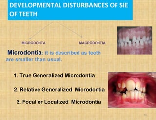
3. CONTENTS:
⢠INTRODUCTION
⢠CLASSIFICATION
⢠DEVELOPMENTAL DISTURBANCES IN
SIZE
⢠DEVELOPMENTAL DISTURBANCES IN
NUMBER
⢠DEVELOPMENTAL DISTURBANCES IN
SHAPE
⢠DEVELOPMENTAL DISTURBANCES IN
STRUCTURE
⢠DEVELOPMENTAL DISTURBANCES IN
ERUPTION.
⢠REVIEW LITERATURE 3
5. INTRODUCTION
⢠DEVELOPMENT
⢠The orderly and sequential changes that occur
with the passage of time as an organism moves
from conception to death.
⢠Also referred to as gradual growth which
indicates changes in character.
5
6. INTRODUCTION
⢠Development of teeth and face is regulated
by genes
⢠Dental developmental anomalies appear as
a result of disruption in the odontogenesis
process by the action of certain disruptive
factors
⢠The disruptive factors can be genetic
factors or environmental factors or even
combinations of these factors.
6
7. INTRODUCTION
⢠But the genetic programme is very sensitive to
disturbances in the environment such as exposure
to infection or toxic chemicals, including drugs
7
DISTURBANCES OF TEETH
ô Environmental
ô Developmental
10. ⢠SIZE OF TEETH
⢠SHAPE OF TEETH
⢠NUMBER OF TEETH
⢠STRUCTURE OF TEETH
⢠GROWTH (ERUPTION) OF TEETH
10
11. CLASSIFICATION
Developmental disturbances in the
size of the teeth
Microdontia
Macrodontia
Developmental disturbances in the
shape of the teeth
Gemination
Fusion
Concresence
Dilaceration
Talonâs cusp
Dens-in-dente
Dens evaginatus
Taurodontism
Supernumerary root
11
12. Developmental disturbances in the number of the teeth
Anodontia
Hypodontia
Supernumerary teeth
Predeciduous dentition
Post permanent dentition
Developmental disturbances in the structure of the teeth
Amelogenesis imperfecta
Dentinogenesis imperfecta
Regional odontodysplasia
Dentin dysplasia
Developmental disturbances in the growth/eruption of
the teeth
Premature eruption
Delayed eruption
Ankylosed teeth
Submerged teeth/ impacted teeth
12
13. Anomalies at different stages of tooth
development
13
Dental lamina formation stage
Anodontia
Initiation and proliferation
Oligodontia
Supernumerary teeth
Geminated / fused teeth
Histodifferentiation -defects in multiple dental tissues
Regional odontodysplasia
14. 14
Morphodifferentiation ( defects in size & shape
Macrodontia / Microdontia
Dens invaginatus / Dens evaginatus
Hutchinson s incisors, mulberry molarsâ
Talon cusp
Taurodontism
Apposition ( defects in structure of enamel &
dentin )
Amelogenesis imperfecta
Enamel Hypoplasia
Dentinogenesis imperfecta
Dentin dysplasia
15. Microdontia: it is described as teeth
are smaller than usual.
1. True Generalized Microdontia
2. Relative Generalized Microdontia
3. Focal or Localized Microdontia
DEVELOPMENTAL DISTURBANCES OF SIE
OF TEETH
15
MICRODONTA MACRODONTIA
16. ď all teeth are smaller than
normal
teeth are well formed
ď occur in some cases of
ďdown syndrome.
pituitary dwarfism
ď exceedingly rare
(1) True Generalized
Microdontia
16
17. ď normal or slightly smaller than
normal teeth.
ď are present in jaws that are
somewhat larger than normal
ď Inheritance of jaw size from one
parent and tooth size from other
parent can lead to this variations.
(2) Relative Generalized
Microdontia
17
18. ď common condition
ď affects most often maxillary
lateral incisior + 3rd
molar
ď these 2 teeth are most often
congenitally missing
(3) Focal/Localized
Microdontia
18
19. ď common forms of localized
microdontia is that which
affects maxillary lateral
incisior
ďź peg lateral
ďź instead of parallel
diverging mesial + distal
surfaces
19
20. ďź sides converge or taper
together incisally
ďź forms cone-shaped crown
ďź root is frequently shorter
than usual
TREATMENT
⢠Composite / Porcelain Veneers
20
21. MACRODONTIA
Also called as megalodontia or megadontia.
It refers to teeth that are larger than usual.
21
ďź(1) True Generalized Macrodontia
ďź (2) Relative Generalize Macrodontia
ďź (3) Focal or Localized Macrodontia
22. ď all teeth are larger than
normal
ď associated with
pituitary gigantism,pineal
hyperplasia with hyperinsulinism.
ď exceedingly rare
(1) True Generalized
Macrodontia
MACRODONTIA
22
23. ď normal or slightly larger than
normal teeth in small jaws
ďź results in crowding of teeth
ďź insufficient arch space
(2) Relative Generalized
Macrodontia
23
24. ď uncommon condition
ď unknown etiology
ďTrue marodontia often confused
with fused teeth.
ď usually seen with
mandibular 3rd
molars
(3) Focal/Localized
Macrodontia
24
25. TREATMENT
25
ô Stripping to reduce tooth size.
ô Can be combined with build â up of
antimere if only one tooth is affected.
ô Extraction & replacement by prosthesis.
26. DEVELOPMENTAL DISTURBANCES IN SHAPE AND FORM
26
ď Crown
ďź Fusion
ďź Gemination
ďź Taurodontism
ďź Talonâs Cusp
ďź Leongâs Cusp
ďź Dens Invaginatus
ďź Peg-shaped Lateral
ďź Hutchinson Incisor
ďź Mulberry Molar
28. DOUBLE TEETH
28
ôjoined by crown,root or both
ďą Gemination and fusion
ô May have very similar clinical appearance
ô Higher frequency in anterior and maxillary
regions
ô Rate is about 0.1% in permanent dentition
and 0.5% in deciduous
ô Bilateral cases more infrequent.
ô Etiology unknown but trauma has been
Suggested.
Clinical importance
ô Crowding / spacing
ô Periodontal problem
ô Caries
ô Eruption disturbance
ô Aesthetics
29. GEMINATION
29
ď an attempt of single tooth bud to divide with resultant
formation of tooth with bifid crown.
ď partial cleavage
ď appearance of 2 crowns
that share same root canal
ď trauma has been suggested
as possible cause.
30. 30
TWINNING:
⢠Designate the production of equivalent
structures by division resulting in one normal
and one supernumerary tooth :
31. FUSION
31
ďFusion of adjacent tooth buds with resultant formation of
joined tooth with confluence of dentin.
ď Fusion may be complete or incomplete based on
the stage of tooth development at the time of fusion.
ď Union of two separate tooth buds.
ďWhen counted ,tooth count reveals
missing tooth when anamalous tooth is counted as one.
Fusion of (a) molar tooth with
supernumerary, (b) maxillary
premolar with fused roots, (c) premolars
32. 32
FUSION
ď Contact occurs before calcification â two teeth may
be completely united to form a single large tooth.
ďContact after calcification of the crowns, the roots
may be united
ďTooth may have fused or seperated root cnals
34. Treatment of Gemination
and fusion
34
Deciduous dentition:
can results in Crowding, abnormal
spacing, delayed eruption of permanent
teeth.
ď Extraction.
Otherwise:
Surgical division & endodontic treatment
is done
35. TAURODONTISM-(tauro bull, donât_ teeth)
35
ďąTaurodontismâ was originated by Sir Arthur
Keith in 1913.
ďąIt is an enlargement of the body and pulp
chamber of a tooth with apical displacement of the
pulpal floor .
ďąShawâ classified taurodont teeth according to
degree of apical displacement of pulpal floor.
ďHypotaurodont:
ďmild
ďMesotaurodont:
ďmoderate
ďHypertaurodont:
ďsevere
36. 36
Hereditary
Mutation resulting from odontoblastic deficiency during
dentinogensis of roots
A primitive pattern.
Failure of Hertwig's epithelial root sheath to invaginate at proper
horizontal level
Clinical Features :
The deciduous or permanent dentition (more common).
Molars.(3rd
,2nd
,1st
)
May be unilateral or bilateral.
Increased frequency in patients with cleft lip/palate,
also associated with syndromes like downs syndrome,
klinefelter syndrome,trichodento-oseous syndrome.
Oculoâdentalâdigital dysplasia.Orofacial digital synd.ectodermal
displasia
â˘Sex chromosomal aberrations.Amelogenesis imperfecta IE,IV
CAUSES
41. ď An accessory cusp located onAn accessory cusp located on
the palatal surface of thethe palatal surface of the
mesiolingual cusp of a maxillary molar.mesiolingual cusp of a maxillary molar.
May be seen in deciduous or permanent teethMay be seen in deciduous or permanent teeth
& varies from cusp to small indented pit or fissure& varies from cusp to small indented pit or fissure..
41
CUSP OF CARABELLI
ď90% in whites and rare in Asians90% in whites and rare in Asians
Requires no therapy unless deep groove is present.Requires no therapy unless deep groove is present.
43. TALON CUSP
43
Eagleâs talon.
â˘A well delineated additional cusp that is located on the surface
of an anterior tooth & extends at least half the distance from the
CEJ to the incisal edge.(lingual side)
â˘Composed of normal enamel and dentin and contains a horn of
pulp tissue.
â˘Prevalence ranges from 1%-8%
⢠Chawla et al-7.7% prevalence- north
Indian children.
44. â˘In primary dentition maxillary central incisor most
common site.
PATTERNS OF TALON CUSPS: three types based
on morphology
⢠Trace talon
â˘Semi-talon
â˘Talon
Forms : T âform ; Y -shaped
â˘Seen in patients with rubinstein teybi
syndrome,struge weber syndrome,mohr syndrome.
â˘Radiographically,the cusp is seen overlying the
central portion of crown & includes enamel and
dentin
â˘Clinical problems include esthetics, caries control
and occlusal accombidation.
44
46. DENS EVAGINATUS
Also called Leongâs premolar, Evaginated odontome
46
â˘It is a developmental condition that appears
clinically as an accessory cusp or a globule of
enamel on the occlusal surface between the
buccal and lingual cusps of premolars,
â˘unilaterally or bilaterally.
â˘Rare in whites 15% in asians.
â˘Occurs in persons of mongoloid ancestryâchinese,
japanese, filipinos, eskimos and american indians.â
â˘Evolves by proliferation and evaginationof an area of the
inner enamel epithelium and subjacent odontogenic
mesenchyme into the dental organ during early tooth
development
47. Extra cusp can lead to:
â˘Incomplete eruption
â˘Displacement of teeth
â˘Pulp exposure following occlusal wear or
fracture.
Radiographic features
â˘Occlusal surface exhibits a
tuberculated apearance, and often
pulpal extension is seen in the cusp
Treatment
Extraction is indicated if tooth is nonvital
Grinding of the tubercle & indirect pulp
capping with calcium hydroxide.
47
48. SHOVEL SHAPED INCISOR
48
â˘Associated with dens evaginatus
â˘Affected incisors demonstrate prominent
lateral margins creating a hallowed lingual
surface that resembles the scoop of shovel.
â˘Maxillary central incisors most affected
â˘Predominantly in asians, native
americans and alaskans
49. DENS INVAGINATUS
49
⢠Also called as Dens In Dente, Dilated
composite odontome
⢠It is a deep surface invagination of the crown
or root by enamel before calcification occurs..
⢠Causes :
â Increased localized external
pressure
â Focal growth retardation
â Focal growth stimulation.
⢠Maxi. Permanent Lateral incisor most
commonly involved while central rare.
⢠Condition is frequently bilateral.
⢠Prevalence 0.04%-10%
50. ďOehlers (1957) divided this condition into 2
forms:
Coronal: Type 1,2,3.
Radicuar
Coronal dens invaginatus has been classified
into three major types:
50
Type I:
An invagination that is confined to the
crown.
Clinical significance
ď§ Predisposition to caries
ď§ Pulpal pathology
ď§ Periapical pathology (Type III)
ď§ Early diagnosis â mandatory
51. Type II
Extends below the CEJ & ends in the blind sac that
may or may not communicate with adjacent dental
pulp.
51
52. Type III:
ďExtends through the root & perforates in the
apical or lateral radicular area without any
immediate communication with pulp.
ďEnamel line often replaced by cementum
close to the radicular perforation.
ďPerforation provides communication from
oral cavity to the intraosseous periradicular
tissue, &often produce inflammatory lesions
52
54. Radiograph:
â˘Affected tooth demonstrate an enlargement of the
root.
â˘Dilated invagination lined by enamel
⢠Radiographs reveal a pear shaped invagination of
enamel and dentin with a narrow constriction at the
opening on the surface of the tooth and closely
approximating the pulp in its depth.
â˘ ďŹ Severe form â an invagination that extends nearly
to the apex of the root
TYPE II CORONAL DENS INVAGINATUS
TYPE III CORONAL DENS
INVAGINATUS
54
55. Treatment:
Small type I opening of the invagination is
restored to prevent carious involvement.
Large type:
Contents of the lumen & any carious dentin
must be removed & calcium hydroxide base
is placed.
Pulpal pathosis or pulpal communication
then endodontic therapy is recommended
55
56. MULBERRY MOLARS (MOONS MOLAR)
56
â˘Molars have multiple poorly developed cusps.
â˘Seen in congenital syphilis.
â˘Mulerry molar shows numerous disorganized globular
projection that resembles like mulberry.
â˘Condition occurs due to spirochete infection of enamel
organ of teeth during amelogenesis.
57. HUTCHINSON INCISOR
57
ď
characteristic of congenital
syphilis
ďlateral incisors are peg-shaped
or screwdriver-shaped
ď widely spaced
ď notched on their biting end
ď with a crescent-shaped defect
ďnamed after Sir Jonathan
Hutchinson
ďź English surgeon +
pathologist who 1st
described it
58. SHAPE AND FORM
58
ROOT
Concrescence
ďŹ Union of two adjacent teeth by
cementum alone.
ďŹ May occur before or after eruption
ďŹ Seen most commonly posterior and
maxillary regions
ďŹ Etiology believed to be trauma or
Overcrowding.
Usually involves two teeth. But involving
three teeth has also been reported.
59. FACTORS ASSOCIATED:
59
LOCAL FACTORS
â˘Abnormal occlusal trauma
â˘Adjacent inflammation
â˘Unopposd tooth;impacted , ,without antagonist
SYSTEMIC FACTORS
Acromegaly and pituitary gigantism
âArthritis
âCalcinosis
âPagetâs disease of bone
âRheumatic fever
âThyroid goiter
âVitamin A deficiency
60. 60
â˘Diagnosed by radiographic
examination
Histopathology
â˘Deposition of excessive cementum over the original layer of
primary cementum.
â˘May be hypocellularor exhibit areas of cellular cementum
resembling bone called osteocementum
.
â˘Polarized light to differentiate dentin and cementum.
61. TREATMENT
61
ďoften requires no therapy
unless union interferes with
eruption; then surgical
removal may be warranted
ďsince with fused teeth,
extraction of one may result in
extraction of the other
62. DILACERATION
62
ďAbnormal angulation or bend in root or less frequently crown of tooth.
ďMajority due to trauma to tooth germ calcified portion.
ďPermanent maxillary incisors are frequently involved, followed by
mandibular incisors.
ďDilaceration in a permanent tooth often follows traumatic injury (avulsion or
intrusion) to the deciduous predecessor in which the tooth is driven apically into
the jaw.
ďCan also develop secondary to adjacent cyst, tumor or odontogenic
hamartoma.
kinked tooth â or â sickle â tooth
63. TREATMENT
63
ďVary according to severity
ď Extraction: if it is non vital then surgical removal is
suggested.
ď Can be problematic during extraction sometimes.
ďDeciduous teeth â Extraction
Permanent teeth
ďMinor â no treatment
64. ECTOPIC ENAMEL
64
ďThought to arise from localized bulging in
the odontoblastic layer with prolonged
contact between HERS and dentin leading
to enamel formation.
ďEnamel in unusual location
Enamel pearlEnamel pearl
ďCommon-roots of max. molars followed by mand.
Molar.
ďDeciduous molar is not rare.
ď1.1-9.7% - highest in Asians
66. CERVICAL ENAMEL PROJECTION
66
ďRepresent dipping of enamel from the CEJ
toward the bifurcation of molar teeth.
ďMandibular molars affected more frequent.
ď prevalence is greater in Asians (8.6%-32.4%)
ďExtensions is correlated with localized loss of
periodontal attachment with furcation
involvement
68. SUPERNUMERARY ROOTS
68
ďRefers to the development of an increased
number of roots on a tooth
compared with that
described in dental anatomy.
â˘May involve any tooth.
â˘Mandibular cuspids and
bicuspidsmay have two roots.
â˘Maxillary and mandibular molars
may exhibit additional roots.
â˘Significant during extraction
71. 71
Clinical features:
Women
Mongoloid
Whites
True total anodontia:
All teeth missing
Can be either deciduous or
permanent
True partial anodontia:
Absence of one or more teeth
Commonly missing teeth â 3rd
molars, max. lateral incisors,
max / mand. 2nd
premolars
73. HYPODONTIA
73
ďlack of development of one or more
teeth.
Common dental anomaly
Etiology:
⢠Familial tendency â point mutations
transmitted in autosomal dominant pattern.
⢠Missing third molars could be an evolutionary
trend towards fewer teeth.
â˘It is associated with hereditary ectodermal
dysplasia
⢠Xray radiation of face during early age â
affecting the sensitive tooth buds
.
74. 74
ďŹ 3.5%-8% (excluding third molars)
ďŹ Female predominance about 1.5:1
ďŹ Uncommon in primary dentition
(<1%)
ďŹ About 20-23% of population
missing third molars.
ďŹ After third molars, second
premolars and laterals most frequent
76. 76
ďŹ Prevalence of supernumerary teeth is
about 1%-3% (higher rate in Asians)
ďŹ Single tooth hyperdontia represent 75%-
85% of cases
ďŹ More common in permanent dentition
ďŹ Almost 90% in maxilla
ďŹ Maxillary incisor region most common site
then 4th molars,premolars and canines
ďŹ If multiples, usually in mandibular premolar
region
HYPERDONTIA
78. 78
Classification of
supernumerary teeth
Modified Howard s classificationâ
ďSupplemental supernumerary teeth
ďAccessory supernumerary teeth
⢠Classified according to
morphology and location, four
different types
⢠Conical
⢠Tuberculate
⢠Supplemental
⢠Odontome
79. 79
Supplemental supernumerary teeth:
These teeth duplicate the typical anatomy of
anterior and posterior teeth
Mesiodens:
â˘Most common Supplemental
supernumerary teeth
â˘Small tooth with cone shaped crown and
short root
â˘Located near the midline in the incisor
region of maxilla between the central
incisors
â˘Occurs single / multiple, erupted /
impacted
â˘Causes retarded eruption, displacement of
adjacent tooth, or resorption of adjacent
root and thus improper alignment of teeth
80. 80
DISTOMOLARS:
ďFound in molar region
ďLocated distal to 3rd
molars
ďGenerally these teeth are smaller
than normal 2nd
& 3rd
molars
ďBut crown morphology is
abnormal
81. 81
PARAMOLARS:
ďIt is an supernumerary molar
ďUsually small and rudimentary
Situated buccally or lingually to one of the maxillary
molars
ďInterâproximally between 1st
, 2nd
, 3rd
maxillary
molars
82. TRANSPOSITION
82
Normal teeth erupting into an
inappropriate position.
â˘Usually involves the canine and
first premolars.
â˘Canine erupting between two
premolars.
â˘Could be confused with
supernumerary teeth
83. 83
Developmental disturbances in
the growth/eruption of the teeth
Premature eruption.
Delayed eruption.
Ankylosed teeth.
Submerged teeth/ impacted teeth
84. 84
NATAL TEETH
ďPremature eruption of teeth like
structures
ďPresent at birth
ďThey are hyper mobile because of
their limited root development
ďConical or normal in size and
shape
ďSome teeth are so mobile that
there is danger of displacement and
possible aspiration â removal is
indicated other wise not !!
85. 85
ďSharp incisal edges
may cause laceration of
lingual surface of tongue
â RIGAâFEDE
DISEASE
ďAssociated with Ellis-
van creveld syndrome
86. 86
Neonatal teeth
These are the teeth or teeth like structures
that erupt prematurely during neonatal
period
From birth to 30 days
87. 87
TREATMENT
Extraction of teeth (if causing
inconvenience)
Or
Rounding of the sharp angles
of the teeth
Extraction should be done
after 10 days of life
If not necessary tooth should
not be removed
88. 88
ERUPTION SEQUESTRUM
ďAnomaly associated with tooth eruption in children.
ďŹ Described by Starkey and Shafer
.
ďŹ It is a tiny, irregular spicule of bone overlying the crown of
an erupting permanent molar, found just prior to or
immediately following the emergence of the tip of the cusps
through the oral mucosa.
Etiology
ďŹ As the molar teeth erupt through the bone, they can
separate a small osseous fragment from the surrounding
bone similar to a cork screw.
ďŹ In most cases, the fragment undergoes complete
resorption before eruption.
ďŹ If the bony spicule is large or the eruption is rapid,
complete resorption cannot occur and hence, it is observed
89. 89
CLINICAL FEATURES
ďŹ The child may complain of slight soreness in the
area during function.
ďŹ The spicule directly overlies the central occlusal
fossa but is within the soft tissue.
ďŹ It may be seen lying in a tiny depression over the
crest of the ridge.
ďŹ As the tooth erupts, the fragment of bone
completely sequesters through the mucosa and is
lostâ
Radiographic features
ďŹ It can be recognized even before the tooth
eruption.
ďŹ Seen as a tiny, irregular opacity overlying the
central occlusal fossa but separated from the tooth
itself.
90. DELAYED ERUPTION
90
In deciduous and permanent teeth, it is
difficult to assess unless a gross variation is
present.
â˘Caused by Systemicconditions like rickets,
cretinism, cleidocranial dysplasia
â˘Localfactors like fibromatosis gingivae.
â˘Treatment of the primary condition may lead
to eruption of teeth
91. 91
IMPACTED TEETH
ďź occurs due to obstruction
from crowding
ďź from some other physical
barrier
ďź occasionally, may be due
to an abnormal eruption
path, presumably because
of unusual orientation of
tooth germ
92. 92
ďAny tooth may be impacted âusually
mandibular third molars(22%),
maxillary third molars(18%) and
maxillary cuspids(0.9%), premolars
and supernumerary teeth.
ďMandibular teeth are more severly
impacted.
93. ANKYLOSIS
93
ďfusion of a tooth to surrounding
bone
ď with focal loss of periodontal
ligament, bone + cementum
become inextricably mixed
ďź cause fusion of tooth to
alveolar bone
94. 94
ďąReported prevalence of clinically
detectable ankylosis- 1.5% to 9%
ďą Primary molars are most commonly
involved teeth with most cases in mandible
ďąEven with extreme root resorption, teeth
lack mobility.
95. 95
â˘Diagnosis is suspected clinically and confirmed
with radiographs.
â˘There is partial absence of the periodontal ligament
with areas of apparent blending between the root of
the tooth and alveolar bone.
complications
Development of malocclusion
â˘Local periodontal disturbance
â˘Dental caries of both the ankylosed tooth and
adjacent teeth.
TREATMENT
Depends on whether tooth are ankylosed or not
permanent successor present or not
96. 96
DEVELOPMENTAL DISTURBANCES
IN THE STRUCTURE OF THE TEETH
Amelogenesis imperfecta
Dentinogenesis imperfecta
Regional odontodysplasia
Dentin dysplasia
Enamel hypoplasia
97. AMELOGENESIS IMPERFECTA
97
ď also known as:
ďź Hereditary Enamel Dysplasia
ďź Hereditary Brown Enamel
ďź Hereditary Brown Opalescent Teeth
98. 98
ďGroup of conditions caused by
defects in the genes encoding
enamel matrix proteins
ď genes that encode for enamel
proteins:
ďź amelogenin mutated in
ďź enamelin in patients
ďź others with this
condition
99. 99
ďąA heterogeneous group of hereditary
disorders that demonstrate developmental
alterations in the structure of enamel in the
absence of a systemic disorder
ďŹ Many subtypes
ďŹ Numerous patterns of inheritance
ďŹ Wide variety of clinical
manifestations
ďŹ Frequency varies between 1:718
and
1:14,000
ďŹ Both dentitions involved
100. 100
Formation of enamel a multistep
process
Formation of enamel matrix
Mineralization of matrix
Maturation of matrix
Hereditary defects of enamel
formation usually classified as:
ďŹ Hypoplastic
ďŹ Hypocalcified
ďŹ Hypomaturative
102. 102
Hypoplastic
Amelogenesis Imperfecta
ď Inadequate formation of matrix
ďFEATURES
ďTeeth erupt with insufficient amounts
of enamel.
ď Enamel present is mineralized
appropriately and contrasts well with
dentin on radiograph.
ď Teeth may have abnormal shape
and open contacts.
ď Open bite may be present
106. 106
HYPOPLASTIC AUTOSOMAL
DOMINANT SMOOTH PATTERN
ďEnamel of all teeth exhibit smooth
surface,thin hard glossy surface.
ďAbsence of appropriate enamel
thickness.
ďColour varies from opaque white to
translucent brown.
ďRadiographically, thin peripheral
radiopaque enamel.
108. HYPOPLASTIC ROUGH PATTERN
108
ďEnamel is thin with hard
rough surface
ďTeeth taper towards
incisal-occlusal surfaces.
ďopen contact points
ďsignificant attrition and anterior
open bite
109. 109
â˘ENAMEL AGENESIS
â˘Demonstrate total lack of enamel
â˘Teeth are shape and colour of dentin with yellow brown hue
â˘Open contacts
â˘Crowns taper towards incisally
â˘Dentin is rough
â˘Radiograph demonstrates no peripheral enamel overlying dentin
â˘A lack of eruption of many teeth occurs frequently
110. HYPOMATURATION TYPE
110
Enamel matrix is laid down properly and
begins to mineralize but there is a
defect in
maturation of enamelâs crystal structure
ďŹ Affected teeth normal in shape
ďŹ Mottled appearance-white, brown or
yellow
ďŹ Enamel soft and chips away from
dentin
ďŹ Enamel has similar radiodensity to
dentin
113. HYPOMATURATIVE-AUTOSOMAL
RECESSIVE
113
ďBoth primary and permanent
dentition
ďEnamel shows milky to shiny
agar brown deeply stained on
contact with exogenous agents
ďChips away around teeth
restoration
ďTeeth may resorbs within
alveolus
115. HYPOCALCIFIED âAMELOGENESIS
IMPERFECTA
115
Proper amount of enamel matrix is
formed but but
it doesnât mineralize properly.
ďŹ Teeth shaped normally upon
eruption but enamel
is soft and easily lost
ďŹ Enamel yellow-brown upon
eruption but quickly
becomes brown to black
ďŹ Accumulate calculus.
116. HYPOMATURATION HYPOPLASTIC TYPE
116
â˘Enamel hypomaturation is the
dominant feature.
â˘Enamel is yellowish white toâ
yellowish brown.â
â˘Pits are seen on buccal surface.
â˘Enamel is similar to dentin in density.
â˘Large pulp chambers with varying
degrees of taurodontism.
118. TRICHO-DENTO-OSSEOUS SYNDROME
118
â˘Autosomal dominant disorder.
â˘Shows hypoplastic hypomaturationâ
with severe taurodontism.
â˘Kinky hairat birth which may
straighten with age.
â˘Osteosclerosisof the base of skull
and mastoid process.
119. TREATMENT
119
ďVary accoring to sub type and severity
ďMain problems associated are
esthetics,sensitivity,and loss of vertical
dimension.
ďFull coverage restoration recommended in
attrition cases
ďLess severe cases can be improved by
placements of full crowns and facial veneers
120. DENTINOGENESIS IMPERFECTA
120
â˘Called Hereditary opalescent dentin,
Capdepontâs teeth.
â˘Only the mesodermal portion of the
odontogenic apparatus is affected.
â˘Dentin is defective.
â˘Could be due to hereditary factors
â˘Can be seen in association with
osteogenesis imperfecta
121. CLINICAL FEATURES
121
Deciduous teeth are more severely affected
â˘Gray to brownish violet or yellowish brown
with a characteristic translucent or
opalescent hue.
â˘Broad crown with constriction of cervical area
constriction â TULIP SHAPE
122. 122
Enamel may be lost earlyon incisal and occlusal
surfaces due to the abnormal DEJ
.
â˘DEJ lacks the usual scalloping.
â˘Dentin is attrited rapidly.
â˘Occlusal surfaces of affected teeth are severely
flattened.
124. CLASSIFICATION BY SHIELDS
124
Type I âDI that always occur with OIâ
autosomal dominant.
â˘Type II âDI that never occurs with OIâ
hereditary opalescent dentin âautosomal
dominant .
â˘Type III âDI âBrandy wine typeâsimilar
autosomal dominant
125. ď 1 in 8000
ďoccurs in families with
Osteogenesis Imperfecta
ď primary teeth are more severely
affected than permanent teeth
125
TYPE I DENTINOGENESIS IMPERFECTA
126. ď Radiographically:
ďź partial or total obliteration
of pulp chambers + root canals
ďź by continued formation
of dentin
ďź roots may be short + blunted
ďź cementum, periodontal
membrane + bone appear
normal
126
Type I DENTINOGENESIS IMPERFECTA
127. ď never occurs in association
with osteogenesis imperfecta
unless by chance
ď most frequently referred to as
hereditary opalascent dentin
ď only have dentin abnormalities
and no bone disease
127
Type II DENTINOGENESIS IMPERFECTA
128. ď Radiographically:
ďź partial or total obliteration
of pulp chambers + root canals
ďź by continued formation
of dentin
ďź roots may be short + blunted
ďź cementum, periodontal
membrane + bone appear
normal
Type II Dentinogenesis Imperfecta
128
129. ď âBradwine typeâ
ď racial isolate in Maryland
ď multiple pulp exposures in
deciduous not seen in type
I or II
ď periapical radiolucencies
Type III Dentinogenesis
Imperfecta
129
130. ď enamel appears normal
ď large size of pulp chamber
is due not to resorption but
rather to insufficient + defective
dentin formation
Type III Dentinogenesis
Imperfecta
130
Witkop describes it as Shell teeth
Radiographic features
Roots are extremely short
131. HISTOLOGIC FEATURES
131
â˘Dentin has irregular tubules with large areas
of uncalcified matrix.
â˘Tubules are larger in diameter and lesser in
number
Cellular inclusions probably odontoblasts can be seen in
dentin.
â˘
132. 132
â˘The odontoblasts with limited ability to
form well organized dentinal matrix,â
appear to degenerate readily and get
entrapped within the matrix.
â˘Pulp chamber is obliterated by
continuous dentin deposition
133. ď Treatment:
DENTINOGENESIS IMPERFECTA
133
oPreservation of vertical dimension of occlusion
o Continued commitment & support to both child &
family
o Stainless steel crowns to protect against attrition
o
o Initial composite build â up for anteriors to be
replaced by veneers later Overdentures / implants
later.
134. ď also known as âRootless Teethâ
ď rare disturbance of dentin
formation
ďatypical dentin formation
DENTIN DYSPLASIA
134
First described by Ballschmiede(1920) âspontaneous
exfoliation of multiple teeth.
â˘Rushton(1939) designated it as dentin dysplasia.
136. CLASSIFICATION
136
Shields:
Type I âdentin dysplasia
Type II âanomalous dysplasia of dentin
â˘Witkops:
Type I âRadicular dentin dysplasia âMore common.
Type II âCoronal dentin dysplasia
138. ď both dentitions are of
normal color
ďMost common type
ď prevalence 1:100000
Type I (Radicular Type)
138
(Rootless
teeth)
ďWide variation in root formation because dentinal
disorganization may occur at different stages of tooth
development
139. ď Radiographically:
ďź roots are extremely short
ďź pulps almost completely
obliterated
ďź periapical radiolucencies:
⢠granulomas
⢠cysts
⢠chronic abscesses
Type I (Radicular Type)
139
140. ď autosomal dominant
ďRoot length is normal.
ďColour blue -amber to brown
translucency
ď coronal pulps are usually large
(thistle tube appearance)
ďź filled with globules of abnormal
dentin
Type II (Coronal Type)
140
141. ď Radiographically:
(Deciduous)
ďź roots are extremely short
ďź pulps almost completely
obliterated
(Permanent)
ďź abnormally large pulp
chambers in coronal portion of
tooth
Type II (Coronal Type)
141
142. ď also known as:
ďź Odontogenic Dysplasia
ďź Odontogenesis Imperfecta
ďź Ghost Teeth
142
REGIONAL
ODONTODYSPLASIA
143. ď one or several teeth in a
localized area are affected
ď maxillary teeth are involved
more frequently than
mandibular area
ď etiology is unknown
143
144. ď teeth affected may exhibit
a delay or total failure in
eruption
ď shape is altered, irregular in
appearance
144
145. ď Radiographically:
ďź marked reduction in
radiodensity
ďź teeth assume a âghostâ
appearance
ďź both enamel + dentin appear
very thin
ďź pulp chamber is exceedingly
large
145
146. ď Treatment:
ďź poor cosmetic appearance
of teeth
ďź extraction with restoration
by prosthetic appliance
146
147. ENAMEL HYPOPLASIA
147
ď§An incomplete or defective formation of the
organic enamel matrix of teeth.
â˘It occurs only when the injury occurs during the
development or more specifically, formative
stage of enamel.
TYPES:
I.Hereditary type enamel
hypoplasia
II.Environmental type
hypoplasia
148. Hereditary type enamel hypoplasia
148
â˘This is an ectodermal disturbance that occurs during the
embryonic development of the enamel.
⢠The mesodermal components are normal. Both the
deciduous and permanent teeth are involved and only the
enamel is affected.
The three types of hereditary type of enamel hypoplasia
are the:
â˘Hypoplastic type, where there is a defective formation of
organic matrix.
â˘Hypocalcification type, in which there is a defective
mineralization of the matrix.
â˘Hypomaturation type, where there is a defective
maturation of the matrix
149. Environmental type enamel hypoplasia-
149
ď§This is caused by the environmental factors that causes
damage to the enamel cells.
ď§Either deciduous or permanent teeth are involved and
sometimes a single tooth is involved
ď§Hypoplasia results only if the injury occurs during the
time the teeth are developing or more specifically during
the formative stage of enamel development.
The environmental factors which produce
enamel hypoplasia can include
151. CLINICAL FEATURES
151
Mild:
Few small grooves, pits or fissures on
enamel surface
Moderate:
Enamel may exhibit rows of deep pits
arranged horizontally across tooth
surface.
Severe:
pitting and brownish staining of the
surface. Tendency of enamel to wear or
fracture
152. TREATMENT
152
Treatment of enamel hypoplasia is always
individualized and depends on the location and
severity of then condition.
The location of enamel hypoplasia also
affects treatment options :
> Anterior teeth -
For sensitive teeth with no wear,
desensitizing agent (such as potassium nitrate)
as needed can be used.
If there are esthetic concerns, composite or
porcelain veneers may be bonded to afected
tooth.
153. 153
Posterior teeth â
1. For sensitive teeth with minimal wear, desensitizing agent
(such as potassium nitrate) can be used.
2. For mildly hypoplastic molars, place pit and fissure sealant
on the occlusal surface. - at 6 month re-evaluation
3. Remove demineralized enamel and restore with composite.
- at 6 month re-evaluation, if composite is lost, either
replace using good isolation techniques or go to step 4
4. Perform minimal reduction of tooth and cement a stainless
steel crown - evaluate clinically and radiographically as
indicated
155. 155
A CASE REPORT OF A PATIENT AFFECTED BY
AMELOGENESIS
IMPERFECTASHOWING A POSSIBLE MINIMAL
AND CONSERVATIVE ADHESIVE TREATMENT
APPROACH.
A 14 years old boy with amelogenesis imperfecta
consulted a dentist for full mouth rehabilitation
After examination ,step by step minimum
conservative approach planned.
S Ardu O Duc I Krejci
R Perroud
Clinical views at the patientâs first
appointment
156. 156
In the posterior area conservative resin composite onlays
were realized , whereas in the anterior area
freehand bonded composite restorations were
performed according to the natural layering technique
proposed.
Intraoral view of the conservative preparations for resin
composite onlays and clinical view after posterior
onlays placement
157. 157
Intraoral view of the preparations for freehand bonded composite
restorations and clinical view after teeth rehydration. Last image
shows the clinical view after the placement of a cantilever bonded bridge
23
158. 158
Clinical views at the end of the treatment, together with the final treatment status at the 3-month recall. Except
for the lithium disilicate
bridge from 21â23, all other teeth are single-unit lithium disilicate bonded crowns.
159. 159
â˘case report describes a possible application of a
minimally invasive approach for treating amelogenesis
imperfecta in a young adolescent.
â˘This conservative
adhesive treatment allows a two-stage
intervention that accompanies the patient during
his adolescence until the complete formation of hard
and soft tissues.
â˘This will allow acceptable esthetics
and lack of pain during eating and will allow the
patient to have a more normal social life.
160. 160
Dan Emil GEORGESCU, Rodica LUCA and Elisa
MÄdÄlina CHIRCA
Conducted study to assess the frequency of development
anomalies of teeth and soft tissue in the anterior region, of a school
children from Bucharest.
cross sectional study is conducted
Material and methods: 226 children from two secondary schools,
aged between 5-8years
â˘Children were examined in the school dental office.
⢠The presence of dental development anomalies or of
other types was recorded.
⢠The frequency of developmental anomalies was
determined.
â˘4.87% of the examined school children had oral
developmental anomalies:
.
Fused teeth
161. 161
Dental size anomalies (0.44%) - one fusion and one
gemination and 9 abnormal maxillary frenum attachment
(3.98%).
Dental anomalies were found in front teeth, in both
dentition.
Diastema was found in permanent dentition, between the
two upper central incisors. The evaluated age group had a
low frequency for front teeth dental development
anomalies.
Follow up of all children is necessary until the eruption of
all permanent teeth
Gemination
Diastema between the two upper
permanent central incisors
162. 162
Endodontic treatment of developmental
anomalies in posterior teeth: Treatment of
geminated/fused teeth Nelly Steinbock
Two cases of complex endodontic treatment of fused/geminated
teeth are presented.
The first is an 11-year-old girl with an anomalous âdoubleâ first
mandibular molar and premolar diagnosed as having necrotic
pulp with chronic apical abscess of endodontic origin;
.
Fused crowns of the mandibular right first molar
and second premolar
163. 163
ď§ Two separate endodontic access cavities were prepared in the
fused crown in the molar
and the premolar area
â˘Five canal orifices were found â two leading to the
distal root canals
(emerging from the molar pulp chamber) and the other
three leading to the mesial roots and
emerging from the floor of the premolar pulp chamber
â˘The canals were instrumented manually using K-files
and irrigated with 3% sodium
hypochlorite solution.
⢠Working length was determined with an electronic
apex locator
Working length radiograph of
the fused mandibular right
first molar and second
premolar
â˘Follow-up 6 months later showed that the tooth
was restored with composite filling
164. 164
The second is a 16-year-old boy with âdoubleâ
second and supernumerary mandibular molars, who
was diagnosed with irreversible pulpitis.
Preoperative radiograph of the geminated second
molar tooth (fusion of molar with supernumerary).
ď§Clinical examination revealed an anomalous
second molar that could have been a fusion of
the mandibular right second molar with a
supernumerary tooth in its mesiobuccal aspect
â˘Radiographic examination
â˘Two separate access cavities were prepared
â˘Five canal orifices were found on the mesial side of the
pulp
chamber floor â two leading to the supernumerary tooth
and three leading to the mesial root
of the molar.
165. 165
Working length determination radiograph
demonstrating five of the six canals.
â˘Irrigated with 3% sodium hypochlorite solution
â˘The tooth was obturated using
the lateral condensation technique with AH-26 as
a sealer
DISCUSSION
â˘Fusion and gemination are developmental
anomalies with inherently unusual and bizarre
anatomy.
â˘A prerequisite for endodontic treatment of
anomalous teeth is a careful examination
of radiographs from various angles
In both the cases, there was a communication between two
parts of the pulp chambers and root canal systems. Thus,
endodontic treatment was performed on both
parts of these teeth.
166. 166
Dental Anomalies in Chinese Children with
Cleft Lip and Palate
Hai Ming Wong1*, Moon Cheung Lai2 and Nigel Martyn
King3
⢠The prevalence of anomalies in children with CLP and to
ascertain if there were any differences between the
prevalence figures for CLP and non-CLP children
⢠The sample consisted of 231 pairs of age and gender matched CLP
and non-CLP southern Chinese aged between 12 and 16 years.
⢠The dental records of the subjects were examined to gather data on
anomalies of tooth number, size and shape.
: Missing permanent maxillary
lateral incisor and canines.
Number of types of dental anomalies per individual
Hypodontia (57.6%)
Microdontia (42.4%),
The cleft type associated with the highest
percentage of supernumerary teeth was CL (25.0%),
167. 167
â˘Approximately 10% of BCLP, UCLP and CP children
were found to have taurodontism
Taurodontism of permanent maxillary first
molars.
â˘This group of Chinese CLP children exhibited the
various types of dental anomalies.
⢠Statistically higher prevalences of hypodontia,
supernumerary teeth and microdontia were
demonstrated in the CLP children than in the non-
CLP children.
A conical shaped supernumerary tooth in
relation to permanent maxillary right canine
168. CONCLUSION
168
Anomalies on tooth have
aesthtic,functional,psychological
impact on the individual.so any
abnormalities or pathologies on
hard or soft tissues in oral cavity
must prompt clinician to think
beyond the local factors for
proper diagnosis and treatment
169. References:References:
ďś BooksBooks
ď Neville, et al: Oral and Maxillofacial PathologyNeville, et al: Oral and Maxillofacial Pathology
33rdrd
EditionEdition
ďShafer, et al: A textbook of Oral Pathology,Shafer, et al: A textbook of Oral Pathology,
33rdrd
EditionEdition
ďAnil govindrao ghom:textbook of oral medicine.Anil govindrao ghom:textbook of oral medicine.
44thth
editionedition
ďCawson, R.A: Cawsonâs Essentials of OralCawson, R.A: Cawsonâs Essentials of Oral
Oral Pathology and Oral Medicine,Oral Pathology and Oral Medicine,
88thth
EditionEdition
169
170. ďą Burkits textbook of oral medicine.11th
edition.
ďą DentalAnomalies inChineseChildrenwithCleft LipandPalate
HaiMingWong1*, MoonCheungLai2andNigelMartynKing3
ďą Endontic treatment of developmentalanomalies inposteriorteeth:treatment
of fusionandgeminationteeth(endodontic journal2003)
170
ďąAmelogenesis Imperfecta:
A Conservative and Progressive Adhesive Treatment Concept
S Ardu O Duc I KrejciR Perroud (operative dentistry 2013)
Numerous dev alterations of teeth can occur.major reported are
Anomaly that occur in dental lamina
Tooth size variable among different races and diff sexes.although hereditary is major factor,both genetic and enironmental factors influences affect developm of size of teeth.females demonstrate higher frequency
Microdontia due to defect during proliferation
Generally associated with hypodontia.
Normal sized teeth appear smaller when widely placed in larger jaws
Maxillary latrl affected appears as peg shaped lateral
Treatment usually consists of reshapingtooth with composite resin.
Associatd with hyperdontia.males have greater prevale
Also max centrals. Can be seen in facial hemi-hypertrophy of the face â the teeth of involved side may be larger than unaffected side.
Morphodiiferentiation stage
Defect during morphodifferentiation stage.other causes endocrinal disturbances,vitamin defciencies,congenital diseas,inflamatory/infec disease
Gemination tooth counnt is normal when anomalous tooth counted as one.fusion tooth count reveals missing when anomalous tooth counted as one
Molar teeth of cud chewing animal
Unilateral or bilateral..This cusp blends smoothly with the lingual tooth surface except for a deep developmental groove
Dens evaginatus associated with other coronal anotomy shovel incisor
Crowding of teeth with resorption of adjacent interdental bone â contact of two roots is fused by cementum deposition.Â
he curve or bend can occur anywhere along the length of the tooth depending on the amount of tooth formed at the time of injury.Â
Primary tooth germ developmental anomaly
Type of ectopic enamel
No treatment requred.bt important when endodontic theraphy isundertaken.
Defect occur during initiation.mutation of pax9msx1 gene
True total may b associated with ectodermal dysplasia.
In hereditary ectodermal dysplasia few cone teeth are seen
Supernumerary teeth reults from continued proliferation of pmnt or primary dental lamina
There is a marked variation in biologic eruption of deciduous and permanent dentition
 Reason is unknown. ďŹ Hormonal influences like hyperthyroidism, adrenal glands and gonadism, for tooth eruption are also considered
Othr terms for this include infraocclusion,submergence
If not ankylosed wait for exfoliation.perodic observation in primary
Vit d-max laterls,centrals cuspid,decay progress rapidly
During this first restorative phase, minimally invasive
dentistry was performed; only the most external,
porous enamel layer was removed before resin
composite placement
Four years later, at 20 years of age, a clinical and radiographic evaluation was
done, and a crown lengthening procedure of the
upper two posterior quadrants was performed. Two months later preparations for lithium disilicate crowns were performed on all the teeth, which
consequently received their crowns bonded with a dual resin cement