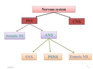
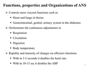
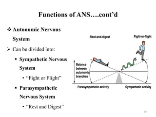
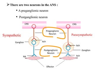
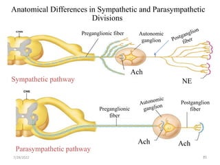
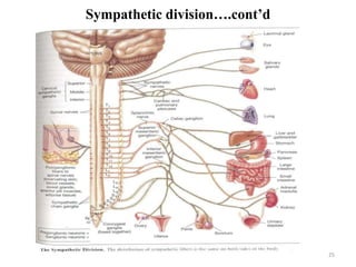
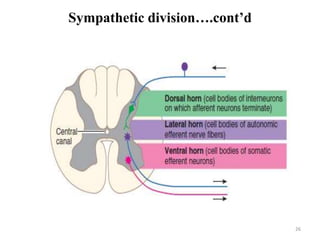
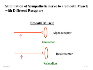
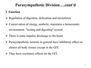
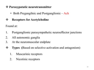
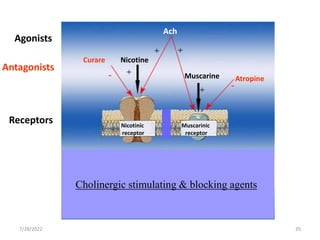
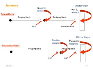
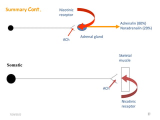
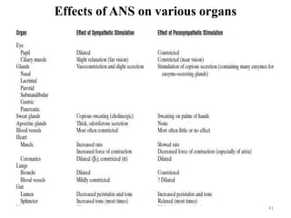
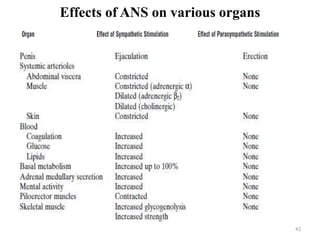
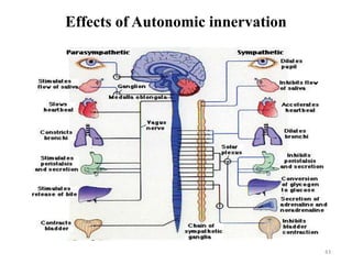
2. Learning objectives
At the end of this lesson, students will be able to:
Identify types of nerves and their action
Describe the function of ANS
Realize the effect of impaired ANS
2
4. Peripheral Nervous System -PNS
Peripheral Nervous System -PNS
The nervous system outside of the brain and spinal cord
Consists of:
• 31 pairs of Spinal nerves
• 12 pairs of Cranial nerves
Carry info to and from the spinal cord and brain
4
5. PNS….cont’d
PNS Can be divided further into:
Somatic nervous system -SoNS
Voluntary (generally)
Conduct impulses from the CNS to skeletal muscles
Autonomic nervous system -ANS
Involuntary (generally)
Conducts impulses from the CNS to smooth muscle,
cardiac muscle, and glands.
5
6. Somatic nerves
Originate in anterior horn cells
A single job; innervates and commands skeletal muscle
fibers
Consists of one neuron from CNS to effectors
Produce only excitation of its effectors with pin point
accuracy
Neurotransmitter (NT) is acetylcholine
Control its effectors via monosynaptic pathway
6
7. Enteric nervous system
Also known as “the little brain /the gut's brain”
It can and does operate independently of the brain and the
spinal cord.
Control peristalsis and churning of intestinal contents and
secretion of enzymes, mucus, electrolyte,and blood flow
Is embedded in the lining of the gastrointestinal system,
beginning in the esophagus and extending down to the anus.
Autorhythimicity
7
8. Comparison of SoNS VS. ANS
SoNS
1. Controls contraction of the
sk/muscle.
2. Nerve fibres are originated
from the anterior horn of
the spinal cord.
3. The motor nerve contains
single, long, thick and
myelinated axon.
4. The NT is always Ach and
the receptor is always
nAchR
ANS
1. Controls involuntary activities
such as CVS, GIT, sweat glands
2. Nerve fibres are originated from
the lateral horn of the spinal
cord.
3. Autonomic fibres contain two
neurons( pre- and
postganglionic)
4. There are both cholinergic and
adrenergic fibres acting on
cholinergic and adrenergic
receptors
8
9. The Cranial Nerves-PSNS components
There are 12 cranial pairs of nerves
Each of them are designated by the Roman numbers (I-XII)
Majority are mixed nerves (sensory, motor, autonomic)
Only few are pure sensory ( Cranial nerve I,II and VIII)
9
10. Cranial nerves (I-VI)
Nerve Name Sensory Motor Autonomic
Parasympathetic
I Olfactory Smell
II Optic vision
III Oculomotor Proprioception
4 Extrinsic Eye
muscles
Pupillary constriction
Accommodation
Focusing
IV Trochlear Proprioception
1 Extrinsic Eye
muscle
(Sup. Oblique)
V Trigeminal
Somatic senses
(Face, tongue),
corneal
sensitivity
Chewing
VI Abducens Proprioception
1 Extrinsic Eye
muscle
(Lateral rectus) 10
11. Cranial nerves (VII-XII)
Nerve Name Sensory Motor
Autonomic
Parasympathetic
VII Facial
Taste
Proprioception
Muscles of facial
expression
Salivary glands
Tear glands
VIII
Auditory
(Vestibulo
cochlear)
Hearing.&
Balance
IX
Glossopharyn
geal
Taste
Blood gases
Swallowing
Gagging
Salivary glands
X Vagus
Blood pressure
Blood gases
Taste
Speech
Swallowing Gagging
Many visceral
organs
(heart, gut, lungs)
XI
Spinal
accessory
Proprioception
Neck muscles:
Sternocleidomastoid
Trapezius
XII Hypoglossal Proprioception
Tongue muscles
Speech 11
12. Cranial nerve ….. cont’d
Cranial Nerves Mediate 5 Special Senses: Smell, Vision,
Hearing, Taste, Equilibrium
Smell: CN-I (Olfactory)
Vision: CN- II (Optic)
Hearing: CN-VIII (Cochlear division)
Equilibrium: CN-VIII (vestibular division)
Taste: CNs -VII, IX, X (minor) (Facial, Glossopharyngeal,
Vagus respectively)
Most of the Nerves Carry Somatic (Skin & Muscle) Sense
The trigeminal (V) is the sensory nerve for the face
&corneal sensitivity.
The nerves which are primarily motor also have fibers for
muscle sense (proprioception): III, IV, VI, VII, XI, XII
12
13. Cranial nerve ….. cont’d
Three Nerves Are Concerned With Eyeball Movements
Oculomotor (III): superior rectus, medial rectus, inferior
rectus, inferior oblique
Trochlear (IV): superior oblique
Abducens (VI): lateral rectus
Several of the Nerves Innervate Skeletal Muscles
The Facial nerve (VII) controls muscles of facial
expression
The Spinal accessory (XI) stimulates the trapezius and
sternocleidomastoid muscles
Chewing muscles (masseter, temporalis) are innervated by
the Trigeminal (V)
Speech muscles (larynx) are under the control of the Vagus
(X)
The Hypoglossal (XII) moves the tongue
13
14. Cranial nerve ….. cont’d
Four of the CNs Carry Parasympathetic Fibers
Oculomotor (III): innervates iris constrictor (causes pupil
constriction); also controls ciliary muscle (focuses the
lens)
Facial (VII) and Glossopharyngeal (IX): stimulate salivary
glands to secrete
Vagus (X): the major nerve of the parasympathetic system:
goes to most visceral organs (heart, lungs, kidneys, liver,
stomach, intestines)
14
15. Spinal nerves
31 pairs of spinal nerves
All contain afferent and efferent
8 cervical
12 thoracic
5 lumbar
5 sacral
1 coccygeal
Afferent
Sensory receptors relay to CNS
Long part of the axon is found out side CNS
Afferent neurons are 1st order neurons
Efferent
Carry signals from CNS to muscles & glands
More complicated than the afferent division
15
17. Functions, properties and Organizations of ANS
Controls most visceral functions such as
Heart and lungs in thorax
Gastrointestinal, genital, urinary system in the abdomen
Orchestrates the continuous adjustments in
Respiration
Circulation
Digestion
Body temperature
Rapidity and intensity of changes on effectors functions
With in 3-5 seconds it doubles the heart rate
With in 10-15 sec it doubles the ABP 17
18. Functions of ANS….cont’d
ANS is controlled by centers located in the
Spinal cord
Brain stem
Hypothalamus
Portions of cerebral cortex specially in Limbic cortex
ANS operates through autonomic reflexes
18
19. Functions of ANS….cont’d
Autonomic Nervous
System
Can be divided into:
Sympathetic Nervous
System
• “Fight or Flight”
Parasympathetic
Nervous System
• “Rest and Digest”
19
20. 20
There are two neurons in the ANS :
• A preganglionic neuron
• Postganglionic neuron
CNS
CNS
Ganglion
Preganglionic
Neuron
Ach
Postganglionic
Neuron
Ganglion
Effector
NA
Ach
Ach
Sympathetic Parasympathetic
21. 21
21
Anatomical Differences in Sympathetic and Parasympathetic
Divisions
Sympathetic pathway
Parasympathetic pathway
Preganglionic
fiber
Postganglion
fiber
Ach Ach
Ach
Autonomic
ganglion
NE
Preganglionic fiber
Ach
7/28/2022
22. 22
Criteria for distinguishing between:
• Sympathetic
and
• Parasympathetic
Origin of preganglionic neuron
Type of neurotransmitters
Location of autonomic gangliaa
23. 23
Originates in lateral horns of T1-T12 and L1-L2 region of
spinal cord
Components of the Sympathetic neurons
1. Cell bodies of preganglionic motor neurons are located in
the thoracic and lumbar part of the spinal cord
2. Preganglionic axons synapse in lateral/collateral ganglia,
which are located near the spinal cord far away from the
organs being innervated
3. Contains short preganglionic neurons which are
cholinergic.
4.Sympathetic postganglionic axons travel from the
lateral/collateral ganglia to the target organ
5. Contains long postganglionic neurons
Sympathetic division
24. 24
T1 head
T2 neck
T3-T6 thorax
T7-T11 abdomen
T12, L1&L2 legs
Sympathetic outflow/ origin (Fig-1 )
Sympathetic neurotransmitters
Pregangilnic – Ach
Postganglionic –NE, except sympathetics innervating sweat
glands, blood vessels in skeletal muscle, and piloerection muscles
are cholinergic.
7/28/2022
27. 27
Function:
Sympathetic mass discharge during emergency" situations (flight or
fight response)
Sympathetic mass discharge: simultaneous activation of various
organs
The sympathetic neurons have excitatory effects on almost all body
tissues. Their effect is excitatory on CVS and inhibitory on the GIT
Catabolic, increases MR and energy output
Tonic discharge of impulse to the heart and blood vessels
Sympathetic division….cont’d
28. 28
Two classes of adrenoceptors are distinguished
• - Receptors
• β - Receptors
7/28/2022 28
Alpha receptors
Constriction of gastric intestinal and bladder sphincter
Vasoconstriction
Contraction of pupillary dilator
Beta receptors
Increase in cardiac rate and strength of contraction
Inhibition of gastric motility
30. Parasympathetic Division
Originates from cranial and sacral regions.
Cranial components are part of CN III (Oculomotor), VII
(Facial), IX (Glossopharyngeal), and X (Vagus)
Sacral components from S1 – S4 segments of the spinal cord
30
32. Components of the parasympathetic neurons
1. Cell bodies of preganglionic motor neurons located in parts of
certain nuclei of cranial nerves and in the sacral part of the
spinal cord
2. Preganglionic axons synapse in terminal ganglia which are
located close to or on the organ being innervated
3. Contains long preganglionic neurons
4. Parasympathetic postganglionic axons travel from the terminal
ganglia to the target organ
5. Contains short postganglionic cholinergic neurons
32
Parasympathetic Division… cont’d
33. Parasympathetic Division….cont’d
3. Function
Regulation of digestion, defecation and micturition
Conservation of energy, anabolic, maintains a homeostatic
environment ”resting and digesting" system
There is tonic impulse discharge to the heart
Parasympathetic neurons in general have inhibitory effect on
almost all body tissues except in the GIT.
They have excitatory effects on the GIT.
33
34. 34
Parasypatatic neurotransmitter
• Both Pregangilnic and Postganglionic – Ach
Receptors for Acetylcholine
Found at:
1. Postganglionic parasympathetic neuroeffector junctions
2. All autonomic ganglia
3. At the neuromuscular endplate
Types (Based on selective activation and antagonism)
1. Muscarinic receptors
2. Nicotinic receptors
38. Autonomic ganglia
A nucleus (nuclei = pl) is a collection of cell bodies within
the CNS
A ganglion (ganglia = pl) is a collection of cell bodies
outside the CNS
Contains
• Cell bodies of postganglionic neurons
• Axon terminals of pregangionic neurons
They are divergence centers (1 Preg.---8 post g.)
38
40. Autonomic ganglia.....cont’d
Types of the autonomic ganglia
1. Lateral (paravertibral) ganglia
Are sympathetic ganglia
Form sympathetic chains on both sides of the vertebral
column
2. Collateral (prevertebral) ganglia
Are also sympathetic ganglia
Located in midway b/n the cord and the viscera
3 types: celiac g., superior mesentric g. and Inferior MG
3. Terminal ganglia: a parasympathetic ganglia, located near/within
the organ that they innervate
40
44. 44
1. Complete autonomic paralysis.
2. Excessive sweating
3. Disturbance of sexual functions
4. Pupillary and salivatory disturbances
5. Disturbance of the bladder
Clinical disorder of ANS
45. 45
1. Complete autonomic paralysis (Dysantonomic
polyneuropathy)
Relatively rare condition
Both sympathetic and parasympathetic systems are
affected mainly at the postganglonic level.
Anhidrosis, hypotension, paralysis of pupillary reflexes.
Loss of lacrimation and salivation impotence,
Reduced gastric acidity (ulcer symptoms may disappear)
Clinical disorder … cont’d
46. 46
2. Excessive Sweating:
It is a sympathetically mediated disorder.
Its cause is not known
One variety, presumably of congenital origin, affects the
palms
In some cases, the hyperhidrosis affects mainly the
face & lower extremities.
Clinical disorder … cont’d
47. 47
Sexual function in the male:
sexual impulse, drive or desire often referred to as libido.
Arousal of libido in man and woman may result from a
variety of stimuli, some purely imaginary.
3. Disturbances of Sexual Function:
Penile erection
By Parasympathetic motor neurones (S3 & S4).
T12 & L1 can mediate psychogenic erections.
Ejaculation
By sympathetic neuron.
7/28/2022 47
48. 48
Assignment (presentation)
Reflex action g1
Autonomic reflexes
Component of reflex
The Baroreceptor reflex
The chemoreceptor reflex
Defecation reflex
Micturition reflex
Water and electrolyte balance g5
Water balance
Electrolyte balance
Special sense
Physiology of hearing and balance g 4
Physiology Vision g2
Physiology Taste
Physiology Smell g3