Austin Ophthalmology is an open access, peer reviewed, scholarly journal dedicated to publish articles covering all areas of Ophthalmology.
The journal aims to promote latest information and provide a forum for doctors, researchers, physicians, and healthcare professionals to find most recent advances in the areas of Ophthalmology. Austin Ophthalmology accepts research articles, reviews, mini reviews, case reports and rapid communication covering all aspects of Ophthalmology.
Austin Ophthalmology strongly supports the scientific up gradation and fortification in related scientific research community by enhancing access to peer reviewed scientific literary works. Austin Publishing Group also brings universally peer reviewed journals under one roof thereby promoting knowledge sharing.
The human eye is an organ that reacts to light in many circumstances. As a conscious sense organ the human eye allows vision; rod and cone cells in the retina allow conscious light perception and vision, including color differentiation and the perception of depth. The human eye can distinguish about 10 million colors.
The human eye is an organ that reacts to light in many circumstances. As a conscious sense organ the human eye allows vision; rod and cone cells in the retina allow conscious light perception and vision, including color differentiation and the perception of depth. The human eye can distinguish about 10 million colors.
he sense organs — eyes, ears, tongue, skin, and nose — help to protect the body. The human sense organs contain receptors that relay information through sensory neurons to the appropriate places within the nervous system.
Each sense organ contains different receptors.
General receptors are found throughout the body because they are present in skin, visceral organs (visceral meaning in the abdominal cavity), muscles, and joints.
Special receptors include chemoreceptors (chemical receptors) found in the mouth and nose, photoreceptors (light receptors) found in the eyes, and mechanoreceptors found in the ears.
Anatomy of Eye by radhika kulvi, M.Sc nursingRadhika kulvi
The eye is a paired organ, the organ of vision. The eye is made up of various components, which enable it to receive light stimuli from the environment, and deliver this stimuli to the brain in the form of an electrical signal. Vision involves all components of the eye.
DEFINITION:The human eye is a sensory organ,that reacts to visible light and allow to use visual information for various purposes including seeing things,keeping our balance and circadian rhythm.
STRUCTURE
The eye is contained within the bony orbit of the head. The bony orbit is a cavity, comprising parts of the lacrimal bone (includes fossa for nasolacrimal duct) and the maxilla (includes caudal foramen of infraorbital canal). It is continuous with the temporal bone and the pterygopalatine fossa caudally. It is situated in the orbital cavity and is supplied by 2nd cranial nerve.
anatomy of an eye, internal structure of eye,layers of an eye ball, features of eye ball, cornea, sclera, retina, choroid, ciliary body lense, macula, blind spot, yellow spot, clinical aspect of eye , drainage of aqueous humor, cataract, glaucoma, causes of cataract and treatment.
structure and fuction of eyes and ears,types of memory,sharpe memory,attentionUmarKhan422
The external covering of the eyeball comprises of a generally intense, white layer called the sclera (or white of the eye). Close to the front of the eye, in the zone secured by the eyelids, the sclera is secured by a slim, straightforward layer (conjunctiva), which rushes to the edge of the cornea.
The eye is our organ of sight. The eye has a number of components which include but are not limited to the cornea, iris, pupil, lens, retina, macula, optic nerve, choroid and vitreous.
Special Senses: Eye | Physiology and Anatomy | Assignment Md. Shakil Sarker
Special Senses: Eye | Physiology and Anatomy | Assignment
Special Senses: Eye | Physiology and Anatomy | Assignment
Special Senses: Eye | Physiology and Anatomy | Assignment
Special Senses: Eye | Physiology and Anatomy | Assignment
Special Senses: Eye | Physiology and Anatomy | Assignment
Special Senses: Eye | Physiology and Anatomy | Assignment
Special Senses: Eye | Physiology and Anatomy | Assignment
Special Senses: Eye | Physiology and Anatomy | Assignment
Special Senses: Eye | Physiology and Anatomy | Assignment
Special Senses: Eye | Physiology and Anatomy | Assignment
Special Senses: Eye | Physiology and Anatomy | Assignment
Special Senses: Eye | Physiology and Anatomy | Assignment
Special Senses: Eye | Physiology and Anatomy | Assignment
Special Senses: Eye | Physiology and Anatomy | Assignment
Special Senses: Eye | Physiology and Anatomy | Assignment
Special Senses: Eye | Physiology and Anatomy | Assignment
Special Senses: Eye | Physiology and Anatomy | Assignment
Special Senses: Eye | Physiology and Anatomy | Assignment
Special Senses: Eye | Physiology and Anatomy | Assignment
Special Senses: Eye | Physiology and Anatomy | Assignment
Special Senses: Eye | Physiology and Anatomy | Assignment
Special Senses: Eye | Physiology and Anatomy | Assignment
Special Senses: Eye | Physiology and Anatomy | Assignment
Special Senses: Eye | Physiology and Anatomy | Assignment
Special Senses: Eye | Physiology and Anatomy | Assignment
Special Senses: Eye | Physiology and Anatomy | Assignment
Special Senses: Eye | Physiology and Anatomy | Assignment
Special Senses: Eye | Physiology and Anatomy | Assignment
Special Senses: Eye | Physiology and Anatomy | Assignment
Special Senses: Eye | Physiology and Anatomy | Assignment
Special Senses: Eye | Physiology and Anatomy | Assignment
Special Senses: Eye | Physiology and Anatomy | Assignment
Special Senses: Eye | Physiology and Anatomy | Assignment
Special Senses: Eye | Physiology and Anatomy | Assignment
Special Senses: Eye | Physiology and Anatomy | Assignment
Special Senses: Eye | Physiology and Anatomy | Assignment
Special Senses: Eye | Physiology and Anatomy | Assignment
Special Senses: Eye | Physiology and Anatomy | Assignment
Special Senses: Eye | Physiology and Anatomy | Assignment
Special Senses: Eye | Physiology and Anatomy | Assignment
Special Senses: Eye | Physiology and Anatomy | Assignment
Special Senses: Eye | Physiology and Anatomy | Assignment
Special Senses: Eye | Physiology and Anatomy | Assignment
Special Senses: Eye | Physiology and Anatomy | Assignment
Special Senses: Eye | Physiology and Anatomy | Assignment
Special Senses: Eye | Physiology and Anatomy | Assignment
Special Senses: Eye | Physiology and Anatomy | Assignment
Special Senses: Eye | Physiology and Anatomy | Assignment
Special Senses: Eye | Physiology and Anatomy | Assignment
Special Senses: Eye | Physiology and Anatomy | Assignment
Special Senses: Eye | Physiology and Anatomy
Palestine last event orientationfvgnh .pptxRaedMohamed3
An EFL lesson about the current events in Palestine. It is intended to be for intermediate students who wish to increase their listening skills through a short lesson in power point.
June 3, 2024 Anti-Semitism Letter Sent to MIT President Kornbluth and MIT Cor...Levi Shapiro
Letter from the Congress of the United States regarding Anti-Semitism sent June 3rd to MIT President Sally Kornbluth, MIT Corp Chair, Mark Gorenberg
Dear Dr. Kornbluth and Mr. Gorenberg,
The US House of Representatives is deeply concerned by ongoing and pervasive acts of antisemitic
harassment and intimidation at the Massachusetts Institute of Technology (MIT). Failing to act decisively to ensure a safe learning environment for all students would be a grave dereliction of your responsibilities as President of MIT and Chair of the MIT Corporation.
This Congress will not stand idly by and allow an environment hostile to Jewish students to persist. The House believes that your institution is in violation of Title VI of the Civil Rights Act, and the inability or
unwillingness to rectify this violation through action requires accountability.
Postsecondary education is a unique opportunity for students to learn and have their ideas and beliefs challenged. However, universities receiving hundreds of millions of federal funds annually have denied
students that opportunity and have been hijacked to become venues for the promotion of terrorism, antisemitic harassment and intimidation, unlawful encampments, and in some cases, assaults and riots.
The House of Representatives will not countenance the use of federal funds to indoctrinate students into hateful, antisemitic, anti-American supporters of terrorism. Investigations into campus antisemitism by the Committee on Education and the Workforce and the Committee on Ways and Means have been expanded into a Congress-wide probe across all relevant jurisdictions to address this national crisis. The undersigned Committees will conduct oversight into the use of federal funds at MIT and its learning environment under authorities granted to each Committee.
• The Committee on Education and the Workforce has been investigating your institution since December 7, 2023. The Committee has broad jurisdiction over postsecondary education, including its compliance with Title VI of the Civil Rights Act, campus safety concerns over disruptions to the learning environment, and the awarding of federal student aid under the Higher Education Act.
• The Committee on Oversight and Accountability is investigating the sources of funding and other support flowing to groups espousing pro-Hamas propaganda and engaged in antisemitic harassment and intimidation of students. The Committee on Oversight and Accountability is the principal oversight committee of the US House of Representatives and has broad authority to investigate “any matter” at “any time” under House Rule X.
• The Committee on Ways and Means has been investigating several universities since November 15, 2023, when the Committee held a hearing entitled From Ivory Towers to Dark Corners: Investigating the Nexus Between Antisemitism, Tax-Exempt Universities, and Terror Financing. The Committee followed the hearing with letters to those institutions on January 10, 202
Model Attribute Check Company Auto PropertyCeline George
In Odoo, the multi-company feature allows you to manage multiple companies within a single Odoo database instance. Each company can have its own configurations while still sharing common resources such as products, customers, and suppliers.
Synthetic Fiber Construction in lab .pptxPavel ( NSTU)
Synthetic fiber production is a fascinating and complex field that blends chemistry, engineering, and environmental science. By understanding these aspects, students can gain a comprehensive view of synthetic fiber production, its impact on society and the environment, and the potential for future innovations. Synthetic fibers play a crucial role in modern society, impacting various aspects of daily life, industry, and the environment. ynthetic fibers are integral to modern life, offering a range of benefits from cost-effectiveness and versatility to innovative applications and performance characteristics. While they pose environmental challenges, ongoing research and development aim to create more sustainable and eco-friendly alternatives. Understanding the importance of synthetic fibers helps in appreciating their role in the economy, industry, and daily life, while also emphasizing the need for sustainable practices and innovation.
How to Make a Field invisible in Odoo 17Celine George
It is possible to hide or invisible some fields in odoo. Commonly using “invisible” attribute in the field definition to invisible the fields. This slide will show how to make a field invisible in odoo 17.
Acetabularia Information For Class 9 .docxvaibhavrinwa19
Acetabularia acetabulum is a single-celled green alga that in its vegetative state is morphologically differentiated into a basal rhizoid and an axially elongated stalk, which bears whorls of branching hairs. The single diploid nucleus resides in the rhizoid.
The Roman Empire A Historical Colossus.pdfkaushalkr1407
The Roman Empire, a vast and enduring power, stands as one of history's most remarkable civilizations, leaving an indelible imprint on the world. It emerged from the Roman Republic, transitioning into an imperial powerhouse under the leadership of Augustus Caesar in 27 BCE. This transformation marked the beginning of an era defined by unprecedented territorial expansion, architectural marvels, and profound cultural influence.
The empire's roots lie in the city of Rome, founded, according to legend, by Romulus in 753 BCE. Over centuries, Rome evolved from a small settlement to a formidable republic, characterized by a complex political system with elected officials and checks on power. However, internal strife, class conflicts, and military ambitions paved the way for the end of the Republic. Julius Caesar’s dictatorship and subsequent assassination in 44 BCE created a power vacuum, leading to a civil war. Octavian, later Augustus, emerged victorious, heralding the Roman Empire’s birth.
Under Augustus, the empire experienced the Pax Romana, a 200-year period of relative peace and stability. Augustus reformed the military, established efficient administrative systems, and initiated grand construction projects. The empire's borders expanded, encompassing territories from Britain to Egypt and from Spain to the Euphrates. Roman legions, renowned for their discipline and engineering prowess, secured and maintained these vast territories, building roads, fortifications, and cities that facilitated control and integration.
The Roman Empire’s society was hierarchical, with a rigid class system. At the top were the patricians, wealthy elites who held significant political power. Below them were the plebeians, free citizens with limited political influence, and the vast numbers of slaves who formed the backbone of the economy. The family unit was central, governed by the paterfamilias, the male head who held absolute authority.
Culturally, the Romans were eclectic, absorbing and adapting elements from the civilizations they encountered, particularly the Greeks. Roman art, literature, and philosophy reflected this synthesis, creating a rich cultural tapestry. Latin, the Roman language, became the lingua franca of the Western world, influencing numerous modern languages.
Roman architecture and engineering achievements were monumental. They perfected the arch, vault, and dome, constructing enduring structures like the Colosseum, Pantheon, and aqueducts. These engineering marvels not only showcased Roman ingenuity but also served practical purposes, from public entertainment to water supply.
Unit 8 - Information and Communication Technology (Paper I).pdfThiyagu K
This slides describes the basic concepts of ICT, basics of Email, Emerging Technology and Digital Initiatives in Education. This presentations aligns with the UGC Paper I syllabus.
Operation “Blue Star” is the only event in the history of Independent India where the state went into war with its own people. Even after about 40 years it is not clear if it was culmination of states anger over people of the region, a political game of power or start of dictatorial chapter in the democratic setup.
The people of Punjab felt alienated from main stream due to denial of their just demands during a long democratic struggle since independence. As it happen all over the word, it led to militant struggle with great loss of lives of military, police and civilian personnel. Killing of Indira Gandhi and massacre of innocent Sikhs in Delhi and other India cities was also associated with this movement.
The French Revolution, which began in 1789, was a period of radical social and political upheaval in France. It marked the decline of absolute monarchies, the rise of secular and democratic republics, and the eventual rise of Napoleon Bonaparte. This revolutionary period is crucial in understanding the transition from feudalism to modernity in Europe.
For more information, visit-www.vavaclasses.com
The French Revolution Class 9 Study Material pdf free download
9 eye
1. The sense organs enable us to be aware of the conditions of our external as well as internal environment.
The major sense organs in our body are the eyes, ears, tongue, nose and skin which are sensitive to light,sound, taste, smell and touch respectively. In addition, there are also the senses of
balance, body movements, hunger, thirst and pain, etc. The actual sensation is perceived by the sensory cells located in these organs — such cells are categorised as receptors.
RECEPTORS
11.1
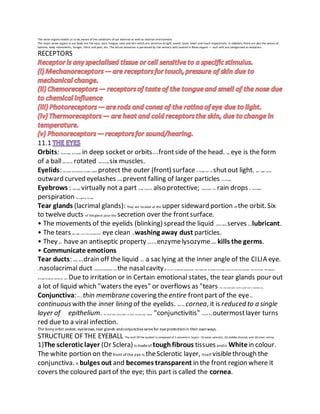
Orbits: Thetwo eyes are located in deep socket or orbitson thefrontside of the head. Each eye is the form
of a ball and can be rotated with thehelp six muscles.
Eyelids: Theupper and thelower movable eyelids protect the outer (front) surface of theeyes and can shutout light. Each eyelid carries
outward curved eyelashes which prevent falling of larger particles into theeye.
Eyebrows : Although virtually not a part of eye, these are also protective; theyprevent the rain drops or thetrickling
perspiration from getting the eyes.
Tear glands (lacrimal glands): They are located at the upper sideward portion of the orbit. Six
to twelve ducts of thé gland pour the secretion over the frontsurface.
• The movements of the eyelids (blinking) spread the liquid which mainly serves as a lubricant.
• The tears also keep the front surfaceof the eye clean by washing away dust particles.
• They also have an antiseptic property due to theenzymelysozymewhich kills the germs.
• Communicate emotions
Tear ducts: These ducts drain off the liquid into a sac lying at the inner angle of the CILIA eye.
A nasolacrimal duct conducts thesecretion into the nasalcavity.All of us of sometimes experienced that medicines dropped in the eyes come into thenose and even into thethroat. This happens
through theabove-mentioned duct. Due to irritation or in Certain emotional states, the tear glands pour out
a lot of liquid which "waters the eyes" or overflows as "tears".You may shed tears both in grief and in extreme joy.
Conjunctiva: It is a thin membrane covering theentire frontpart of the eye.It is
continuous with the inner lining of the eyelids. Over the cornea, it is reduced to a single
layer of epithelium. You must have often heard of avery common eye disease "conjunctivitis" in which this outermostlayer turns
red due to a viral infection.
The bony orbit socket, eyebrows, tear glands andconjunctiva servefor eyeprotectionin their ownways.
STRUCTURE OF THE EYEBALL The wall Of the eyeball is composed of 3 concentric layers : (l) outer sclerotic, (2) middle choroid, and (3) inner retina.
1)The sclerotic layer (Or Sclera)is madeof toughfibrous tissuesandis Whitein colour.
The white portion on thefront of the eye is theSclerotic layer, itself visiblethrough the
conjunctiva. It bulges out and becomes transparent in the front region where it
covers the coloured partof the eye; this part is called the cornea.
2. Sometimes,thecornea ofsomepatients turns opaque(white) andnon-functional.In suchcases, the defectivecornea can bereplacedby a
healthycornea froma donated eye. Donationofeye
Cornea remains alive up to nearly 40 hours after the death ofa person.If donated,the eye can be removed soon
within about 4 hours) after death.It is stored in an eye bank at a very low temperature in a suitable
medium such as blood plasma or certain other sterile (germ-free) liquid medium. Forgrafting, thecornea
part is taken outfrom the eyeandis fixed inplace ofthedefective cornea and thevision is restored.
According to a recent report, AllMS (New Delhi) has developed the technique of slicing the cornea into 2 -3 layers that can be grafted on as many defective eyes.
(2) The choroidlayer is richly supplied with blood vessels for providing nourishment to the
eye. It contains a dark black pigment (melanin) which prevents lightrays from reflectingand
scatteringinsidethe eye. In the front ofthe eye,the choroid expands to form the ciliary body (containing
circular muscles). The smoothmuscles in theciliary body alter the shape of the lens.
The iris (Latin iris : rainbow) is also anextension of the choroid, partially covering the
lens and leaving a circular opening in the center, the pupil. The blue, brown or black colour of
the eye refers to the colour of the iris. ("PUPIL"namehas been derivedfrom the Latinword"pupa"meaning
"doll", which in this contextrefers to thetiny image ofoneselfseen reflectedin another's eye). The iris contains radial muscles to
widen and circular muscles to constrictthe pupil. This adjustmentof the sizeof
the pupil regulates the amount of light entering the eye. You can easily observethis by throwing a
torch light intotheeyes whilelooking in a mirror. In dim ordark light,thepupilis dilated, whilein brightlight,it is constricted. The
pattern and arrangementof the iris muscles is unique for every individual and
therefore these are also a source of an individual's identification.
(3) The retinaor the innermostlayer is sensitive tolight. Itcontains two types of
sensecells called rods and cones.
— The rodcells (inner ends rod-like) are sensitive todimlight but do not respond
to colour. They contain the pigment rhodopsinor visual purple. The rod cells are
distributed almostthroughout theretina.
— The cones (inner ends conical) are sensitivetobright light and areresponsible
for colour vision. They contain the pigment iodopsin. The cone cells are mostly
confinedto the yellowspot.
YELLOW SPOT - The area of best vision the distribution ofrods and cones is notuniform.A particular
spotcalled the macula lutea(macula: pit; luteum: yellow) or simply yellowspot
or fovea centralis liesatthe backof the eye almost at the center onthe horizontal axis
of the eyeball. Thisspotcontainsthe maximum number of sensory cells and particularly
the cones. As a result,this is the region of brightestvision and also of the colour vision. The rest ofthe
retina has fewer cones and more rods. Yellow spotis theplace ofbestvision of thenormaleye. This is the reason why you moveyoureyes from
word to word as youread a linethrougha printedpage.
BLIND SPOT - The area of no vision Lateral to the yellowspoton the nasal side is
the blind spot. Thereare no sensory cells here and,therefore, this is thepointof no vision. This is thepointatwhich the
nerve fibres from all the sensory cells of the retina converge and bundle together to
leave the eyeball in the form ofthe optic nerve.
3. LENS
The lens is a transparent biconvex crystalline body locatedjust behindthe pupil. It contains
transparent lens fibres (long thincells which have lost theirnuclei).The lens is collectively held inposition by
fibres calledthe suspensory ligament, which attaches it to the ciliary body. The ciliary
body lies at the junction of the choroid and the iris, and is itselfa part of the choroid. The
ciliary body contains muscles which on contractionand relaxation, changetheshape of the lens
(being somewhat elastic) for viewing objects atdifferentdistances.
TWO CHAMBERS OF THEEYE — AQUEOUS AND VITREOUS CHAMBERS
The lens, together with its suspending structures, divides the inner cavity of the
eyeball into two chambers: aqueous chamberin frontofthelens and vitreous chamber behindthelens
(l) Aqueous chamber is the front chamber between the lens and the cornea.It is filled with a
clear watery liquidcalledaqueous humour (aqueous : watery; humour : fluid).
The aqueous humour serves in twoways :
(i) Keeps the lens moist and protects it from physical shock,
(ii) Itrefracts light.
(2) Viltreous chamber is the larger cavity ofthe eyeballbehind the lens.It is filled with a transparent
jelly-like thicker fluid called vitreoushumour (vitreous : glassy;t: fluid).
The vitreous humour serves two functions:
(i) It helps inkeeping the shape ofthe eyeball,
(ii) It protects the retinaand its nerve endings.
The four majorsteps in seeing anobject are as follows:
(1) of light rays: Light rays from the object enter the eyes through the transparent
structures (conjunctiva, cornea, aqueous humour, lens, vitreous humour).
(2) of image: First, the curvature of the cornea converges thelight rays tosome
extent and the lens converges them further to form an image onthe retina. The image
on the retina is invertedandreal.
(3) ofnerve impulses fromretina tobrain: The light energy ofthe imageproduces chemical
changes in the sensitivecells (rods and cones). These changes generatenerve impulses which
travel through the Optic nerveand reachthevisual areaof the cerebrum, wherethey givethe
sensation of sight.
(4) by the brain : Our brain interprets the imageinmany ways, e.g.,it "sees" theobjects
upright even ifthe imageformedinthe eyeis inverted.
(viewing objects in sharpfocus). To seeanobject clearly,its image should be insharpfocus in
each eye. The process of focusing the eye tosee objects atdifferent distances is called accommodation.
This is mainly brought about by a change inthe curvature of the elastic lens making it thinnerorfatter.
• For distant vision, the lens is more flattened or thinner.
• For near vision (nearer than 6 metres), the lens becomes more convex or
rounded. These changes in the shape of the lens is broughtabout by the ciliary
muscles.
4. _
In the normal condition (ciliary muscles relaxed), thelens remains stretched by
the suspensory ligaments and it is less convex, suited for viewing distant objects.
_
When we look at nearby objects, the ciliary muscles (which arecircular) contract
and tendto pull the ciliary body slightly forward. This releasesthe tension on
the suspensory ligament making itloose and the lens, on account of its elasticity,
becomes thicker and morerounded or convex (Fig. 11.4.B).
When you pass from a lighted area to a room (such as a cinema hall), you experience
difficulty in seeing objects for a shortwhile. Slowly, your vision is improved. This improvementis
called dark adaptation. This change is due to
(a) regenerationof the visual purple or rhodopsin, the pigment of the rods,
which was earlier broken down due to bright light, and
(b) dilationof the pupil permitting more light to enter the eyes.
When a person with adapted eyes moves a lightedarea, as in coming out ofa cinema hall afterthenoon show,
he experiences a dazzling effect for a short period.After a few seconds, hecomes back to normal
viewing through light adaptation. The adaptationis dueto reverseof the previous changes,
(a) the visualpurple of the rods is bleached, reducing their sensitivity, and
(b) the pupil constricts (gets narrower), to reduce the amount of light entering the eyes.
The partial closureof the eyelids in dazzling light also serves the samepurpose.
Colour Vision is possibleonly through cones of the retina which are stimulated
only in bright light. You cannot make out the red, violet or purple flowers in a
garden on a moonlit night, because then only the rods are in action and not the cones.
11.2 COMMONDEFECTS OF THE EYE
1. Near or short-sightedness (Myopia) is a condition in whichthe near objects can be seen clearly while the
distant objects appear blurred. In it, the imageofdistant objects is formed in frontof the
retina. Reasons for myopia: The two possiblereasonsare
(i) the eye ball is lengthened fromfrontto back OR
(ii) the lens is too curved (even both reasons may occur together).
Correction of myopia: This defect can be corrected by suitableconcave(diverging) lens which
causes the lightrays todiverge beforethey strikethe lens ofthe eye. Most ofyourclassmates using spectacles may be
suffering from myopia (power of glasses used is mentioned in minus "—").
2. Far or long-sightedness (Hyperopia, oldterm-Hypermetropia) is a condition in whichthereis a difficulty in seeing
near objects. In it, the image ofnearobject falls behindthe retina.
Reasons for hyeropia : This defect results on accountof either
1)shortening of the eyeball fromfrontto back 2)or the lens is too flat.
Correction of hyperopia: A convex (converging) lens is required to correct it
(power of the glasses used is mentioned in plus "+").
5. 3. Astigmatismis a defect in which some parts of the object are seen in focus
while others are blurred. Itarises dueto the unevencurvature of the cornea. This
is corrected by cylindrical lenses.
4. Presbyopiais a condition affecting older people who cannot see near objects
clearly. Their lens loses flexibility resulting in a kind of far- sightedness. This
again is corrected by a convex lens.
5. Cataract is a condition in which the lens turns opaque and the vision is cut
down even to total blindness. Itcan be corrected by surgically removing the lens,
and by using spectacles with highly convex lenses, compensating for themissing
lens, or in a newer technique, a small plastic lens is implanted behind or in front
of the iris.
6. Night-blindness is a condition in which a person feels difficulty in seeing in dim
light as during the night. This is due to non-formationof the pigment visual
purple of the rods. Only rods function in dim light and in the absence of the
pigment, they cannot function. This is usually due to the deficiency of vitaminA
which is required for the synthesis of the pigment.
7. Colour blindness is a condition in which somepeople by birth cannot
discriminate between certain colours such as red and green. This is due to a
genetic defect. Mostly males suffer fromthis defect, whereas it rarely occurs in
females.
8. Corneal opacities : The cornea of some patients gets scarredandturns opaque
(white) and non- functional. Such defects can cause anything fromminor irritation
to vision problems and even
After-images the basis of motion pictures.
If one looks at a bright object for a moment and then closes the eyes, the
sensation of light persists for a shortperiod. In the same way, if one looks at a
brightly coloured object and then looks at a dark surface, an image of the object
in the same colour will persist. This is known as persistenceimage or the after-
image. Itlasts for about one-tenth of a second. This is the principle on which the
technique of motion pictures is based.
In a movie, pictures are projected on a screen at the rate of about 24 pictures per
second, but we cannot see the individual frames on account of the after-images in
our eyes. The life-like continuous movement on the screen is an illusion.
Television too is similar, wherethe scanning beams a picture frame of the TV
camera moves so rapidly on the viewing screen of the TV set that our eyes cannot
keep pace with it. Outof numerous other optical illusions, two are shown at the
end of this chapter.