Downloaded 450 times


![Physiology of vision
The main mechanisms are
[ ]Initiation of vision (Phototransduction)
[ ]Processing and transmission of visual sensations
[ ]Visual perception](https://image.slidesharecdn.com/retina-140816061721-phpapp01/85/Retina-3rd-mbbs-ophthalmology-29-320.jpg)

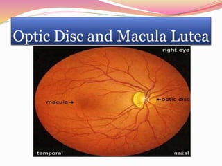
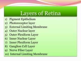
The document summarizes the anatomy and physiology of the retina. It describes the retina as having multiple layers that contain light-sensitive cells. These cells convert light rays into electrical signals that travel along the optic nerve to the brain. The retina contains two main areas - the posterior pole with the optic disc and macula lutea, and the peripheral retina. The macula lutea contains the fovea centralis, which has the highest concentration of light receptors and is responsible for sharp central vision. The document further details the layers of the retina, blood supply, phototransduction process of vision initiation, and dark adaptation.


