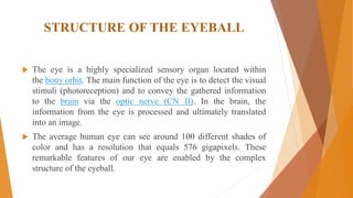
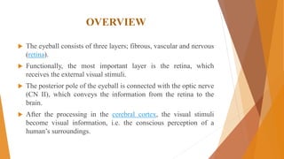
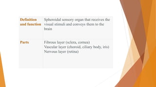
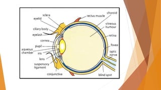
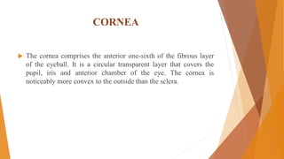
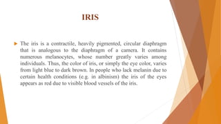
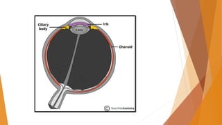
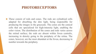
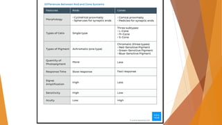
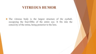
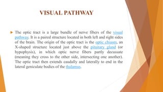
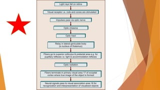
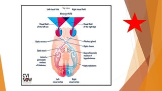
Unit:1
Topic: Special Sense
VISION
Prepared by: Dr. Khushboo Dutt (PT)
MPT- Neurology
STRUCTURE OF THE EYEBALL
 The eye is a highly specialized sensory organ located within
the bony orbit. The main function of the eye is to detect the visual
stimuli (photoreception) and to convey the gathered information
to the brain via the optic nerve (CN II). In the brain, the
information from the eye is processed and ultimately translated
into an image.
 The average human eye can see around 100 different shades of
color and has a resolution that equals 576 gigapixels. These
remarkable features of our eye are enabled by the complex
structure of the eyeball.
OVERVIEW
 The eyeball consists of three layers; fibrous, vascular and nervous
(retina).
 Functionally, the most important layer is the retina, which
receives the external visual stimuli.
 The posterior pole of the eyeball is connected with the optic nerve
(CN II), which conveys the information from the retina to the
brain.
 After the processing in the cerebral cortex, the visual stimuli
become visual information, i.e. the conscious perception of a
human’s surroundings.
Definition
and function
Spheroidal sensory organ that receives the
visual stimuli and conveys them to the
brain
Parts Fibrous layer (sclera, cornea)
Vascular layer (choroid, ciliary body, iris)
Nervous layer (retina)
 The eyeball sits within the orbit, surrounded by the adipose tissue.
 The eyeball consists of three distinct layers. From superficial to deep,
they include:
 The fibrous layer, which consists of the sclera and cornea. The sclera is
an opaque layer which surrounds the posterior five-sixths of the
eyeball. The cornea is a transparent layer that is anteriorly continuous
with the sclera, occupying the anterior one-sixth of the eyeball.
 The vascular layer, also known as the uvea or uveal tract. It consists of
three parts that are continuous with each other. From posterior to
anterior, they are the choroid, ciliary body, and iris.
 The nervous layer, also known as the retina, which is the innermost
layer of the eyeball. The retina itself is divided into two layers; an outer,
pigmented layer, and an inner neurosensory layer.
 These three layers comprise the circular outline of the eyeball.
The inside of the eye contains the two refractive structures of the
eye called the lens and vitreous body. Together with the cornea
and aqueous humor, the vitreous body and lens belong to the
refractive media of the eyeball. The role of refractive structures to
bend the direction of the light that falls onto the eye and focus it
onto the retina.
SCLERA
 The sclera is an opaque, white, outer layer that surrounds the
posterior five-sixths of the eyeball. The sclera is thickest
posteriorly, becoming progressively thinner anteriorly.
Function
 The function of the sclera is to protect the inner contents of the
eye from the mechanical trauma. Moreover, its rigid structure
contributes to maintaining the shape of the eyeball and keeping
the eye structures in place, especially during the contractions of
the extraocular muscles.
CORNEA
 The cornea comprises the anterior one-sixth of the fibrous layer
of the eyeball. It is a circular transparent layer that covers the
pupil, iris and anterior chamber of the eye. The cornea is
noticeably more convex to the outside than the sclera.
FUNCTION OF CORNEA
 The main function of the cornea is to participate in the refraction of
light. In fact, the cornea is the most important refractory structure of the
eye as it has the highest optical power (42 diopters). The refraction of
light occurs in the center of the cornea, where its refractive power is
significantly higher than that of the atmospheric air.
 The preservation of corneal transparency is very important for
maintaining its proper function. In case of any damage to the corneal
epithelium, the fluid from the anterior chamber may enter the corneal
stroma and cause the cloudiness of the cornea. Being part of the
outermost (fibrous) layer of the eye the cornea also has a protective
role, protecting the eye's delicate components from foreign particles
IRIS
 The iris is a contractile, heavily pigmented, circular diaphragm
that is analogous to the diaphragm of a camera. It contains
numerous melanocytes, whose number greatly varies among
individuals. Thus, the color of iris, or simply the eye color, varies
from light blue to dark brown. In people who lack melanin due to
certain health conditions (e.g. in albinism) the iris of the eyes
appears as red due to visible blood vessels of the iris.
 The iris represents the border between the anterior and posterior
chambers of the eye. It is located anterior to the lens and posterior
to the cornea, being immersed in the aqueous humor. The
periphery of the iris is marked as its root or the ciliary margin
 The iris contains two smooth muscles that enable its contractile
property. These are the sphincter pupillae muscle and dilator
pupillae muscle. The center of the iris features a circular opening
called the pupil. The inner margin of the iris that bounds the pupil
is called the pupillary margin. The size of the pupil can change by
the action of the two pupillary muscles and usually varies from 1-
8 millimeters. The purpose of these changes in the size of the
pupil is to control the amount of light that enters the eye.
FUNCTION AND PUPIL MOVEMENTS
 The function of the iris is to control the size of the pupil through
the actions of the sphincter and dilator pupillae muscles.
 The dilation of the pupil is called mydriasis. The mydriasis is a
result of dilator pupillae muscle contraction. It occurs when the
intensity of light is low, and also in the states of fear and
excitement (sympathetic predominance).
 The contraction of the pupil is called miosis. It is a result of the
sphincter pupillae muscle contraction. The miosis occurs in
conditions of high light intensity, convergence (simultaneous
inward movement of both eyes toward each other) and sleeping.
NERVOUS LAYER (RETINA)
 The retina is the innermost layer of the eyeball that extends from
the site of exit of the optic nerve to the posterior margin of the
ciliary body. It is a site where the image of the environment is
converted to the neural impulses that are transmitted to the brain
via optic nerve for interpretation and analysis.
 The neural retina contains 6 types of cells which are distributed
throughout its nine layers.
PHOTORECEPTORS
 These consist of rods and cones. The rods are cylindrical cells
adapted for absorbing the dim light, being responsible for
producing the images in the grayscale. The cones are the conical
cells that are specialized for high-intensity light, enabling the
color vision. The distribution of the cones and rods varies across
the retinal surface; the rods are absent within fovea centralis,
increasing in density going to the periphery of the retina. The
cones, however, are the most abundant at the fovea, decreasing in
number towards the periphery.
VITREOUS HUMOR
 The vitreous body is the largest structure of the eyeball,
occupying the four-fifths of the entire eye. It fits into the
concavity of the retina, being posterior to the lens.
AQUEOUS HUMOR
 The aqueous humor is a nutrient-rich fluid that fills the anterior
and posterior chambers of the eye. The amount of aqueous humor
in a healthy human eye is 200 milliliters. The aqueous humor is
produced by the ciliary processes and delivered into the posterior
chamber of the eye.
CATARACT
 Some people experience cloudiness or loss of transparency of the
lens from areas of opaqueness, e.g. as seen in cataracts. An
operation to extract a cataract is a common treatment option for
individuals affected a cataract condition.
PAPILLEDEMA
 This condition may occur if there is an abnormal increase in the
pressure of cerebrospinal fluid (CSF) flow in the extension of
the subarachnoid space around the optic nerve. This CSF pressure
slows down venous return from the retina leading to fluid
accumulation in the retina (edema of the retina). Edema of the
retina can occur as swelling of the optic disc, and this is referred
to as papilledema.
GLAUCOMA
 Glaucoma is a condition of increased pressure within the eyeball,
which results in gradual loss of sight. Such pressure usually
builds up within the anterior and posterior chambers of the
eyeball due to obstruction in the aqueous humor drainage. There
can be primary or secondary glaucoma. Chronic raised pressure in
the eye usually cause direct mechanical damage or affects the
blood supply subsequently leading to blindness.
VISUAL PATHWAY
 The optic tract is a large bundle of nerve fibers of the visual
pathway. It is a paired structure located in both left and right sides
of the brain. The origin of the optic tract is the optic chiasm, an
X-shaped structure located just above the pituitary gland (or
hypophysis), in which optic nerve fibers partly decussate
(meaning they cross to the other side, intersecting one another).
The optic tract then extends caudally and laterally to end in the
lateral geniculate bodies of the thalamus.
 Almost all axons of the left and right optic tracts synapse with the cells
of their corresponding lateral geniculate nucleus (LGN). However,
some fibers of the optic tract, particularly the fibers concerned with the
light reflexes, bypass the lateral geniculate nucleus and continue to the
pretectal nucleus and superior colliculus of the midbrain.
 The organization of the fibers that carry visual information in the optic
tract and the lateral geniculate nucleus is specific, which is known
as retinotopy:
 In the optic tract, the fibers from the superior retinal quadrants lie
laterally, while inferior retinal fibers run medially.
 The two lateral geniculate nuclei receive input from both eyes.
However, each nucleus only receives information from one half of the
visual field, due to the decussation of fibers in the chiasm. The axons of
the ganglion cells from the outer half of the retina (temporal side/field)
remain on the same side of the brain, and the axons from the inner half
(nasal side/field) cross over to the contralateral side. Within one
nucleus, the visual information is divided among the various layers.
 The efferent fibers from the lateral geniculate nucleus emerge as
the optic radiation and travel to the primary visual cortex located
in the occipital cortex of the brain (Brodmann's area 17) where
the visual processing continues.
 The blood supply to the optic tract is variable, but typically arises
from anastomotic branches of the posterior
communicating and anterior choroidal arteries, together with
branches from the middle cerebral arteries.
 The venous drainage is from the superior aspect through
the anterior cerebral veins and from the inferior aspect through
the basal vein.
Retina Retinal pigmented epithelium, photoreceptor layer, outer (external) limiting
membrane, outer nuclear layer, outer plexiform layer, inner nuclear layer, inner
plexiform layer, ganglion cell layer, nerve fiber layer, inner limiting membrane
Retinal layers mnemonic 'My Nerves Get In Knots Outside Our Easy Practice Review'
Optic nerve (CN II) Formed by the axons of ganglion cells coming together at the optic disc, it exists
the orbit via the superior orbital fissure.
Optic chiasm It represents the point of decussation of the optic nerves, where the nasal fibers of
each eye cross the midline to join the temporal fibers of the contralateral eye.
Optic tracts Each tract carries fibers that are stimulated from the contralateral visual field.
Visual reflexes Accomodation, light reflexes, saccades - they are mediated by the axons of the
ganglion cells synapsing on the oculomotor and Edinger-Westphal nuclei.
Lateral geniculate body Part of the thalamus
Optic radiation Each one contains six layers that receives retinotopic input from the corresponding
visual field and is separated into two loops.
Primary visual cortex Brodmann area 17
REFRACTIVE ERRORS
 Myopia – This is the inability of the eyes to focus light on the
retina but in front of the retina. Symptoms are inability to view
far objects but close ones.
 Presbyopia – This is the inability of the eyes to focus near
objects, it happens progressively and is common with elderly.
Symptoms are eye strain, inability to focus on fine prints and
small objects. The ability to focus on near objects declines
throughout life, from an ability to focus at 50 mm away in
children, to an ability to focus at 100 mm at 25 years of age, to an
ability to focus at only about 2 m away by 60 years of age.
 Hypermetropia is also referred to as hyperopia or long-
sightedness or far-sightedness. Hypermetropia is the condition of
the eyes where the image of a nearby object is formed behind the
retina. Here, the light is focused behind the retina instead of
focusing on the retina.
 Astigmatism is a type of refractive error due to rotational
asymmetry in the eye's refractive power. This results in distorted
or blurred vision at any distance. Other symptoms can
include eyestrain, headaches, and trouble driving at night.
Astigmatism often occurs at birth and can change or develop later
in life. If it occurs in early life and is left untreated, it may result
in amblyopia.
VISION.pdf

VISION.pdf

  • 1.
    Unit:1 Topic: Special Sense VISION Preparedby: Dr. Khushboo Dutt (PT) MPT- Neurology
  • 2.
    STRUCTURE OF THEEYEBALL  The eye is a highly specialized sensory organ located within the bony orbit. The main function of the eye is to detect the visual stimuli (photoreception) and to convey the gathered information to the brain via the optic nerve (CN II). In the brain, the information from the eye is processed and ultimately translated into an image.  The average human eye can see around 100 different shades of color and has a resolution that equals 576 gigapixels. These remarkable features of our eye are enabled by the complex structure of the eyeball.
  • 3.
    OVERVIEW  The eyeballconsists of three layers; fibrous, vascular and nervous (retina).  Functionally, the most important layer is the retina, which receives the external visual stimuli.  The posterior pole of the eyeball is connected with the optic nerve (CN II), which conveys the information from the retina to the brain.  After the processing in the cerebral cortex, the visual stimuli become visual information, i.e. the conscious perception of a human’s surroundings.
  • 4.
    Definition and function Spheroidal sensoryorgan that receives the visual stimuli and conveys them to the brain Parts Fibrous layer (sclera, cornea) Vascular layer (choroid, ciliary body, iris) Nervous layer (retina)
  • 6.
     The eyeballsits within the orbit, surrounded by the adipose tissue.  The eyeball consists of three distinct layers. From superficial to deep, they include:  The fibrous layer, which consists of the sclera and cornea. The sclera is an opaque layer which surrounds the posterior five-sixths of the eyeball. The cornea is a transparent layer that is anteriorly continuous with the sclera, occupying the anterior one-sixth of the eyeball.  The vascular layer, also known as the uvea or uveal tract. It consists of three parts that are continuous with each other. From posterior to anterior, they are the choroid, ciliary body, and iris.  The nervous layer, also known as the retina, which is the innermost layer of the eyeball. The retina itself is divided into two layers; an outer, pigmented layer, and an inner neurosensory layer.
  • 7.
     These threelayers comprise the circular outline of the eyeball. The inside of the eye contains the two refractive structures of the eye called the lens and vitreous body. Together with the cornea and aqueous humor, the vitreous body and lens belong to the refractive media of the eyeball. The role of refractive structures to bend the direction of the light that falls onto the eye and focus it onto the retina.
  • 8.
    SCLERA  The sclerais an opaque, white, outer layer that surrounds the posterior five-sixths of the eyeball. The sclera is thickest posteriorly, becoming progressively thinner anteriorly. Function  The function of the sclera is to protect the inner contents of the eye from the mechanical trauma. Moreover, its rigid structure contributes to maintaining the shape of the eyeball and keeping the eye structures in place, especially during the contractions of the extraocular muscles.
  • 9.
    CORNEA  The corneacomprises the anterior one-sixth of the fibrous layer of the eyeball. It is a circular transparent layer that covers the pupil, iris and anterior chamber of the eye. The cornea is noticeably more convex to the outside than the sclera.
  • 10.
    FUNCTION OF CORNEA The main function of the cornea is to participate in the refraction of light. In fact, the cornea is the most important refractory structure of the eye as it has the highest optical power (42 diopters). The refraction of light occurs in the center of the cornea, where its refractive power is significantly higher than that of the atmospheric air.  The preservation of corneal transparency is very important for maintaining its proper function. In case of any damage to the corneal epithelium, the fluid from the anterior chamber may enter the corneal stroma and cause the cloudiness of the cornea. Being part of the outermost (fibrous) layer of the eye the cornea also has a protective role, protecting the eye's delicate components from foreign particles
  • 11.
    IRIS  The irisis a contractile, heavily pigmented, circular diaphragm that is analogous to the diaphragm of a camera. It contains numerous melanocytes, whose number greatly varies among individuals. Thus, the color of iris, or simply the eye color, varies from light blue to dark brown. In people who lack melanin due to certain health conditions (e.g. in albinism) the iris of the eyes appears as red due to visible blood vessels of the iris.
  • 13.
     The irisrepresents the border between the anterior and posterior chambers of the eye. It is located anterior to the lens and posterior to the cornea, being immersed in the aqueous humor. The periphery of the iris is marked as its root or the ciliary margin  The iris contains two smooth muscles that enable its contractile property. These are the sphincter pupillae muscle and dilator pupillae muscle. The center of the iris features a circular opening called the pupil. The inner margin of the iris that bounds the pupil is called the pupillary margin. The size of the pupil can change by the action of the two pupillary muscles and usually varies from 1- 8 millimeters. The purpose of these changes in the size of the pupil is to control the amount of light that enters the eye.
  • 14.
    FUNCTION AND PUPILMOVEMENTS  The function of the iris is to control the size of the pupil through the actions of the sphincter and dilator pupillae muscles.  The dilation of the pupil is called mydriasis. The mydriasis is a result of dilator pupillae muscle contraction. It occurs when the intensity of light is low, and also in the states of fear and excitement (sympathetic predominance).  The contraction of the pupil is called miosis. It is a result of the sphincter pupillae muscle contraction. The miosis occurs in conditions of high light intensity, convergence (simultaneous inward movement of both eyes toward each other) and sleeping.
  • 15.
    NERVOUS LAYER (RETINA) The retina is the innermost layer of the eyeball that extends from the site of exit of the optic nerve to the posterior margin of the ciliary body. It is a site where the image of the environment is converted to the neural impulses that are transmitted to the brain via optic nerve for interpretation and analysis.  The neural retina contains 6 types of cells which are distributed throughout its nine layers.
  • 17.
    PHOTORECEPTORS  These consistof rods and cones. The rods are cylindrical cells adapted for absorbing the dim light, being responsible for producing the images in the grayscale. The cones are the conical cells that are specialized for high-intensity light, enabling the color vision. The distribution of the cones and rods varies across the retinal surface; the rods are absent within fovea centralis, increasing in density going to the periphery of the retina. The cones, however, are the most abundant at the fovea, decreasing in number towards the periphery.
  • 19.
    VITREOUS HUMOR  Thevitreous body is the largest structure of the eyeball, occupying the four-fifths of the entire eye. It fits into the concavity of the retina, being posterior to the lens.
  • 20.
    AQUEOUS HUMOR  Theaqueous humor is a nutrient-rich fluid that fills the anterior and posterior chambers of the eye. The amount of aqueous humor in a healthy human eye is 200 milliliters. The aqueous humor is produced by the ciliary processes and delivered into the posterior chamber of the eye.
  • 21.
    CATARACT  Some peopleexperience cloudiness or loss of transparency of the lens from areas of opaqueness, e.g. as seen in cataracts. An operation to extract a cataract is a common treatment option for individuals affected a cataract condition. PAPILLEDEMA  This condition may occur if there is an abnormal increase in the pressure of cerebrospinal fluid (CSF) flow in the extension of the subarachnoid space around the optic nerve. This CSF pressure slows down venous return from the retina leading to fluid accumulation in the retina (edema of the retina). Edema of the retina can occur as swelling of the optic disc, and this is referred to as papilledema.
  • 22.
    GLAUCOMA  Glaucoma isa condition of increased pressure within the eyeball, which results in gradual loss of sight. Such pressure usually builds up within the anterior and posterior chambers of the eyeball due to obstruction in the aqueous humor drainage. There can be primary or secondary glaucoma. Chronic raised pressure in the eye usually cause direct mechanical damage or affects the blood supply subsequently leading to blindness.
  • 25.
    VISUAL PATHWAY  Theoptic tract is a large bundle of nerve fibers of the visual pathway. It is a paired structure located in both left and right sides of the brain. The origin of the optic tract is the optic chiasm, an X-shaped structure located just above the pituitary gland (or hypophysis), in which optic nerve fibers partly decussate (meaning they cross to the other side, intersecting one another). The optic tract then extends caudally and laterally to end in the lateral geniculate bodies of the thalamus.
  • 27.
     Almost allaxons of the left and right optic tracts synapse with the cells of their corresponding lateral geniculate nucleus (LGN). However, some fibers of the optic tract, particularly the fibers concerned with the light reflexes, bypass the lateral geniculate nucleus and continue to the pretectal nucleus and superior colliculus of the midbrain.  The organization of the fibers that carry visual information in the optic tract and the lateral geniculate nucleus is specific, which is known as retinotopy:  In the optic tract, the fibers from the superior retinal quadrants lie laterally, while inferior retinal fibers run medially.  The two lateral geniculate nuclei receive input from both eyes. However, each nucleus only receives information from one half of the visual field, due to the decussation of fibers in the chiasm. The axons of the ganglion cells from the outer half of the retina (temporal side/field) remain on the same side of the brain, and the axons from the inner half (nasal side/field) cross over to the contralateral side. Within one nucleus, the visual information is divided among the various layers.
  • 28.
     The efferentfibers from the lateral geniculate nucleus emerge as the optic radiation and travel to the primary visual cortex located in the occipital cortex of the brain (Brodmann's area 17) where the visual processing continues.  The blood supply to the optic tract is variable, but typically arises from anastomotic branches of the posterior communicating and anterior choroidal arteries, together with branches from the middle cerebral arteries.  The venous drainage is from the superior aspect through the anterior cerebral veins and from the inferior aspect through the basal vein.
  • 29.
    Retina Retinal pigmentedepithelium, photoreceptor layer, outer (external) limiting membrane, outer nuclear layer, outer plexiform layer, inner nuclear layer, inner plexiform layer, ganglion cell layer, nerve fiber layer, inner limiting membrane Retinal layers mnemonic 'My Nerves Get In Knots Outside Our Easy Practice Review' Optic nerve (CN II) Formed by the axons of ganglion cells coming together at the optic disc, it exists the orbit via the superior orbital fissure. Optic chiasm It represents the point of decussation of the optic nerves, where the nasal fibers of each eye cross the midline to join the temporal fibers of the contralateral eye. Optic tracts Each tract carries fibers that are stimulated from the contralateral visual field. Visual reflexes Accomodation, light reflexes, saccades - they are mediated by the axons of the ganglion cells synapsing on the oculomotor and Edinger-Westphal nuclei. Lateral geniculate body Part of the thalamus Optic radiation Each one contains six layers that receives retinotopic input from the corresponding visual field and is separated into two loops. Primary visual cortex Brodmann area 17
  • 31.
    REFRACTIVE ERRORS  Myopia– This is the inability of the eyes to focus light on the retina but in front of the retina. Symptoms are inability to view far objects but close ones.  Presbyopia – This is the inability of the eyes to focus near objects, it happens progressively and is common with elderly. Symptoms are eye strain, inability to focus on fine prints and small objects. The ability to focus on near objects declines throughout life, from an ability to focus at 50 mm away in children, to an ability to focus at 100 mm at 25 years of age, to an ability to focus at only about 2 m away by 60 years of age.
  • 32.
     Hypermetropia isalso referred to as hyperopia or long- sightedness or far-sightedness. Hypermetropia is the condition of the eyes where the image of a nearby object is formed behind the retina. Here, the light is focused behind the retina instead of focusing on the retina.  Astigmatism is a type of refractive error due to rotational asymmetry in the eye's refractive power. This results in distorted or blurred vision at any distance. Other symptoms can include eyestrain, headaches, and trouble driving at night. Astigmatism often occurs at birth and can change or develop later in life. If it occurs in early life and is left untreated, it may result in amblyopia.