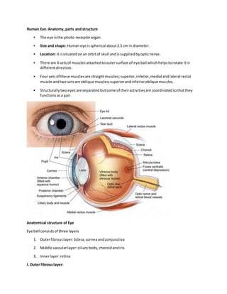
1. Human Eye:Anatomy, parts and structure
The eye isthe photo-receptororgan.
Size and shape: Human eye isspherical about2.5 cm indiameter.
Location: it issituatedonan orbitof skull andissuppliedbyopticnerve.
There are 6 setsof muscles attachedtooutersurface of eye ball whichhelpstorotate itin
differentdirection.
Four setsof these musclesare straightmuscles;superior,inferior,medial andlateral rectal
muscle andtwo setsare oblique muscles;superiorandinferiorobliquemuscles.
Structurallytwoeyesare separatedbutsome of theiractivitiesare coordinatedsothatthey
functionsasa pair.
Anatomical structure of Eye
Eye ball consistsof three layers
1. Outerfibrouslayer:Sclera,corneaandconjunctiva
2. Middle vascularlayer:ciliarybody,choroidandiris
3. Innerlayer:retina
I. Outer fibrouslayer:
2. It consistsof followingparts.
1. Sclera:
It isoutermostsupportinglayerconsistsof thickmembraneof toughfibrousconnective tissue.
It covers5/6 parts of eye ball.
It maintainsthe shape of eye andprovide attachmenttothe extrinsicmuscle of eye
2. Cornea:
It isa thintransparentfrontpart of sclera.
It formsa slightbulge atthe frontand coversan anterior1/6 part of sclera.
Corneaisavascular andabsorbsoxygen fromair.
It refractslightto focuson retina.
3. Conjunctiva:
It isa thintransparentlayerthatcoversthe cornea.
It isformedof single layerof stratifiedsquamousepithelium
It protectsthe cornea.
II. Middle vascular layer:
It consistsof followingparts:
1. Choroid:
It isthick vascularandpigmentedlayersituatedbelow sclera.
The pigmentedcellsabsorbslightandpreventitfrombeingreflected.
The functionof choroidisto provide nutritionandtopreventreflectionof light.
2. Ciliarybody:
These are attachedto choroidand presentatthe junctionof scleraandcornea.
It consistsof twosetsof ciliarymuscle andsuspensoryligament.
Ciliarybodyisattachedto lensandholdsitinposition
Its functionistochange the shape of lensbycontractionor relaxationof muscle
3. Iris:
It ismuscular,pigmentedandopaque diaphragmwhichhangsinthe eye ball infrontof lens.
It has small circularopeningcalledpupil.
3. It has twotypesof muscles;circularand radial muscle.The movement of these musclescontrol
the size of pupil.
Pigmentinirisgivescolortoeye.
Iriscontrol the amountof lightenteringintoeye bycontrollingthe size of pupil.
III. Inner layer:
It consistsof photoreceptorcellsandphotosensitive elements.
1. Retina:
Retinaisinnermostlayer.
Neuroretinacontainshighlyspecializedphotoreceptornerve cells;rodsandcones
Each eye ball has 125 millionsof rodcellsand7 millionsof cone cells.
Small depressioninretinal walliscalledFoveacentraliswhich containsonlycone cells.
Foveacentralisishighlysensitive tolightandformsmagnifiedimage andgive sharpandacute
vision.
The optic nerve enterretinaata pointcalledblindspot.Itdoesnotcontainsanyrods or cone
cells.Itisleastsensitivetolightandformsno image whenlightfallsonblindspot
Rod cell:
rods are sensorsforperceptionof blacktowhite shades
Nightvisionisalmostrodvision.
It functionindimlight
Containsa photosensitive pigmentrhodopsinformedfromvitaminA.
Cone cell:
Conesare sensorsforperceptionof colors.
It functionsinbrightlightanddifferentiate colors.
Containsa photosensitive pigmentiodopsin.
Eye lensand chambers
1. Eye Lens:
It isa large,flexible,transparentbiconvex andfibrouscrystalline bodysituatedbehindiris.
Lensis enclosedinatransparentelasticcapsule.
Ciliarymusclescontrol the thicknessof lensanditspowerof accommodation.
It formsthe image of the objectonretina.
4. Lensseparatesthe eye ball intotwochamber
i.Aqueous chamber
ii.Vitreouschamber
Aqueouschamber:
It isa smallerfluidfilledchamberbetweencorneaandlens.
It isfilledwithaqueoushumourcontainingaminoacids,glucose,ascorbicacid,hyaluronicacid
and respiratorygases.
The aqueoushumournourishes the lensandcorneaandrefractslightraysto focus onretina.
Vitreouschamber:
It isa largerfluidfilledchamberbetweenlensandretina.
It isfilledwithgelatinousvitreoushumourcontainingsaltsandmucoproteins
It supportsretinaandrefractslightto focuson retina.