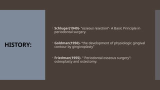
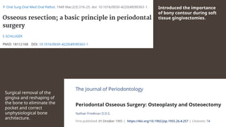
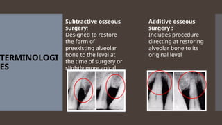
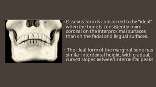
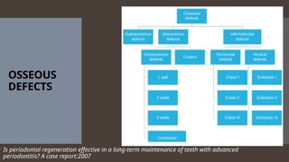
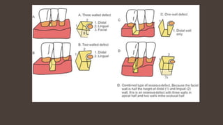
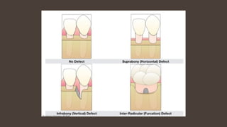
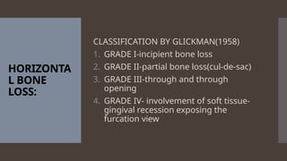
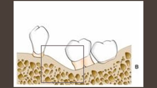
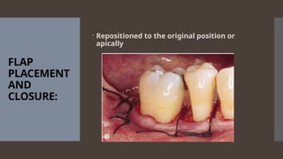
Resective
Osseous
Surgery
Presented by:
Dr Divjot
Guided by: Dr
Kumar Saurav
Singh
CONTENTS:
ď‚– INTRODUCTION
ď‚– HISTORY
ď‚– TERMINOLOGY
ď‚– FACTORS IN SELECTION OF TYPE OF ROS
ď‚– EXAMINATION
ď‚– METHODS
ď‚– TECHNIQUES
ď‚– CONCLUSION
ď‚– REFERENCES
HISTORY:
 Schluger(1949)- “osseous resection”- A Basic Principle in
periodontal surgery.
 Goldman(1950)- “the development of physiologic gingival
contour by gingivoplasty”
 Friedman(1955)- “ Periodontal osseous surgery”:
osteoplasty and ostectomy.
Introduced the importance
of bony contour during soft
tissue gingivectomies.
Surgical removal of the
gingiva and reshaping of
the bone to eliminate the
pocket and correct
unphysiological bone
architecture.
INTRODUCTIO
N:
ď‚– Periodontal destruction often leads to alveolar bone
destruction.
 This bone loss has been classified as “vertical” or
“horizontal”
ď‚– Osseous surgery may be defined as the procedure by
which changes in the alveolar bone can be accomplished
to rid it of deformities induced by the periodontal disease
process or other related factors, such as exostosis and
tooth supraeruption.
TERMINOLOGI
ES
Subtractive osseous
surgery:
Designed to restore
the form of
preexisting alveolar
bone to the level at
the time of surgery or
slightly more apical
Additive osseous
surgery :
Includes procedure
directing at restoring
alveolar bone to its
original level
ARCHITECTUR
E
INTERDENTAL BONE
RADICULAR BONE
RADICULAR BONE
INTERDENTAL BONE
RADICULAR BONE
INTERDENTAL BONE
POSITIVE
FLAT NEGATIVE
 Osseous form is considered to be “ideal”
when the bone is consistently more
coronal on the interproximal surfaces
than on the facial and lingual surfaces.
ď‚– The ideal form of the marginal bone has
similar interdental height, with gradual,
curved slopes between interdental peaks
OSSEOUS
DEFECTS
ď‚– Is periodontal regeneration effective in a long-term maintenance of teeth with advanced
periodontitis? A case report:2007
HORIZONTA
L BONE
LOSS:
CLASSIFICATION BY GLICKMAN(1958)
1. GRADE I-incipient bone loss
2. GRADE II-partial bone loss(cul-de-sac)
3. GRADE III-through and through
opening
4. GRADE IV- involvement of soft tissue-
gingival recession exposing the
furcation view
VERTICAL
BONE
DEFECTS:
CLASSIFICATION BY TARNOW AND
FLETCHER (1984)
1. SUB-CLASS A= 0-3mm
2. SUB-CLASS B = 4-6mm
3. SUB-CLASS C = >7mm
1) BONE
GRAFTS
2)GTR
OSSEOUS
SURGERY
ADDITIVE RESECTIVE
1)OSTEOPLAST
Y
2)OSTECTOMY
INDICATION
S:
1. pocket elimination
2. Tori reduction
3. Intrabony defects adjacent to
edentulous ridge
4. Incipient furcation involvement
5. Thick heavy ledges and exostoses
6. Shallow osseous craters
7. Small intra bony defects
CONTRA-
INDICATION
S:
1. Areas of insufficient remaining
attachment
2. Anatomic limitations(prominent ridge)
3. Aesthetic limitations(anteriorly, high
smile line)
OSTEOPLASTY:
1. It is a plastic
procedure by
which non
supporting bone
is reshaped to
achieve a
physiologic
gingival and
osseous contours.
2. Osteoplasty
includes the
techniques of
radicular blending
OSTETECTOMY:
1. It is the plastic
removal of
radicular and
interradicular
supporting bone
to eliminate
osseous
deformities
2. Done by the
technique of
spheroidng or
parabolizing
RATIONALE:
1) PERIOODNTAL DISEASE
2) DISCREPENCIES IN LEVEL AND
SHAPE OF BONE
3)POCKET RECURRENCE
EXAMINATION:
Periodontal probing and exploration
• pocket depth greater than that of a normal
gingival sulcus,
• the location of the base of the pocket
relative to the mucogingival junction and
attachment level on adjacent teeth, the
number of bony walls
• the presence of furcation defects.
• Transgingival probing, or sounding, under
local anesthesia confirms the extent and
configuration of the intrabony component
of the pocket and of furcation defects
Instrument
s:
Rongeurs
Interproximal
files
Ochsenbein
chisels
Friction grip
Burs
Sequential
steps :
Vertical grooving
Radicular blending
Flattening interproximal
bone
Gradualizing marginal bone
Vertical
Grooving:
1)reduce the thickness of the
alveolar housing
2)relative prominence to the
radicular aspects of the teeth
3)provides continuity from the
interproximal surface onto the
radicular surface.
Vertical grooves
ď‚–Instruments: Round carbide or
diamond burs
ď‚–Indications: bony margins,
shallow crater formations with
thick bony margins.
ď‚–Contraindications : close roots
or thin alveolar housing
RADICULAR
BLENDING:
ď‚–To gradualize the bone over
the entire radicular surface
to provide the best results
from vertical grooving.
ď‚–This provides a smooth,
blended surface for good
flap adaptation
Radicular
blending
ď‚–Indications:
1) same as vertical grooving
ď‚–Contraindications:
1)vertical grooving is very minor
2) radicular bone is thin or
fenestrated
Flattening
interproxim
al bone:
ď‚–Removal of very small
amounts of vertical bones
ď‚–Used in defects that have a
coronally placed, one-walled
edge of a three-walled
angular defect
ď‚–Improves flap closure and
healing in three-walled
defects
Flattening
interproximal
bone:
ď‚–Indications:
1)one-walled interproximal defects
or hemiseptal defects.
ď‚–Contraindications:
1) inter-proximal crater formations
2) flat interproximal defects
Gradualizin
g marginal
bone:
•Ostectomy step
•No bony discrepancies should be
left in the interdental or marginal
or interradicular area otherwise the
pocket reduction process remains
incomplete
•A widows peak should be left and
blend on the radicular root surface
area
The reduction should be made to remove the least
amount of alveolar bone required to
(1) produce a satisfactory form,
(2) prevent the therapeutic invasion of furcations
(3) blend the contours with the adjacent teeth.
The selective reduction of bony defects by “ramping” the
bone to the palatal or lingual to avoid involvement of the
furcations has been advocated by Ochsenbein and
Bohannan18 Tibbetts et al.
SPECIFIC
OSSEOUS
RESHAPING
SITUATIONS
:
ď‚–Correction of one wall hemi
septal defects is by reducing the
bone level as apical as possible.
ď‚– If one-walled defects occur next
to an edentulous space, the
edentulous ridge is reduced to
the level of the osseous defect:
RAMPING
ď‚–The walls of the crater may be
reduced at the expense of the
buccal, lingual, or both walls.
CRATERS:
FLAP
PLACEMENT
AND
CLOSURE:
ď‚– Repositioned to the original position or
apically
POST
OPERATIVE
MAINTENECE:
ď‚–Nonresorbable sutures such as silk are
usually removed after 1 week of healing
ď‚–Resorbable sutures maintain wound
approximation for varying periods of 1 to
3 weeks or more
ď‚–After suture removal the surgical site is
examined carefully, and any excessive
granulation tissue is removed with a
sharp curette.
ď‚–the surgical site is gently cleansed of
debris with a cotton pellet dampened
with saline
BASIC
RULES OF
OSSEOUS
SURGERY:
ď‚–When properly performed, resective
osseous surgery achieves a
physiologic architecture of marginal
alveolar bone for to gingival flap
adaptation with minimal probing
depth.
RULE 1:
ď‚–A full thickness mucoperiosteal
flap should be raised whenever
osseous resective surgery is
planned.
RULE 2:
1) The scalloping of the flap should
anticipate the final underlying osseous
contour, which is most prominent
anteriorly and decreases posteriorly.
2) The scalloping of the flap should reflect
the patients own healthy gingival
architecture.
3) The degree of the tissue and bone
scalloping is reduced as the
interproximal area becomes broader as
a result of bone loss.
RULE 3:
Osteoplasty generally
precedes ostectomy
RULE 4:
Osseous resective surgery
should result in a positive
architecture
RULE 5:
1) High speed rotatry
instruments should never be
used adjacent to the teeth.
2) Copious irrigation should be
maintained
RULE 6:
The final bony contours should
approximate the expected healthy
postoperative gingival form, with no
attempt to improve on it.
ď‚– FibReORS aims to shift the base of the intrabony defect to
a more coronal position, thus making it shallower and
more easily eliminated with minimal resection of
supporting bone.
ď‚– In addition, the apical migration of the gingival margin is
reported to be less pronounced with FibReORS as
compared with traditional ORS
CONCLUSIO
N
ď‚– Although osseous surgical techniques
cannot be applied to every bony
abnormality or topographic modification,
it has been clearly demonstrated that
properly used osseous surgery can
eliminate and modify defects, as well as
gradualize excessive bony ledges,
irregular alveolar bone, early furcation
involvement, excessive bony exostosis,
and circumferential defects.
TAKE HOME
MESSAGE:
ď‚– The goal is to establish contours that
existed naturally (physiologically), with
the assumption that this will facilitate
hygiene and long-term maintenance.
REFERENCES:
ď‚– Carranza 10th
edition
ď‚– Cohen 4th
edition
ď‚– Schluger S: Osseous resection: a basic
principle in periodontal surgery, Oral
Surg Oral Med Oral Pathol 2:316, 1949.
ď‚– David F. Levine & Greg Filippelli (1999) A
Review of Osseous Resective Surgery,
Journal of the California Dental
Association, 27:2, 125-132, DOI:
10.1080/19424396.2016.12221105
THANK
YOU!

RESECTIVE OSSEOUS SURGERY............................................

  • 1.
  • 2.
    CONTENTS: ď‚– INTRODUCTION ď‚– HISTORY ď‚–TERMINOLOGY ď‚– FACTORS IN SELECTION OF TYPE OF ROS ď‚– EXAMINATION ď‚– METHODS ď‚– TECHNIQUES ď‚– CONCLUSION ď‚– REFERENCES
  • 3.
    HISTORY:  Schluger(1949)- “osseousresection”- A Basic Principle in periodontal surgery.  Goldman(1950)- “the development of physiologic gingival contour by gingivoplasty”  Friedman(1955)- “ Periodontal osseous surgery”: osteoplasty and ostectomy.
  • 4.
    Introduced the importance ofbony contour during soft tissue gingivectomies. Surgical removal of the gingiva and reshaping of the bone to eliminate the pocket and correct unphysiological bone architecture.
  • 5.
    INTRODUCTIO N:  Periodontal destructionoften leads to alveolar bone destruction.  This bone loss has been classified as “vertical” or “horizontal”  Osseous surgery may be defined as the procedure by which changes in the alveolar bone can be accomplished to rid it of deformities induced by the periodontal disease process or other related factors, such as exostosis and tooth supraeruption.
  • 6.
    TERMINOLOGI ES Subtractive osseous surgery: Designed torestore the form of preexisting alveolar bone to the level at the time of surgery or slightly more apical Additive osseous surgery : Includes procedure directing at restoring alveolar bone to its original level
  • 7.
    ARCHITECTUR E INTERDENTAL BONE RADICULAR BONE RADICULARBONE INTERDENTAL BONE RADICULAR BONE INTERDENTAL BONE POSITIVE FLAT NEGATIVE
  • 8.
     Osseous formis considered to be “ideal” when the bone is consistently more coronal on the interproximal surfaces than on the facial and lingual surfaces.  The ideal form of the marginal bone has similar interdental height, with gradual, curved slopes between interdental peaks
  • 9.
    OSSEOUS DEFECTS ď‚– Is periodontalregeneration effective in a long-term maintenance of teeth with advanced periodontitis? A case report:2007
  • 12.
    HORIZONTA L BONE LOSS: CLASSIFICATION BYGLICKMAN(1958) 1. GRADE I-incipient bone loss 2. GRADE II-partial bone loss(cul-de-sac) 3. GRADE III-through and through opening 4. GRADE IV- involvement of soft tissue- gingival recession exposing the furcation view
  • 13.
    VERTICAL BONE DEFECTS: CLASSIFICATION BY TARNOWAND FLETCHER (1984) 1. SUB-CLASS A= 0-3mm 2. SUB-CLASS B = 4-6mm 3. SUB-CLASS C = >7mm
  • 14.
  • 15.
    INDICATION S: 1. pocket elimination 2.Tori reduction 3. Intrabony defects adjacent to edentulous ridge 4. Incipient furcation involvement 5. Thick heavy ledges and exostoses 6. Shallow osseous craters 7. Small intra bony defects
  • 16.
    CONTRA- INDICATION S: 1. Areas ofinsufficient remaining attachment 2. Anatomic limitations(prominent ridge) 3. Aesthetic limitations(anteriorly, high smile line)
  • 17.
    OSTEOPLASTY: 1. It isa plastic procedure by which non supporting bone is reshaped to achieve a physiologic gingival and osseous contours. 2. Osteoplasty includes the techniques of radicular blending OSTETECTOMY: 1. It is the plastic removal of radicular and interradicular supporting bone to eliminate osseous deformities 2. Done by the technique of spheroidng or parabolizing
  • 18.
    RATIONALE: 1) PERIOODNTAL DISEASE 2)DISCREPENCIES IN LEVEL AND SHAPE OF BONE 3)POCKET RECURRENCE
  • 19.
    EXAMINATION: Periodontal probing andexploration • pocket depth greater than that of a normal gingival sulcus, • the location of the base of the pocket relative to the mucogingival junction and attachment level on adjacent teeth, the number of bony walls • the presence of furcation defects. • Transgingival probing, or sounding, under local anesthesia confirms the extent and configuration of the intrabony component of the pocket and of furcation defects
  • 20.
  • 21.
    Sequential steps : Vertical grooving Radicularblending Flattening interproximal bone Gradualizing marginal bone
  • 22.
    Vertical Grooving: 1)reduce the thicknessof the alveolar housing 2)relative prominence to the radicular aspects of the teeth 3)provides continuity from the interproximal surface onto the radicular surface.
  • 23.
    Vertical grooves ď‚–Instruments: Roundcarbide or diamond burs ď‚–Indications: bony margins, shallow crater formations with thick bony margins. ď‚–Contraindications : close roots or thin alveolar housing
  • 24.
    RADICULAR BLENDING: ď‚–To gradualize thebone over the entire radicular surface to provide the best results from vertical grooving. ď‚–This provides a smooth, blended surface for good flap adaptation
  • 25.
    Radicular blending ď‚–Indications: 1) same asvertical grooving ď‚–Contraindications: 1)vertical grooving is very minor 2) radicular bone is thin or fenestrated
  • 26.
    Flattening interproxim al bone: ď‚–Removal ofvery small amounts of vertical bones ď‚–Used in defects that have a coronally placed, one-walled edge of a three-walled angular defect ď‚–Improves flap closure and healing in three-walled defects
  • 27.
    Flattening interproximal bone: ď‚–Indications: 1)one-walled interproximal defects orhemiseptal defects. ď‚–Contraindications: 1) inter-proximal crater formations 2) flat interproximal defects
  • 29.
    Gradualizin g marginal bone: •Ostectomy step •Nobony discrepancies should be left in the interdental or marginal or interradicular area otherwise the pocket reduction process remains incomplete •A widows peak should be left and blend on the radicular root surface area
  • 31.
    The reduction shouldbe made to remove the least amount of alveolar bone required to (1) produce a satisfactory form, (2) prevent the therapeutic invasion of furcations (3) blend the contours with the adjacent teeth. The selective reduction of bony defects by “ramping” the bone to the palatal or lingual to avoid involvement of the furcations has been advocated by Ochsenbein and Bohannan18 Tibbetts et al.
  • 32.
    SPECIFIC OSSEOUS RESHAPING SITUATIONS : ď‚–Correction of onewall hemi septal defects is by reducing the bone level as apical as possible. ď‚– If one-walled defects occur next to an edentulous space, the edentulous ridge is reduced to the level of the osseous defect: RAMPING ď‚–The walls of the crater may be reduced at the expense of the buccal, lingual, or both walls.
  • 33.
  • 36.
  • 37.
    POST OPERATIVE MAINTENECE: ď‚–Nonresorbable sutures suchas silk are usually removed after 1 week of healing ď‚–Resorbable sutures maintain wound approximation for varying periods of 1 to 3 weeks or more ď‚–After suture removal the surgical site is examined carefully, and any excessive granulation tissue is removed with a sharp curette. ď‚–the surgical site is gently cleansed of debris with a cotton pellet dampened with saline
  • 38.
    BASIC RULES OF OSSEOUS SURGERY: ď‚–When properlyperformed, resective osseous surgery achieves a physiologic architecture of marginal alveolar bone for to gingival flap adaptation with minimal probing depth.
  • 39.
    RULE 1: ď‚–A fullthickness mucoperiosteal flap should be raised whenever osseous resective surgery is planned.
  • 40.
    RULE 2: 1) Thescalloping of the flap should anticipate the final underlying osseous contour, which is most prominent anteriorly and decreases posteriorly. 2) The scalloping of the flap should reflect the patients own healthy gingival architecture. 3) The degree of the tissue and bone scalloping is reduced as the interproximal area becomes broader as a result of bone loss.
  • 41.
  • 42.
    RULE 4: Osseous resectivesurgery should result in a positive architecture
  • 43.
    RULE 5: 1) Highspeed rotatry instruments should never be used adjacent to the teeth. 2) Copious irrigation should be maintained
  • 44.
    RULE 6: The finalbony contours should approximate the expected healthy postoperative gingival form, with no attempt to improve on it.
  • 46.
    ď‚– FibReORS aimsto shift the base of the intrabony defect to a more coronal position, thus making it shallower and more easily eliminated with minimal resection of supporting bone. ď‚– In addition, the apical migration of the gingival margin is reported to be less pronounced with FibReORS as compared with traditional ORS
  • 47.
    CONCLUSIO N ď‚– Although osseoussurgical techniques cannot be applied to every bony abnormality or topographic modification, it has been clearly demonstrated that properly used osseous surgery can eliminate and modify defects, as well as gradualize excessive bony ledges, irregular alveolar bone, early furcation involvement, excessive bony exostosis, and circumferential defects.
  • 48.
    TAKE HOME MESSAGE: ď‚– Thegoal is to establish contours that existed naturally (physiologically), with the assumption that this will facilitate hygiene and long-term maintenance.
  • 49.
    REFERENCES: ď‚– Carranza 10th edition ď‚–Cohen 4th edition ď‚– Schluger S: Osseous resection: a basic principle in periodontal surgery, Oral Surg Oral Med Oral Pathol 2:316, 1949. ď‚– David F. Levine & Greg Filippelli (1999) A Review of Osseous Resective Surgery, Journal of the California Dental Association, 27:2, 125-132, DOI: 10.1080/19424396.2016.12221105
  • 50.

Editor's Notes

  • #7 “positive” if the radicular bone is apical to the interdental bone. “negative” architecture if the interdental bone is more apical than the radicular bone. Flat architecture is the reduction of the interdental bone to the same height as the radicular bone.
  • #10 Three walled defects – three osseous walls are intact and one tooth surface is exposed Two walled defects- two osseous walls are intact and two tooth surfaces are exposed One wall defects- one osseous wall is intact and three tooth surfaces are exposed Combined-
  • #12 2- involvement of interradicular bone without a through and through 4
  • #21 , Rongeurs: Friedman (top) and 90-degree Blumenthal (bottom). B, Carbide round burs. Left to right, Friction grip, surgical-length friction grip, and slow-speed handpiece. C, Diamond burs. D, Interproximal files:.E, Back-action chisels. F, Ochsenbein chisels.
  • #25 EXTENSION OF VERTICLE GROOVING
  • #26 These are osteoplastic procedures Used to treat crater like furcations class1 and class2
  • #30 Bone removal is minimal but provides a sound regular base for the gingival tissue
  • #31 If any nicks or grooves are left in this area, selective recession is seen and hence proper blenindg in these areas are required