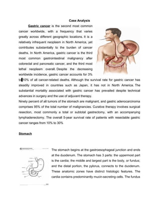
Gastric cancer is the second most common cancer worldwide and the third most lethal neoplasm in North America. It often presents with nonspecific symptoms and is usually diagnosed at advanced stages. Risk factors include H. pylori infection, smoking, and diet high in pickled/salted foods. Diagnosis involves endoscopy with biopsy. Staging utilizes endoscopic ultrasound, CT, and endoscopy to determine tumor depth and lymph node involvement. Prognosis depends on stage, with 5-year survival of 10-30% for resectable gastric cancer.

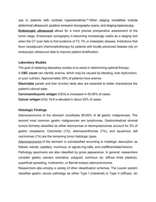
![appealing feature of classifying patients according to the Lauren system is that the
descriptive pathologic entities have clinically relevant differences.
Intestinal, expansive, epidemic-type gastric cancer is associated with chronic atrophic
gastritis, retained glandular structure, little invasiveness, and a sharp margin. The
pathologic presentation classified as epidemic by the Lauren system is associated with
most environmental risk factors, carries a better prognosis, and shows no familial
history.
The second type, diffuse, infiltrative, endemic cancer, consists of scattered cell clusters
with poor differentiation and dangerously deceptive margins. Margins that appear clear
to the operating surgeon and examining pathologist often are determined retrospectively
to be involved. The endemic-type tumor invades large areas of the stomach. This type
of tumor is also not recognizably influenced by environment or diet, is more virulent in
women, and occurs more often in relatively young patients. This pathologic entity is
associated with genetic factors (such as E-cadherin), blood groups, and a family history
of gastric cancer.
Staging
The 2006 American Joint Committee on Cancer (AJCC) Cancer Staging
Manual presents the following TNM classification system for staging gastric carcinoma:
[16]
Primary tumor
• TX - Primary tumor (T) cannot be assessed
• T0 - No evidence of primary tumor
• Tis - Carcinoma in situ, intraepithelial tumor without invasion of lamina propria
• T1 - Tumor invades lamina propria or submucosa
• T2 - Tumor invades muscularis propria or subserosa
• T3 - Tumor penetrates serosa (ie, visceral peritoneum) without invasion of
adjacent structures
• T4 - Tumor invades adjacent structures
Regional lymph nodes
• NX - Regional lymph nodes (N) cannot be assessed](https://image.slidesharecdn.com/62167148-case-analysis-gastro-150904191037-lva1-app6891/85/62167148-case-analysis-gastro-11-320.jpg)


![Health 10 Ppt[1]. Stomach Cancer](https://cdn.slidesharecdn.com/ss_thumbnails/health10ppt1-stomachcancer-091223081620-phpapp02-thumbnail.jpg?width=640&height=640&fit=bounds)


![MALIGNANT CANCER OF THE STOMARCH [Autosaved].pptx](https://cdn.slidesharecdn.com/ss_thumbnails/malignantcancerofthestomarchautosaved-250510054659-d24799df-thumbnail.jpg?width=640&height=640&fit=bounds)


