The document provides information about the digestive system. It discusses:
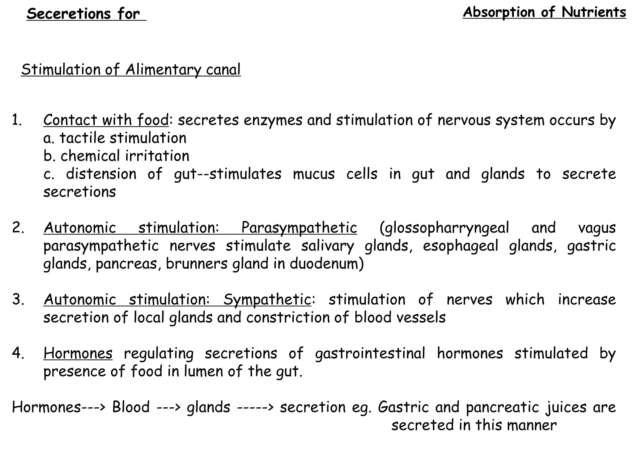
1. The movement of food through the alimentary tract and the secretion of digestive juices and absorption of nutrients.
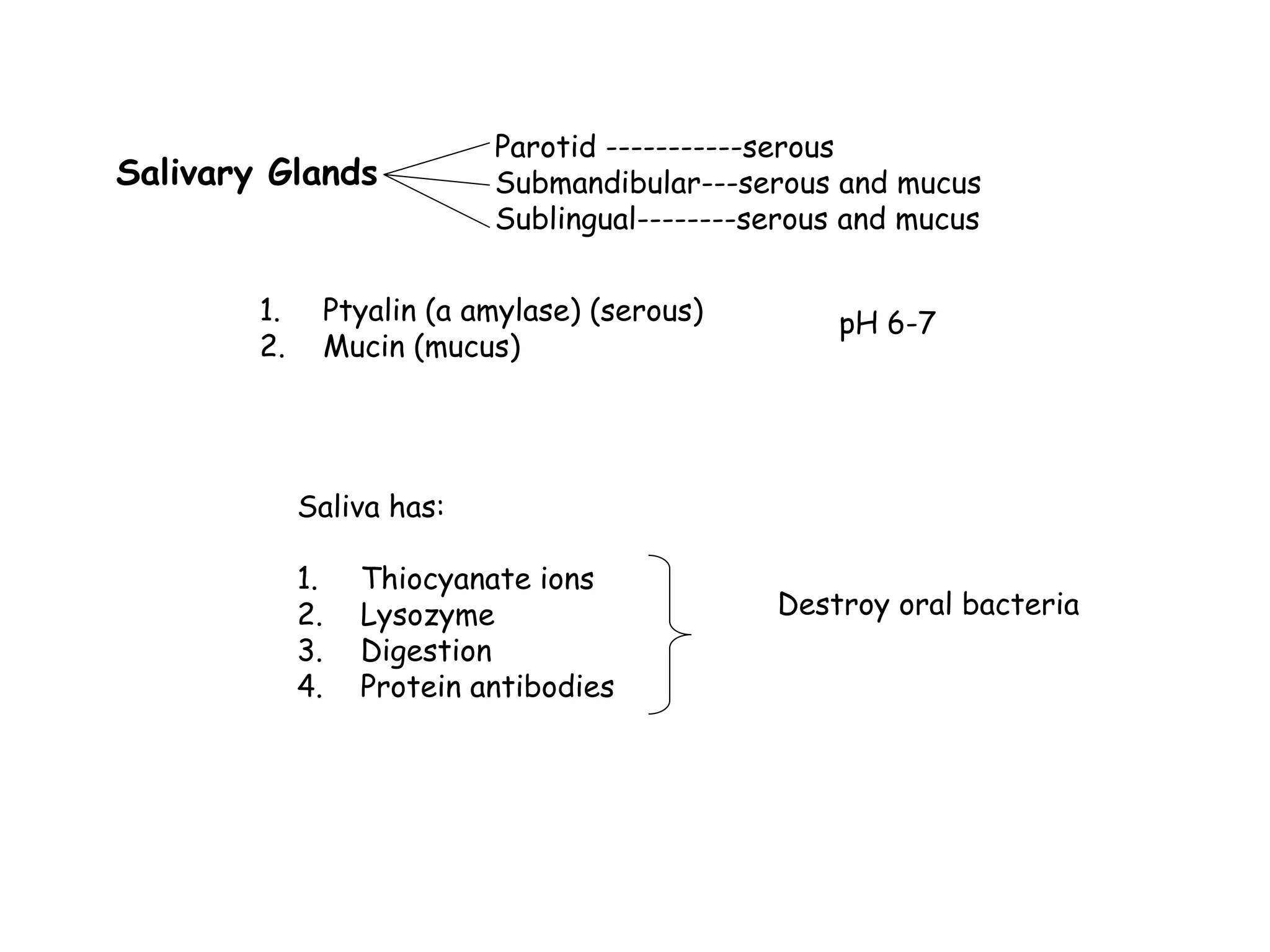
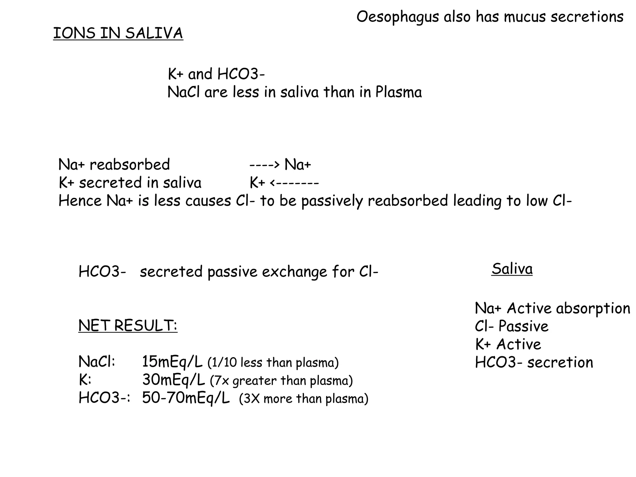
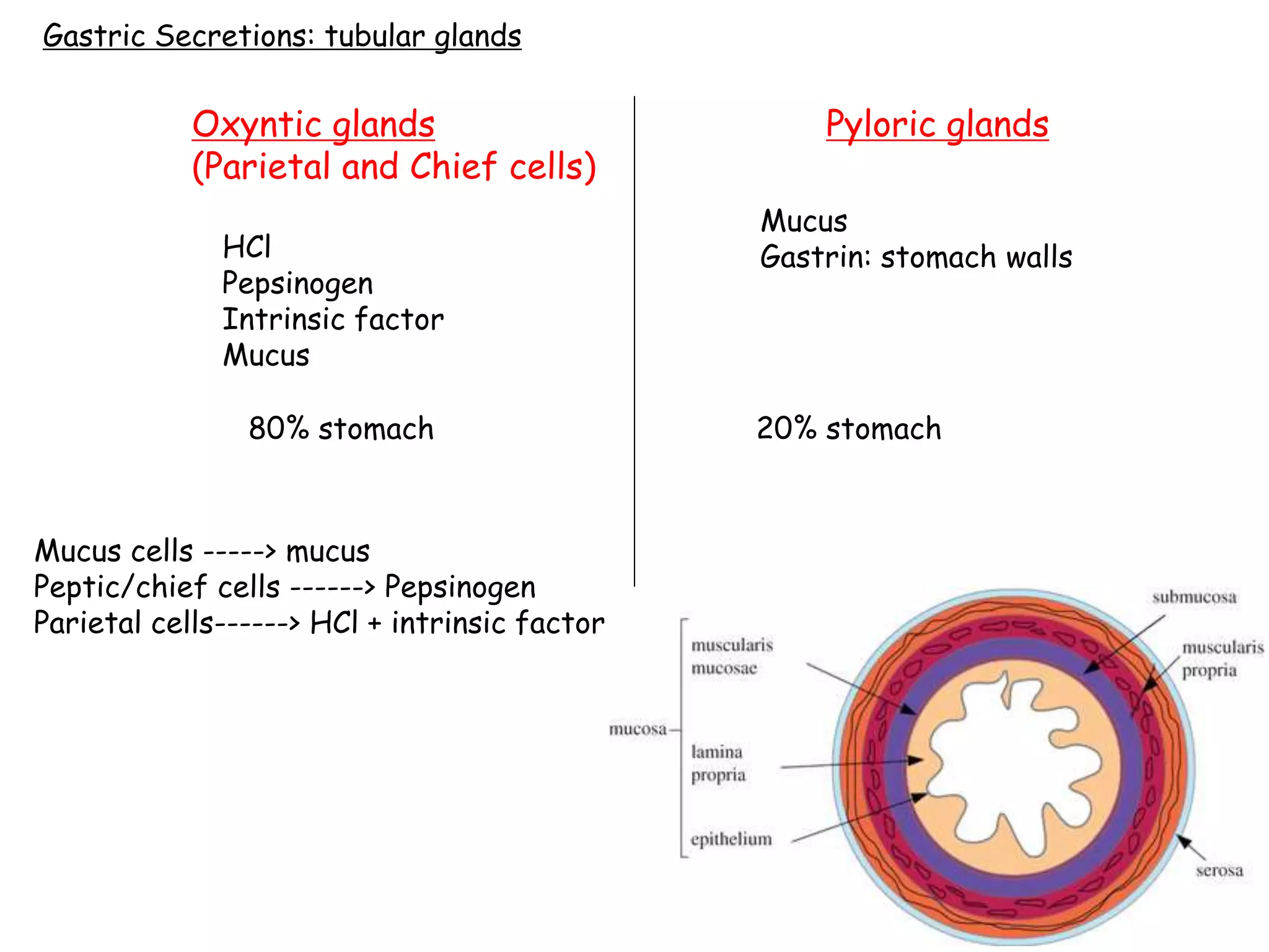
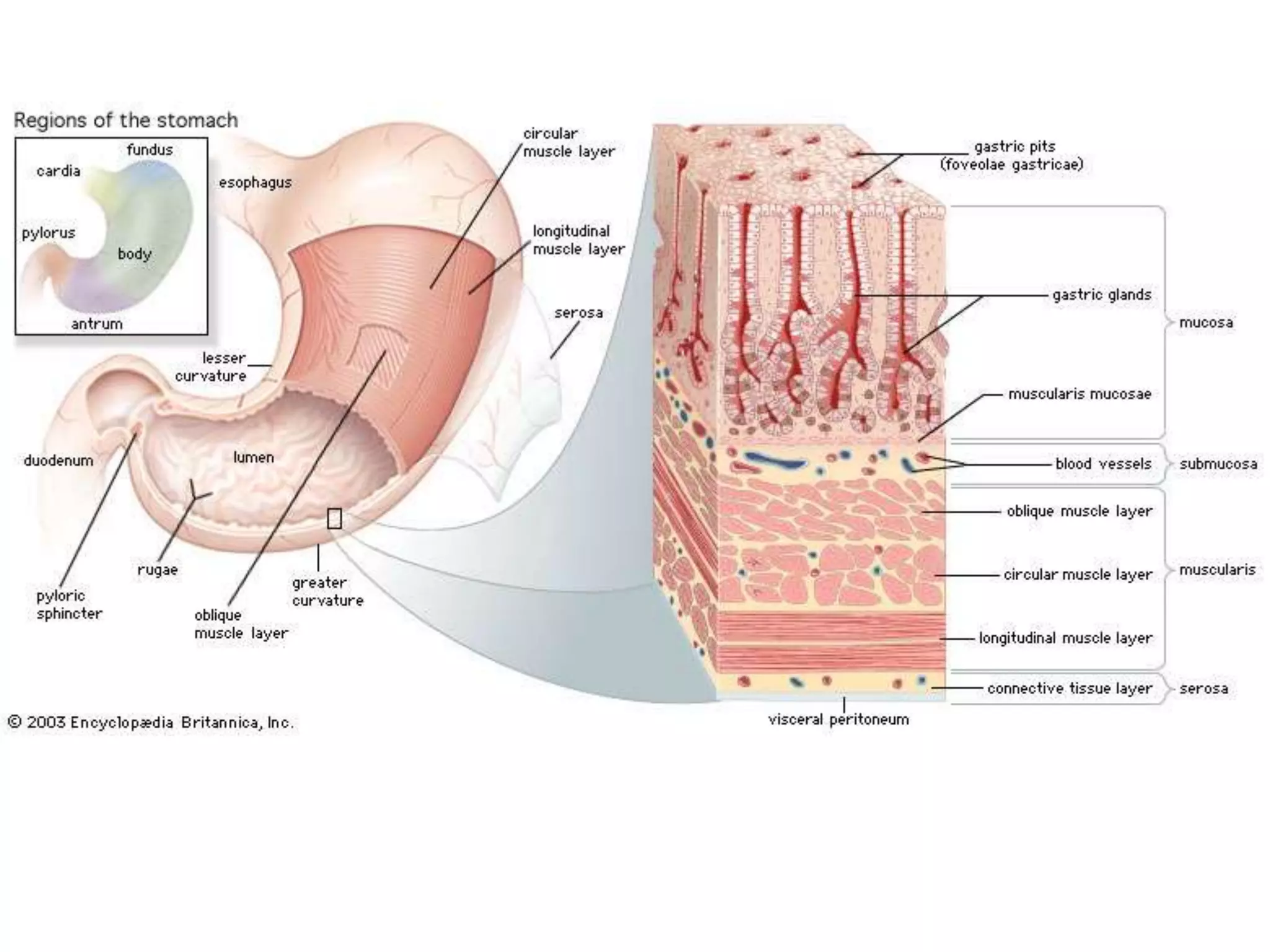
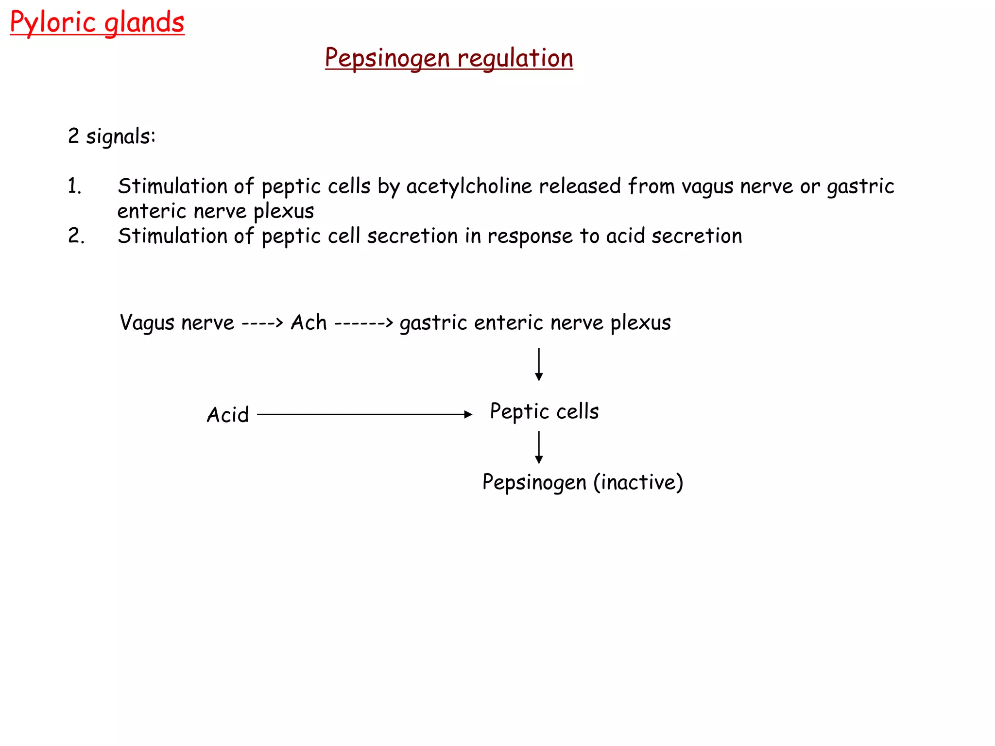
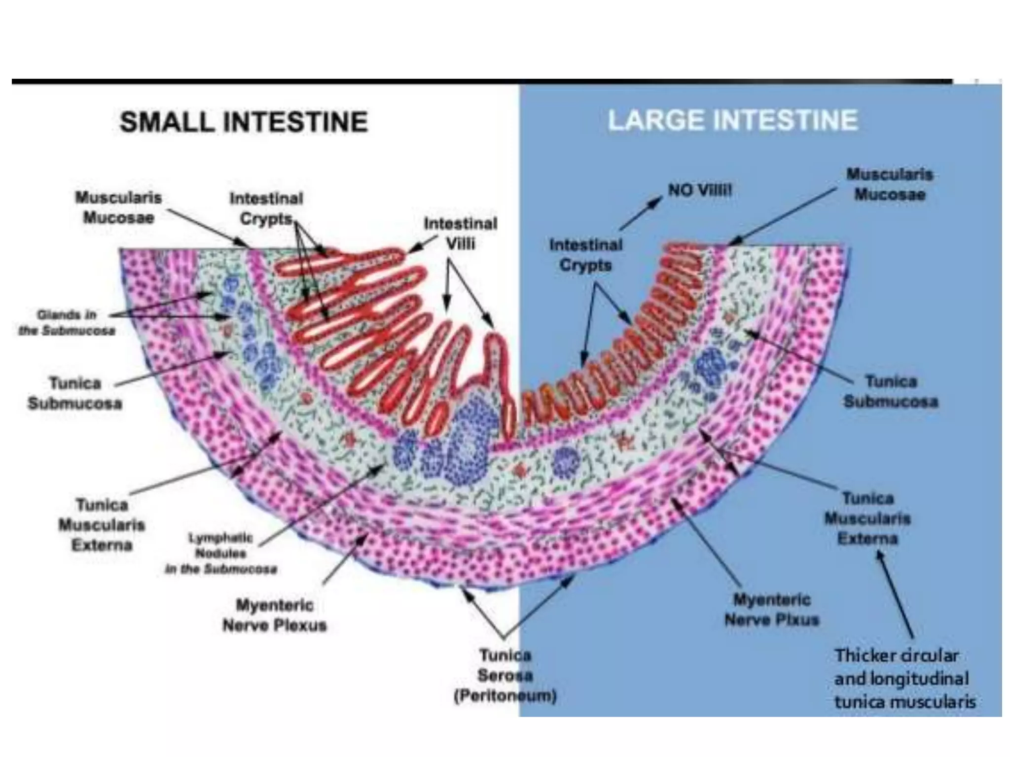
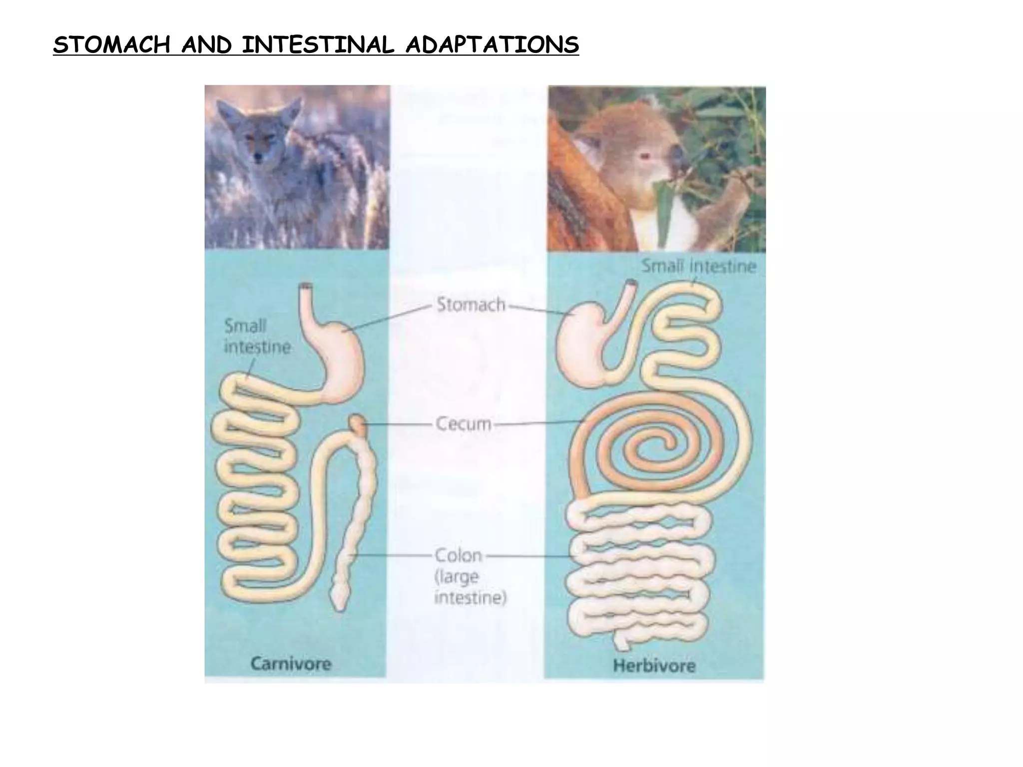
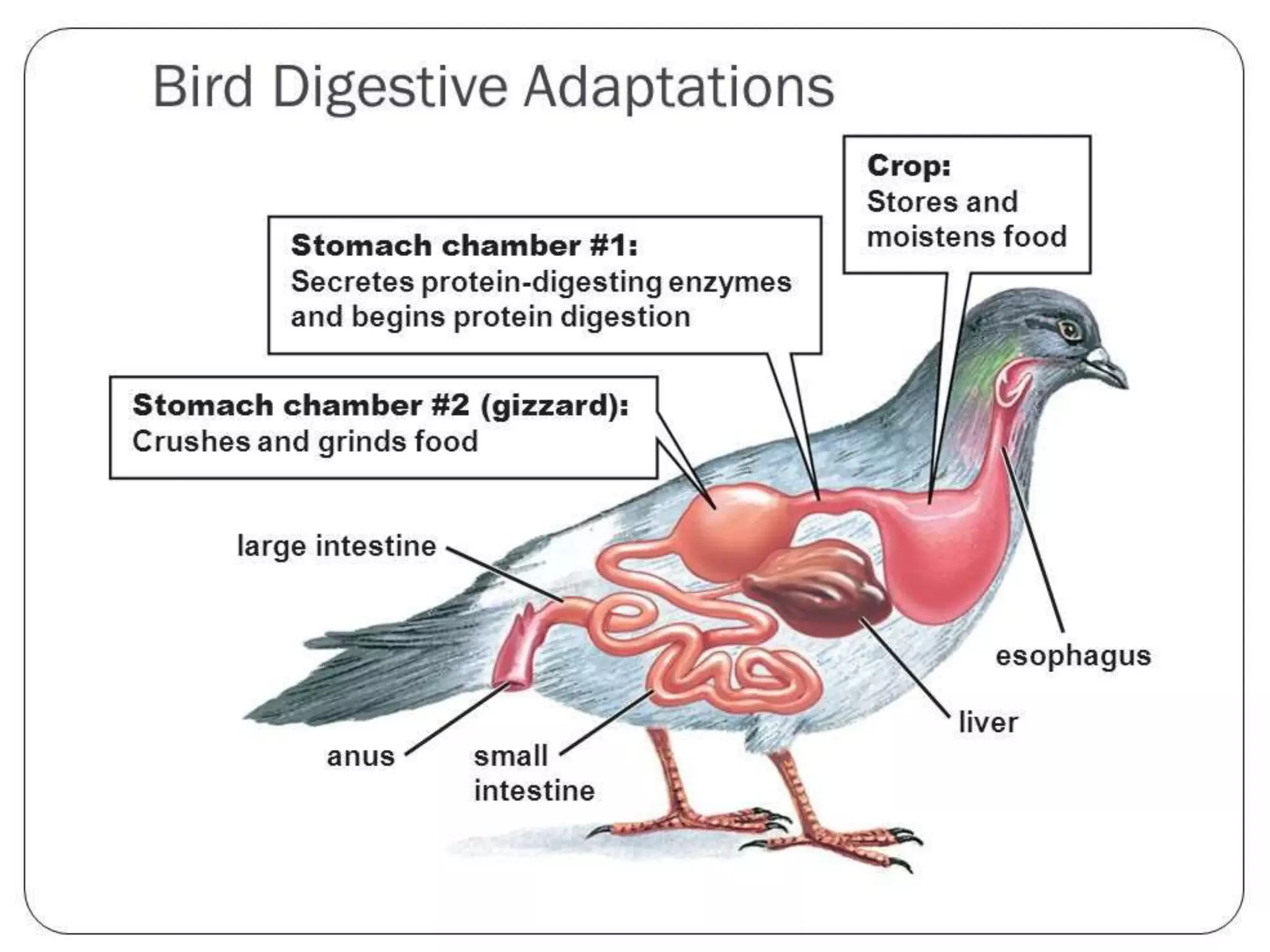
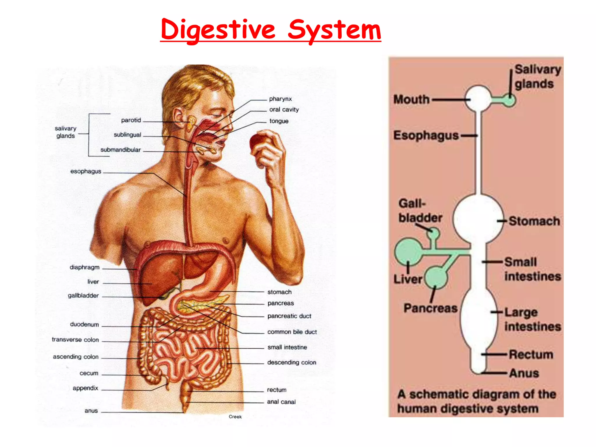
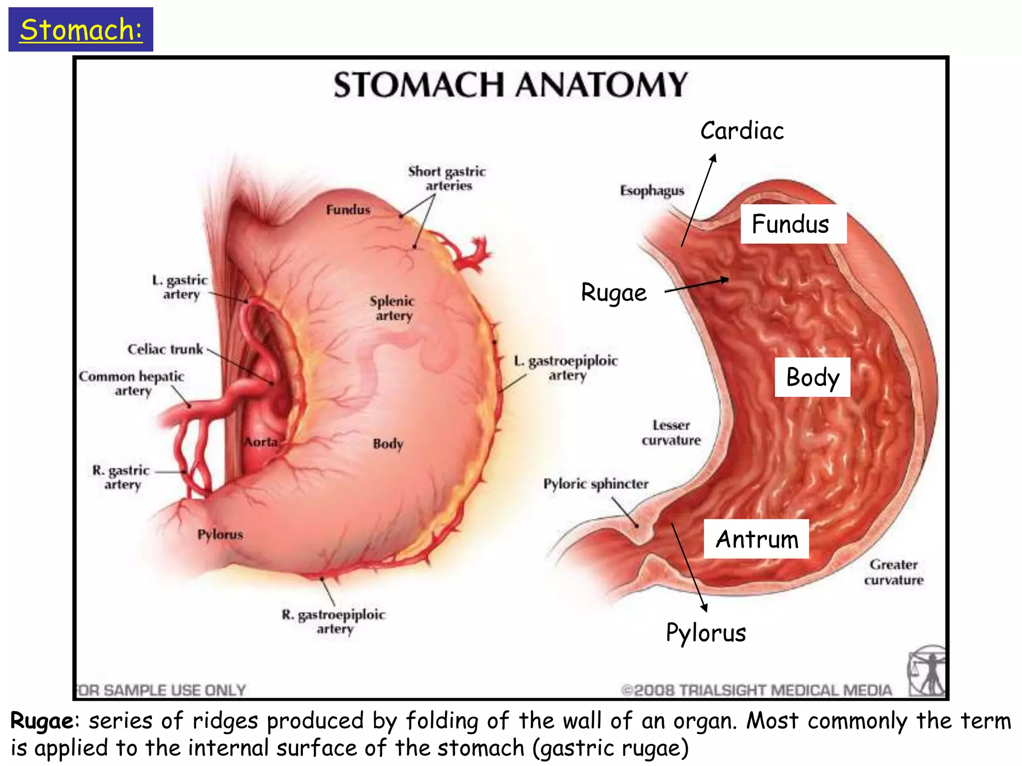
2. It describes the structure and function of different components of the digestive system like the mouth, stomach, small intestine, large intestine.
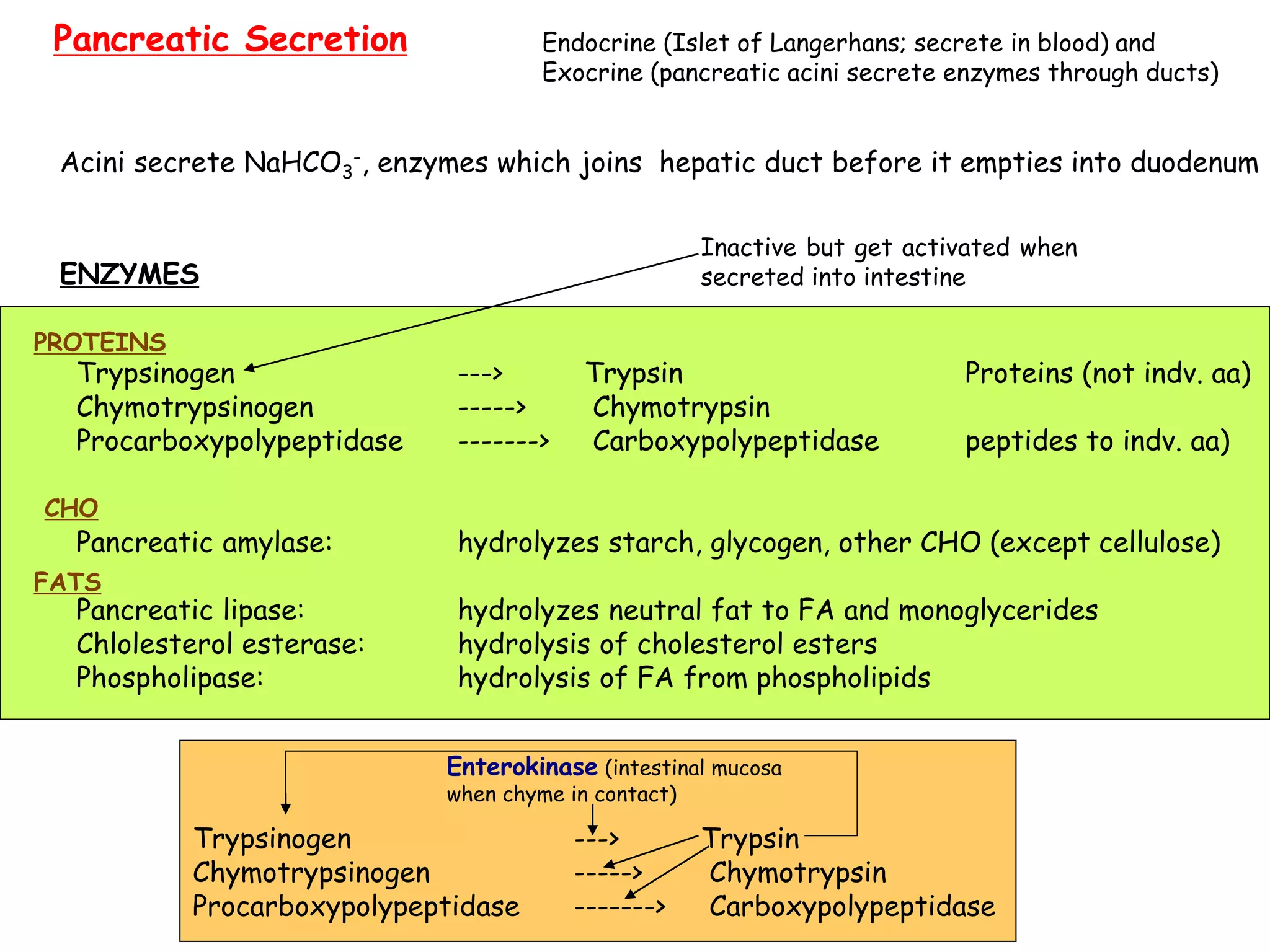
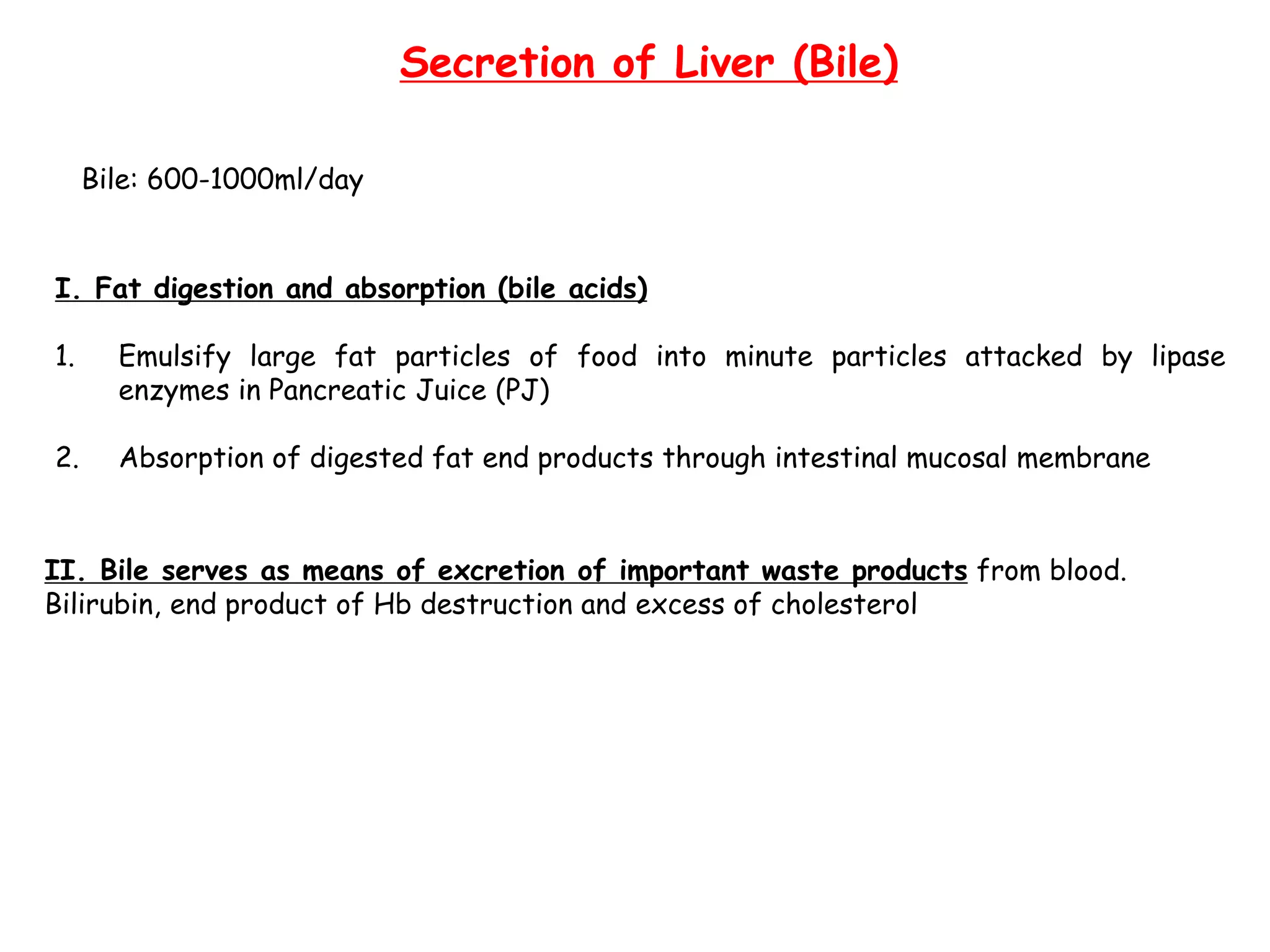
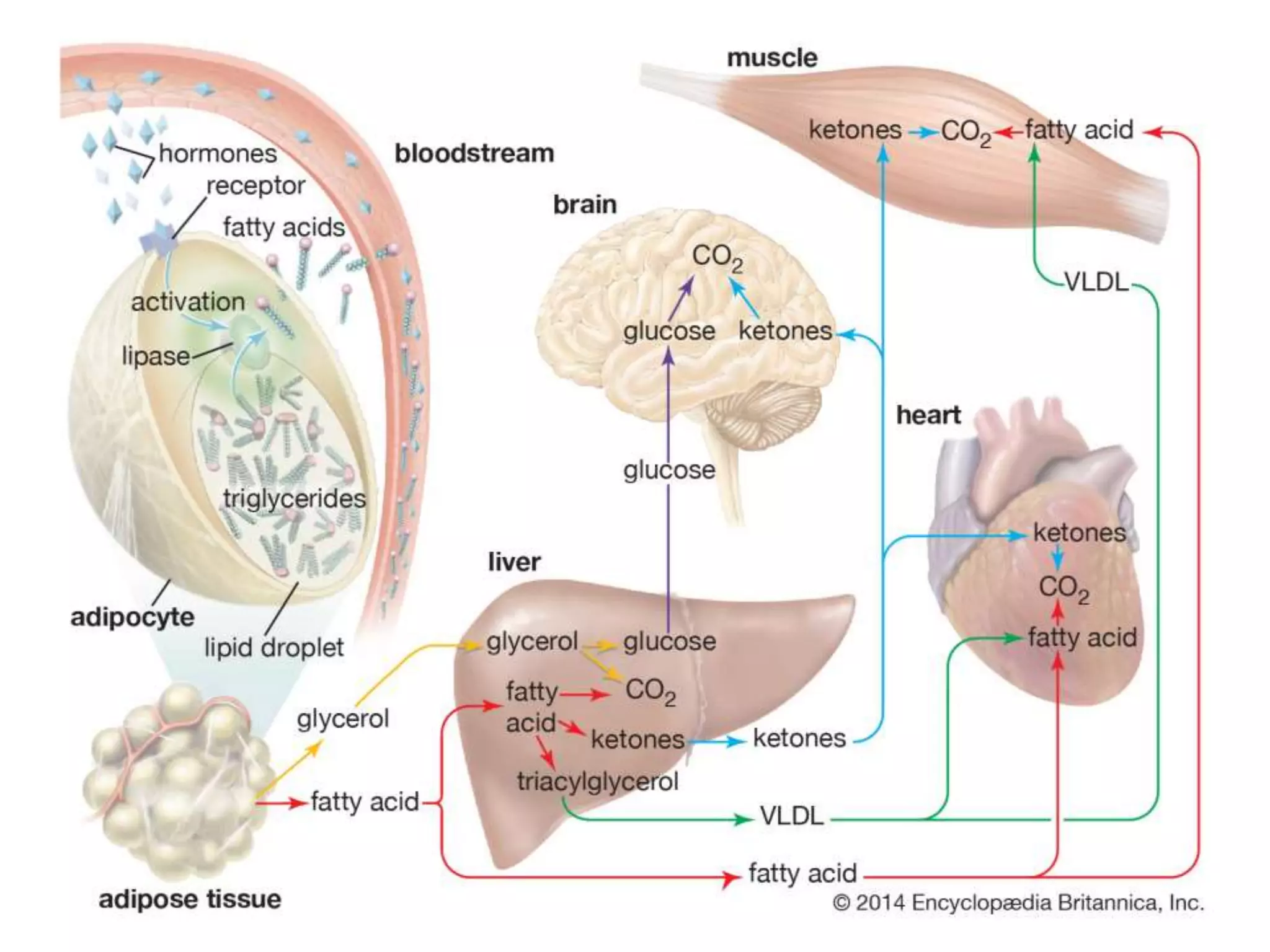
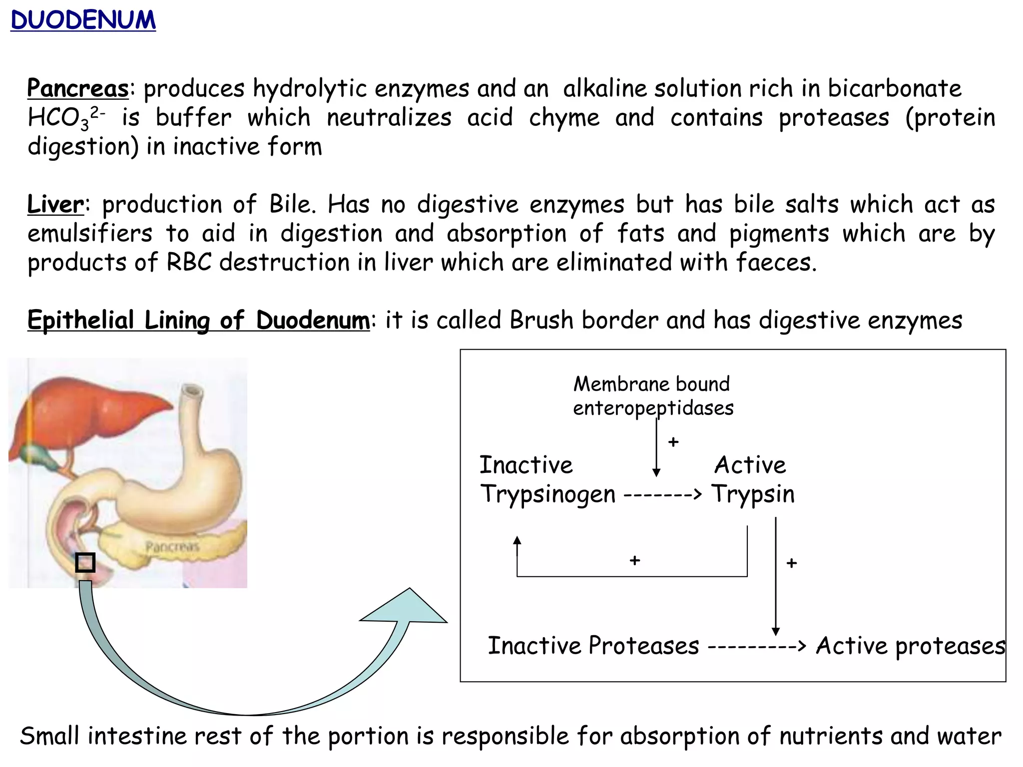
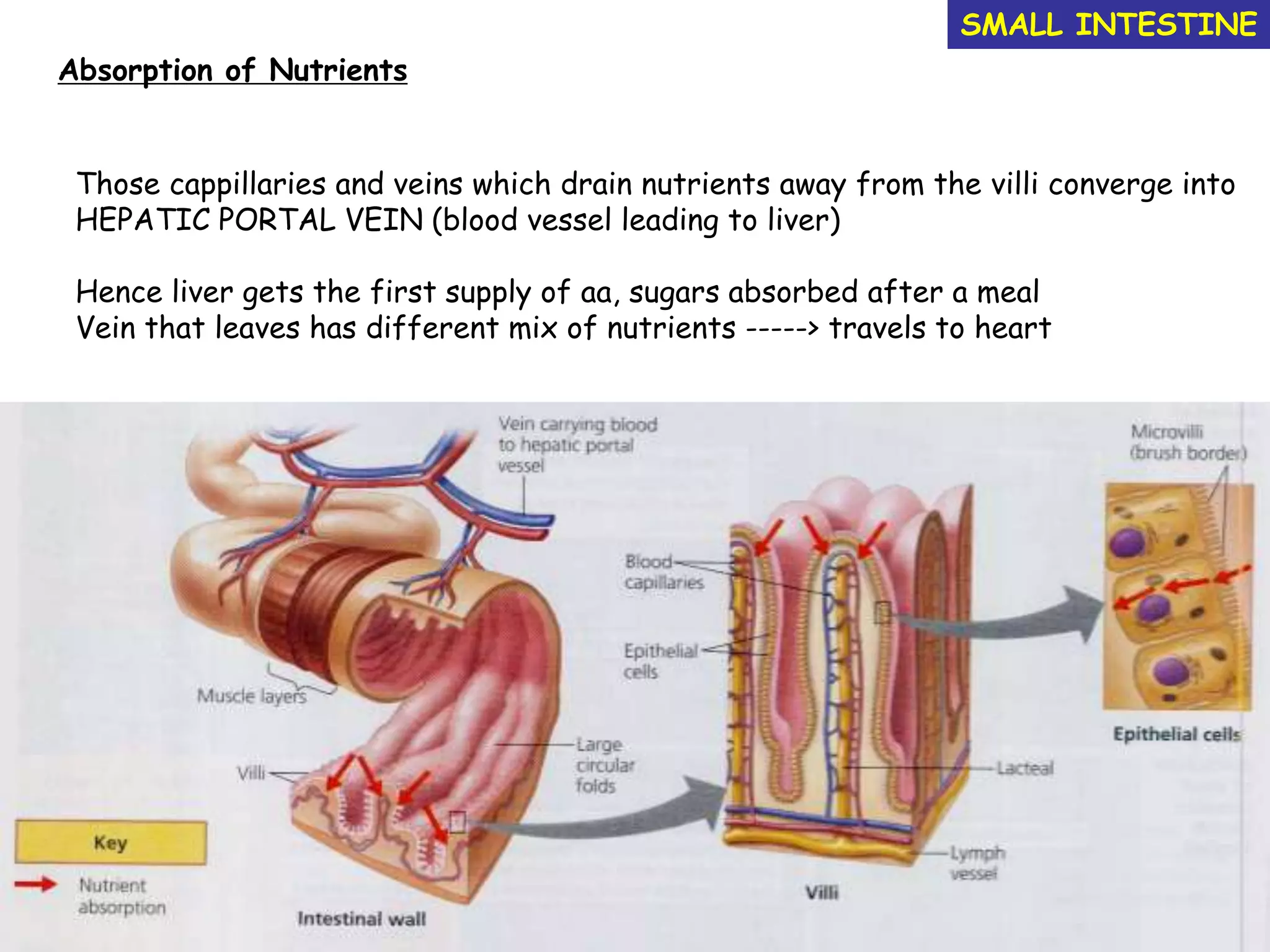
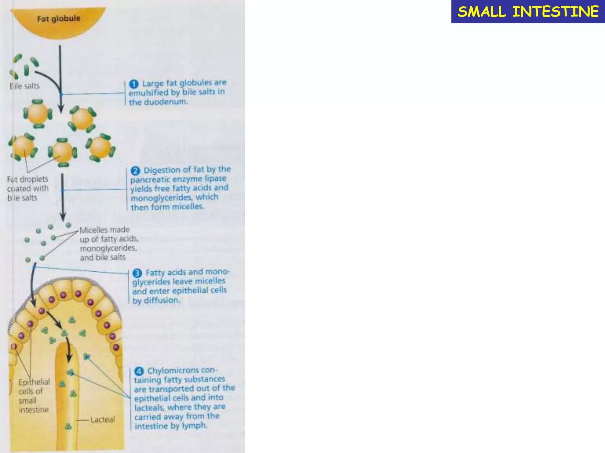
3. It discusses the role of organs like the liver, pancreas and gallbladder in digestion and the absorption of nutrients like carbohydrates, fats and proteins in the small intestine.


![LARGE INTESTINE
colon
Ileo-caecal sphincter
Ileum connects to the cecum which has finger like projections like appendix (has
lymphoidal tissue)
Absorption of water which is not absorbed by small intestine: 90% of water is
reabsorbed by SI and LI
Wastes become more solid as water is absorbed and as they move by peristalsis takes
12-24h to travel
[if bacterial or viral infection is the lining of colon is irritated and less water is
absorbed resulting in diahorrea
If peristalsis moves the faeces too slowly and excess water is absorbed making feaces
more compact resulting in constipation]
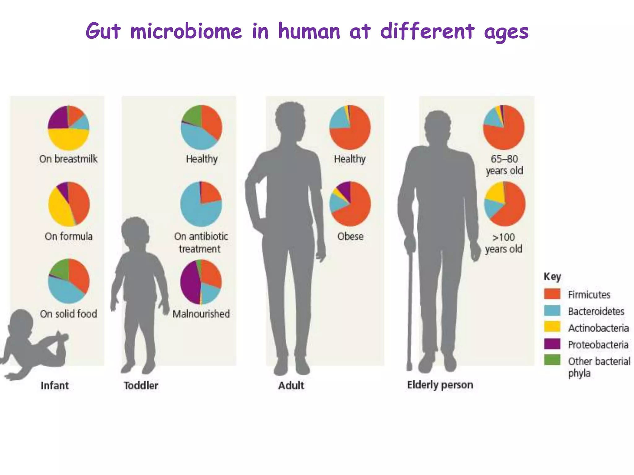
Gut microflora
Rich in micro-organisms which are harmless bacteria eg. E.coli.
They live on undigested organic material and as by products produce gases like
methane, H2S. Some produce important vitamins (B and K) which are absorbed by
humans as essential nutrients.](https://image.slidesharecdn.com/5-d00712b260-2-210523160050/75/5-d00712b260-2-digestive-system-27-2048.jpg)