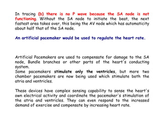
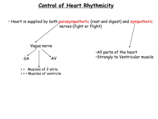
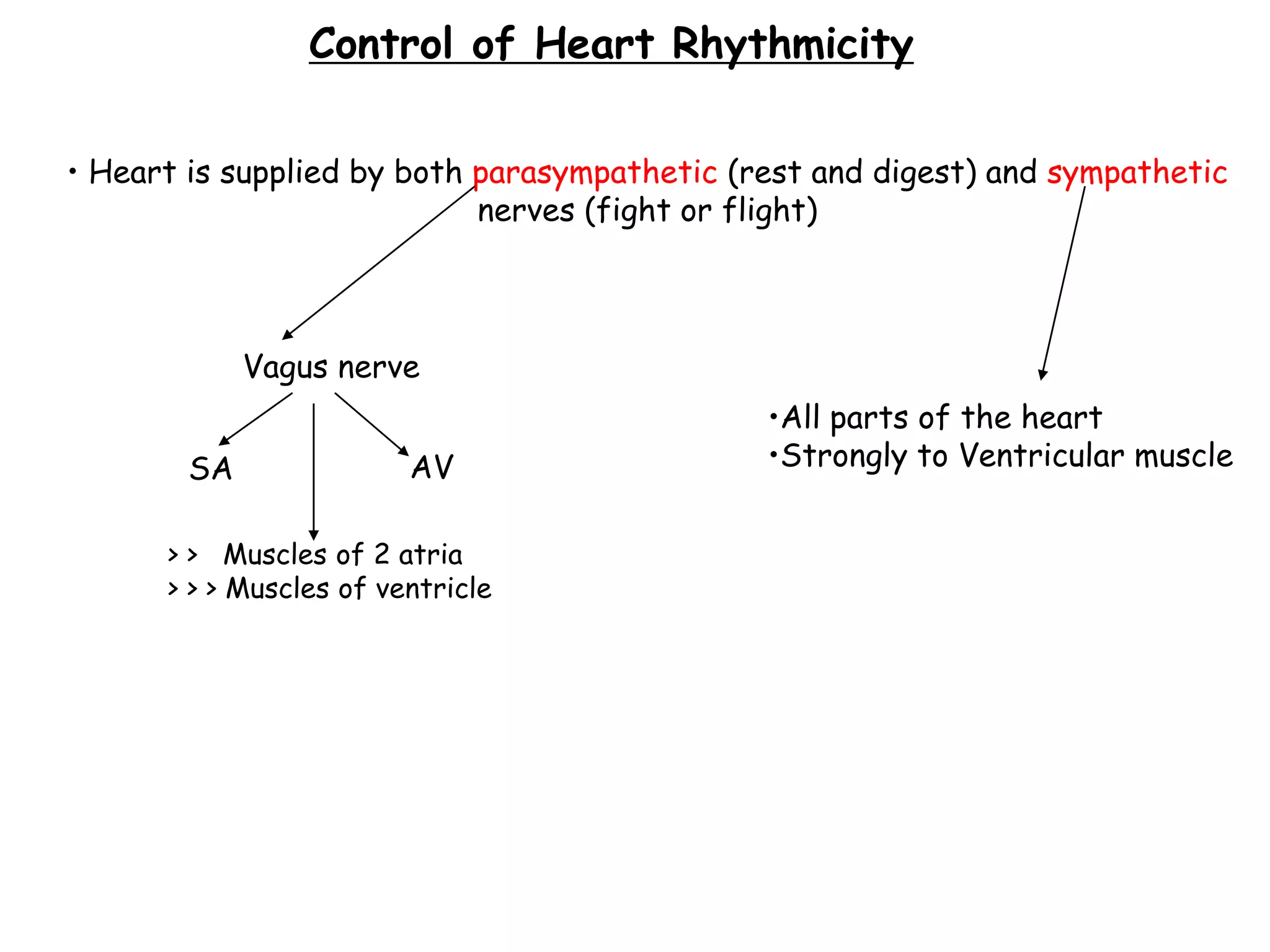
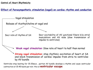
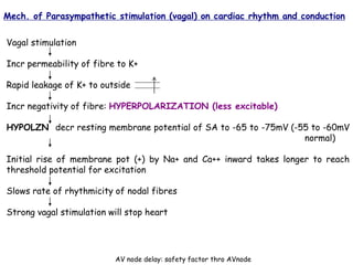
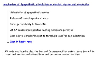
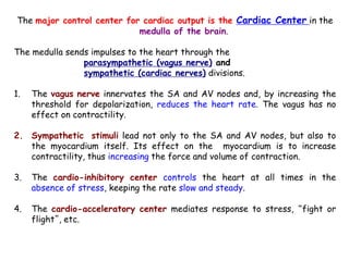
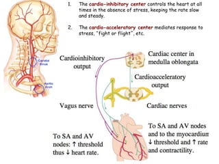
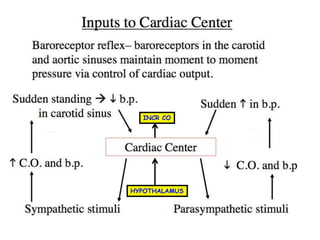
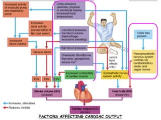
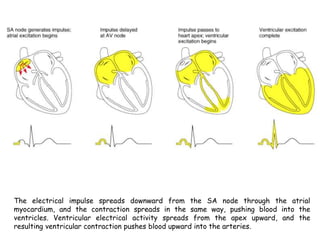
The document discusses how the heart's rhythm is controlled by the autonomic nervous system, with the parasympathetic nervous system slowing the heart rate and the sympathetic nervous system increasing it. It explains in detail the effects of vagal and sympathetic stimulation on cardiac rhythm and conduction. The role of the cardiac center in the medulla in regulating heart rate and the factors that can affect cardiac output are also covered.

![Orientation of the 12 Lead ECG
It is important to remember that the 12-lead ECG provides spatial information
about the heart's electrical activity in 3 approximately orthogonal directions:
Right ⇔ Left
Superior ⇔ Inferior
Anterior ⇔ Posterior
Each of the 12 leads represents a particular orientation in space, as indicated below
(RA = right arm; LA = left arm, LL = left foot):
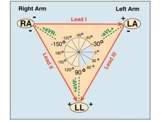
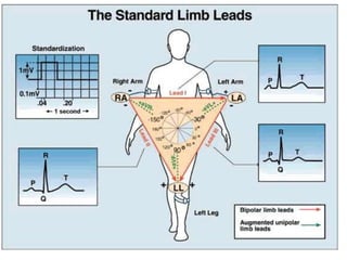
Bipolar limb leads (frontal plane):
Lead I: RA (-) to LA (+) (Right Left, or lateral)
Lead II: RA (-) to LL (+) (Superior Inferior)
Lead III: LA (-) to LL (+) (Superior Inferior)
Augmented unipolar limb leads (frontal plane):
Lead aVR: RA (+) to [LA & LL] (-) (Rightward)
Lead aVL: LA (+) to [RA & LL] (-) (Leftward)
Lead aVF: LL (+) to [RA & LA] (-) (Inferior)
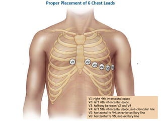
Unipolar (+) chest leads (horizontal plane):
Leads V1, V2, V3: (Posterior Anterior)
Leads V4, V5, V6:(Right Left, or lateral)](https://image.slidesharecdn.com/4-210523170418/85/4-control-regulation-and-disorders-heart-14-320.jpg)