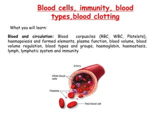
1. Blood cells, immunity, blood
types,blood clotting
Blood and circulation: Blood corpuscles (RBC, WBC, Platelets),
haemopoiesis and formed elements, plasma function, blood volume, blood
volume regulation, blood types and groups, haemoglobin, haemostasis,
lymph, lymphatic system and immunity
What you will learn:
2. Blood corpuscles: RBCs
* RBCs (Erythrocytes): transport Hemoglobin which carries O2 from lungs to tissues
* RBCs contain large quantities of carbonic anhydrase
CO2 + H2O H2CO3 H+ + HCO3- tissues
* Hb is an excellent acid base buffer for blood
Lower animals free Hb not inside RBCs
In humans it is free in plasma it leaks to capillary membranes and kidney filtrates
Lungs
Converted to CO2
Expelled as waste
Blood
3. Blood corpuscles: RBCs
5, 200,000 (+300,000) MEN
4,700,00 (+300,000) WOMEN
RBCs/ mm3
At high altitudes have more numbers
Quantity of concentrated Hb in cell fluid: 34g/100ml of cells
Blood Hematocrit:% of blood that constitutes RBC cells
Men: 15gHb/100ml of cells
Women: 14gHb/100ml of cells
1g of Hb is capable of combining with 1.34ml of O2
MAN: 20ml of O2 + Hb in 100ml of blood
WOMAN: 19ml of O2 + Hb in 100ml of blood
48% for men and 38% for women
Packed cell volume
Hematocrit
Erythrocyte volume fraction
4. Blood corpuscles: Haematopoiesis Formation of blood cellular
components
PLURIPOTENT HEMATOPOIETIC STEM CELLS (HSC) or
HEMOCYTOBLAST
Bone marrow
Common
Myeloid progenitor (stem cell)
Common
Lymphoid progenitor (stem cell)
Blast
CFU-B
CFU-E
Erythrocytes
CFU-S
CFU-GM
Grannulocytes
(Neutrophiles,
Eosinophils,
Basophils)
Monocytes
(Macrocytes)
Spleen
CFU-M
Megakaryocytes
(Platelets)
Growth inducers and differentiation inducers
Interleukin-3 Factors outside bone marrow
Disease etc
Lymphoid
stem cell
Natural
Killer (NK)
cells
T lymphocyte B lymphocyte
Plasma cell
Myelocytes Lymphocytes
CFU-L: lymphoblast
5. Blood corpuscles: Regulation of RBC production
Proerythroblast
Erythrocytes
Heamatopoietic stem cells
TISSUE OXYGENATION
ERYTHROPOIETIN
kidneys
1. Low blood volume
2. Anemia
3. Low hemoglobin
4. Poor blood flow
5. Pulmonary disease
1. An adequate no. of
red cells is available
to provide sufficient
transport of O2
from lungs to tissues
2. Cells do not become
so much that they
impede blood flow
At high altitudes (O2 is less) RBC production is increased: O2 transported to tissues and
demand for RBC (hematocrit)
HYPOXIA
ERYTHROPOIETIN
RBCs prodn.
6. Blood corpuscles: Maturation of RBCs- Vitamin B12
Vitamin B12 (cyanacobalmin)and folic acid, Vit B9 (pteroyl-L-glutamic acid) is required
for maturation of RBCs, synthesis of DNA (TTP)
Deficiency leads to failure of nuclear maturation and cell division
Leads to rapid proliferation of larger RBCs called Macrocytes which have
flimsy membrane and irregular, large (megaloblastic anemia) and oval instead
of biconcave. Highly fragile hence short life span.
Megaloblastic anemia: It is caused by loss of gastric parietal cells
(intrinsic factor), and subsequent inability to absorb vitamin B12.
1. Intrinsic factor binds to Vit B12, hence protected from digestion by
GI secretions
2. IF + Vit B12 binds to receptors in brush border of mucosa of ileum
3. IF + Vit B12 transported to blood by pinocytosis
4. Lack of IF causes loss of absorption of vitB12
7. Blood corpuscles: RBCs- Hb
Hemoglobin is involved in the transport of other gases:
A. Hemoglobin has an oxygen binding capacity of 1.34 ml O2 per gram of
hemoglobin, which increases the total blood oxygen capacity seventy fold
B. it carries some of the body's respiratory carbon dioxide (about 10% of the
total) as carbaminohemoglobin, in which CO2 is bound to the globin protein.
C. The molecule also carries the important regulatory molecule nitric oxide
bound to a globin protein thiol group, releasing it at the same time as oxygen.
Occurence
Hemoglobin is also found outside red blood cells and their progenitor lines. Other
cells that contain hemoglobin include the A9 dopaminergic neurons in the
substantia nigra, macrophages, alveolar cells, and mesangial cells in the kidney.
In these tissues, hemoglobin has a non-oxygen-carrying function as an
antioxidant and a regulator of iron metabolism
8. Blood corpuscles: RBCs- Hb formation
2succinyl CoA + 2glycine ------> pyrrole
4pyrrole ------------> protoporphyrin IX
Protoporphyrin IX + Fe++ ----------> heme
Heme + polypeptide -------------> hemoglobin chain ( or )
2 chains + 2 chains -------------> hemoglobin A (MW 64,458)
Krebs cycle
One Hb molecules has 4 chains (2and 2)
4 Fe++ in one Hb molecule
Binds loosely to 4O2 molecules (8 atoms of O)
Abnormalities in the chains of Hb can alter binding affinity of Hb for O2
SICKLE CELL ANEMIA: val is substituted for glu
(crystals of Hb diff to pass thro capillaries; rupture
cell membrane; anemia
9. Iron Metabolism Hemoglobin
Myoglobin
Cytochromes
Cyt oxidase
Peroxidase
Catalase
Fe
Fe++ absorbed
(Small intestine)
Fe++ excreted
(0.6mg daily;faeces
1.3mg/day; menstruation in women)
TISSUE
Free Fe + Apoferritin
Ferritin (storage iron) Hemosiderin (insoluble)
Heme
Enzymes
Erythroblasts
Mitochondria
Heme synthesis
Hemoglobin
RED CELLS
Fe++ + Plasma + apotransferrin -------> Transferrin** (Fe++ loose bind)
(b globulin)
PLASMA (transferrin)
Excess in liver
hepatocytes and RE
syst
RE: reticuloendothelial system (lymph/spleen)
**Hypochromic anaemia (less Hb)
MACROPHAGES
Degraded Hb ------> free Fe
Bilirubin
IN LIVER
BILE
120 days
10. ANEMIA
Microcytic hypochromic anemia: chronic blood loss, small RBCs
Aplastic Anemia: bone marrow Aplasia i.e.Lack of functioning bone marrow
mabe because of exposure to gamma rays (nuclear bomb), X rays, drugs,
industrial chemicals
Megaloblastic anemia: vitB12, folic acid and IF deficiency leads to odd shaped
RBSc. Patients with loss of entire stomach can also lead to MA
Hemolytic Anemia: hereditary acquired abnormalities of make cells rupture
faster than they are formed; leading to serious anemia.
a. Hereditary spherocytosis: RBCs are spherical, rupture when pass
thro spleenic pulp and capillaries
b. Sickle cell anemia: defective form of Hb S (faulty chain) instead
of Hb A. Hb + low O2-----> conc of Hb into crystals, membrane
rupture, anemia
c. Erythroblastosis foetalis: Rh+ve cells of fetus are attacked by
antibodies from Rh-ve mother. Rh+ve cells rupture and child born
with serious anemia.
11. EFFECT OF ANEMIA ON CIRCULATORY SYSTEM
Viscosity ∝ concentration of RBCs
Severe anemia viscosity is 1.5 times lower than water (normal value is 3)
Hence more blood flows in peripheral blood vessels to tissues and return
Results in greater cardiac output
HYPOXIA (less O2 in blood)
Dilation of peripheral blood vessels
More blood flows
Increased cardiac output (3-4X times than normal)
Increased pumping workload on heart
Reduced O2 carrying capacity
Extreme tissue hypoxia
Acute cardiac failure
Exercise
12. EFFECT OF ANEMIA ON CIRCULATORY SYSTEM
SECONDARY POLYCYTHEMIA
Tissue hypoxia
High altitude/cardiac failure/ failed O2 delivery to tissues
Extra quantities of RBCs
Physiologic Polycythemia (14,000-17,000 feet; 6-7milion/mm3)
Reasons--->
POLYCYTHEMIA VERA (ERYTHEMIA)
Pathological polycythemia: high hematocrit (60-70%; normal is 40-45%)
RBC count is 7-8million/mm3
Genetic abberation in hemocytoblastic cells that produce blood cells
RBCs are produced indefinitely, WBCs and platelets are also produced similarly
Blood volume is increased twice as normal; blood capillaries are plugged by viscous blood
And viscosity is 10 times that of water (normal is 3 times)
Blood flow is sluggish; Decreased return of venous blood;
blood volume is increased; increase return of venous blood
Arterial pressure is elevated; results in hypertension
Bluish tint in skin: large amt of RBCs are deoxygenated masking oxygenated RBCs
13. Blood corpuscles: WBCs: resistance of body to infection
Prevention of disease:
1. Destroying invading bacteria or viruses by phagocytosis
2. Formation of antibodies and sensitized lymphocytes (may destroy or inactivate )
1. Destroying invading bacteria or viruses by phagocytosis
White blood Cells or leucocytes
Formed in the bone marrow (grannulocytes, monocytes, lymphocytes) and
in lymph tissue (lymphocytes and plasma cells)
Transported in blood or specific areas of infection and inflammation
14. Blood corpuscles: WBCs
Polymorphonuclear neutrophils
Polymorphonuclear eosinophils
Polymorphonuclear basophils
Monocytes
Lymphocytes
Plasma cells
Megakaryocyte ------> platelets
Granular
appearance Phagocytosis
Immune system
Normally: 7000 WBCs /ul of blood
62%
2.3%
0.4%
5.3%
30%
300,000
Per ul of
blood
BONE
MARROW
LYMPHOID TISSUES:
Lymph glands
Spleen
Thymus
Tonsils
Peyer’s Patches
BONE
MARROW
Blood
Clotting
Grannulocytes: 4to 8 hr (blood)
4 -5 days in tissues
infection few hrs; get destroyed
Monocytes: 10-20hr (blood); in tissues become tissue macrophages and live for months
Lymphocytes: from lymph few hr in blood; enter tissues by diapedesis; reenter lymph and
in continuous circulation; weeks or months
Platelets: replaced after 10 days; 30,000 platelets per ul of blood per day
LIFE SPAN
15. Blood corpuscles: WBCs NEUTROPHILS AND MACROPHAGES
Attack and destroy bacteria, viruses in the circulating blood: neutrophils
Monocytes inside tissues swell and become: tissue macrophages
Nu and Mac enter from capilliary pores into tissues by DIAPEDESIS
Amoeboid motion
Nu and Mac move to inflamed sites by CHEMOTAXIS
Bacterial/viral toxins
Degenerate products
Complement system components
Plasma clotting substances
Reasons:
PHAGOCYTOSIS
1. Tissues (smooth) resist phagocytosis; rough surface promote
2. Natural substances have protective coat to repel Phago. As opposed to dead tissue
and foreign particles
3. Immune system develops antibodies which adhere to bacterial membranes; make
them suceptible to phago.
4. Antibody + complement (C3)-----> attach to receptors on phagocyte membrane-->
phago. opsonization
16. PHAGOCYTOSIS: neutrophils
Blood corpuscles: WBCs NEUTROPHILS AND MACROPHAGES
Pseudopodia
Phagosome (3-20 bacteria can be engulfed)
Neutrophil inactivates
PHAGOCYTOSIS: macrophages
Phagocytize 100 bacteria in tissues
Engulf large particles
RBCs, malarial parasites
Extrude residual particles
Survive for many more months
Fuse contents with lysosomes (proteolytic enzymes), lipases (digest lipids membranes of
TB bacteria )
18. Blood corpuscles: WBCs NEUTROPHILS AND MACROPHAGES
Contain anti bactericidal agents
Powerful oxidizing agents: PEROXISOMES
superoxide (O2-), H2O2, OH-
myeloperoxidases (H2O2 + Cl- ----> HClO; hypochlorite)
Mycobacterium can resist lysosomal digestion and can survive within macrophages to cause
Tuberculosis
19. Blood corpuscles: WBCs MONOCYTE MACROPHAGE OR
RETICULOENDOTHELIAL SYSTEM
Mobile macrophages phagocytize foreign particles and bacteria, viruses….
Respond to chemotactic stimuli/inflammatory processess
Present in all tissues
Monocyte-macrophage system
Monocytes
Mobile macrophages
Fixed Macrophages
Reticuloendothelial system
Endothelial cells in BM
Spleen and lymph nodes
GENERALIZED PHAGOCYTIC SYSTEM IN ALL TISSUES
WHERE TOXINS AND UNWANTED SUBSTS ARE DESTROYED
Present in:
1. Skin: Histiocytes; inflammed or broken skin; tissue macrophages
2. Lymph Nodes: if not destroyed in skin pass in lymph and trapped by tissue macro. in
sinuses
3. Lungs: alveolar macrophages in alveolar walls; trapped in alveoli and digested products
passed to lymph [if not digestible macrophage forms “giant cell”and forms tubercles or
capsules eg. TB bacteria, carbon particles
4. Liver sinusoids: Kupffer cells; thro GI tract; prevents bacteria to pass into systemic
circulation
5. Spleen and Bone Marrow: bacteria in general circulation are phagocytized by a
meshwork of tissue macro.
20. Blood corpuscles: WBCs NEUTROPHILS AND MACROPHAGES
INFLAMMATION
Vasodilation of Bl vess
Excess blood flow
Incr permb of capillaries
Leakage of fludi in interstitial spaces
Clotting : fibrinogen/other prts
Migration of grannulocytes/monocytes
Swelling of tissue cells
Histamine
Bradykinin
Serotonin
Prostaglandins
Products of complement system
Products of blood clotting syst
Lymphokines (T cells)
Release of :
“Walling off” effect : inflamed areas are separated from local tissues
By blocking by fibrinigen clots
Delaying the spread of bacteria
21. Blood corpuscles: WBCs NEUTROPHILS AND MACROPHAGES
INFLAMMATION
Tissue macrophages
Neutrophil Invasion
of inflammed area
Second Macrophage invasion
Grannulocytes and Monocytes
By Bone marrow
phagocytosis
Margination, diapedesis, chemotaxis
Neutrophilia
Ist line
of defense
IInd line
of defense
IIIrd line
of defense
IVth line
of defense
Increased production
Tumour necrosis factor (TNF)
Interleukin-1 (IL-1)
Grannulocyte-monocyte colony stimulating factor (GM-CSF)
Grannulocyte colony stimulating factor (G-CSF)
Monocyte colony stimulating factor (M-CSF)
Feed back of macrophages to produce more
Formation of PUS
Necrotic tissue
Dead neutrophils
Dead macrophages
Tissue fluid
autolysis
Absorbed into lymph
23. Blood corpuscles: WBCs EOSINOPHILS
Weak phogocytes
Exhibit chemotaxis
Produced in great numbers in parasitic infections and migrate to tissues damaged by parasites
Attach to surface of parasites and release substances to kill them
Schistisomiasis: eosinophils attach and kill by
releasing hydrolytic enzymes from grannules (lysosomes)
Reactive oxygen species
Major basic protein(larvicidal polypeptide)
BASOPHILS
Basophils and Mast cells outside capillaries; release HEPARIN (prevents blood coagulation)
HISTAMINES, BRADYKININ, SEROTONIN
Play a role in allergic reactions: IgE attached to mast cells and basophills
antigen
Rupture and release
Histamine, bradykinin, serotonin, heparin, anaphylactic substance, lysosomal enzymes
Local vascular and tissue reactions
24. Blood corpuscles: WBCs
DISORDERS
Leukopenia: bone marrow produces few WBCs
Body unprotected against many bacteria and invasion of tissues
Many symbiotic bacteria; reduction in WBCs level; invasion of tissues by already present bacteria
2 days
BM stops producing WBCs
Ulcers in mouth/colon/rspiratory infection
Bacteria from ulcers invade surrounding tissues/blood
X rays
Drugs (thiouracil)
Industrial chemicals
Antibiotic (Chloroamphenicol)
Barbiturates
Hypnotics
Bone marrow Aplasia
TREATMENT
Undestroyed cells, stem cells, myleoblasts
Regenerate BM
Proper treatment, drugs, antibiotics
Normal
25. Blood corpuscles: WBCs
DISORDERS
LEUKEMIA: BLOOD CANCER
Lymphocytic luekemia
Myelogenous Leukemia
Cancerous production of
lymphoid cells (lymph node,
lymphoid tissue) and spreads.
Cancerous production of young
myelogenous cells in bone
marrow and spreads to lymph
nodes, spleen and liver.
Partially differentiated cells
Neutrophilic leukemia
Eosinophilic leukemia
Basophilic leukemia
Undifferentiated and bizzare; non functional
Acute leukemia
Chronic develops slowly over years (20)
26. Blood corpuscles: WBCs
DISORDERS
LEUKEMIA: BLOOD CANCER
EFFECTS
Metastatic growth
BM produces so much that invade bones-----> fracture
Spread to spleen, lymph nodes, liver, vascular regions
Infection, severe anemia, bleeding tendency (thrombocytopenia; lack of platelets)
Excessive use of metabolic substrates by cancerous cells
Energy depleted
Deterioration of protein tissues
Metabolic starvation
Death
Normal to non functional bone marrow
27. PLURIPOTENT HEMATOPOIETIC STEM CELLS
HEMOCYTOBLAST
Bone marrow
Common
Myeloid progenitor (stem cell)
Common
Lymphoid progenitor (stem cell)
Blast
CFU-B
CFU E
Erythrocytes
CFU-S
CFU-GM
Grannulocytes
(Neutrophiles,
Eosinophils,
Basophils)
Monocytes
(Macrocytes)
Spleen
CFU-M
Megakaryocytes
(Platelets)
Lymphoid
stem cell
Natural
Killer cells
T lymphocyte B lymphocyte
Plasma cell
Myelocytes Lymphocytes
Phagocytosis Clotting O2, CO2 Immune System
Lymph glands
Spleen
Thymus
Tonsils; payers patches
28. Immunity
Resistance to infection
INNATE IMMUNITY
Phagocytosis
Acid/digestive enzymes
Skin
Blood corpuscles
lysozymes: CW of bact.
Basic polypep: G+ve bact.
complement sys: 20 prts
natural killer cells
ACQUIRED IMMUNITY
adaptive
Develops on exposure to antigens
Immune system
Antibodies
Lymphocytes
immunization
Humoral
Immunity
B cell
antibodies
Cell Mediated
Immunity
T cell or activated
lymphocytes
Lymph nodes
ANTIGENS: foreign organisms, toxin or large polysaccharides and initiate acquired immunity
Must have MW 8000 or more and possess recurring molecular groups, Epitopes on their
surface
29. LYMPHOCYTES Provide acquired immunity
Genetic lack of lymophocytes : no immunity
Lymph nodes----> peripheral tissues
Peyers patches----> GI tract
Spleen
Thymus circulating blood
Bone marrow
Tonsils---> throat and pharynx
Activated T lymphocytes are preprocessed in Thymus (hence T)
B lymphocytes (antibodies) are preprocessed in liver (mid fetal) and bone
marrow (late fetal and after birth) [bursa of Fabricius in birds; hence B]
Mucosal immunity
30. SPECIFICITY OF T AND B CELLS
LYMPHOCYTES
Production of a Clone of lymphocytes
Original stem cell
Functional immune cells
Gene segments (not whole)
Mixed in random combinations
Whole genes
Mature T and B cells clones
Specific for a specific antigen
Antigen
T cell activation B cell activation
Helper T cells
Surface receptor proteins
(T cell markers)
Specific for an antigen
lymphokines
Macrophages
Phagocytosis
Antigen left
Recognized by specific
Lymphocyte clone
IL-1
Lymphocytes
31. B LYMPHOCYTES: humoral immunity and antibodies
Clone of B lymphoctes
Dormant in lymphoid tissue
B lymphocyte
antigen
Macrophage
phagocytize
present
T lymphocyte
present
T helper cells
(TH)
activation
-globulin antibodies
To lymph and
circulating blood
Lymphoblasts
Plasmablasts
Plasma cells
Memory
cells
Primary response
Secondary response
ANTIBODIES BY PLASMA CELL
Concentration
of
antibody
Weeks
0 2 4 6 8
Concept of immunization
32. B LYMPHOCYTES: humoral immunity and antibodies
ANTIBODIES -globulin: Immunoglobulins ; Ig MW 160,000-970,000
20% of plasma proteins are Ig
33. Specificity: unique structure in variable region of both light and heavy chains
differential bonding sites
1. Hydrophobic bonding
2. Hydrogen bonding
3. Ionic interactions
4. Van der waals forces
34. Ig M: primary response; pentavalent
Ig G: bivalent; 75%;cross placental passive immunity
Ig A: mucosa, gut, respiratory and UG tract; secretions
Ig D: Ag receptor; basophils and mast cells
Ig E: allergy
Classes:
36. B LYMPHOCYTES: humoral immunity and antibodies
ANTIBODIES MECHNISM OF ACTION 1. Direct attack on invader
2. Activation of complement system
Agglutination: clumping of bacteria with red cells
Precipitation: soluble antigen and antibody become insoluble and precipitate
Neutralization: Ab cover toxic sites of Ag
Lysis: attack and rupture of Ag
Amplification of response by activation of
COMPLEMENT SYSTEM
20 proteins
Enzyme precursors (inactive
But activated by classical
pathway)
C1-----C9
B and D
Plasma proteins in blood
and tissue spaces
Opsonization and phagocytosis
Lysis
Agglutination
Neutralization of viruses
Chemotaxis
Activation of mast cells and basophils
Inflamatory effects
Effects
37. T LYMPHOCYTES: cell mediated immunity
antigen
Antigen
presenting
cell
Clonal proliferation of
activated T lymphocytes
Lymph
T lymphocyte
memory cells
T lymphocyte
Present antigen
T helper cells
Activation of B
lymphocytes
MHC protein
Macrophages, B lymphocytes,
dendritic cells
Ag
Cell adhesion proteins
Major histocompatibility complex (MHC)
Cell surface
MHC I
Cytotoxic T cells
MHC II
T helper cells
CD4+ T
CD8+ T
T cell receptor
T cell
38. T LYMPHOCYTES: cell mediated immunity Types of T cells
Helper T cells Cytotoxic T cells Suppressor T cells
Lymphokines killer cells suppress fns of HT&CT
IL-2: growth of CT & ST own cells also killed prevent excessive
IL-3 perforins damage to own tissues
IL-4 release cytotoxic give immune tolerance
IL-5 activation of B cells substances
IL-6 B cell growth factors destruction of
GM-CSF: phagocytosis cancer cells, transplant
Interferon- cells, foreign cells
T helper inactivated
in AIDS
HT CT ST
+ ve
F B
Next slide
CD4+ T cells
HTL
TH
CD8+ T
CTL
TC
Treg
39. Immune tolerance: tolerance of acquired immunity: AUTOIMMUNITY
Destruction of own body tissues and organs
Recognition of own body as foreign like bacteria and viruses (normally distinguishes)
Reasons: Thymus Bone marrow
T lymphocytes B lymphocytes
preprocessing
Clone of lymphocytes
Clonal selection
Strong Antigen
During fetal stage
Prevention of development of clones
of lymphocytes in lymphoid tissue
Antigen combines with specific immature lymphocytes
Cells them selves destroyed by thymic epithelial cells
before they migrate and colonize body lymphiod tissue
40. Tolerance of acquired immunity: AUTOIMMUNE DISORDERS
Occurs more in older people
Release of self antigens circulate in body
1. Rheumatic fever: body is immunized against tissues in joints and heart (heart
valves) after exposure to streptococci toxin whose epitope is similar to self
antigens
2. Glomerulonephritis: immunize against basement membrane of glomerulus
3. Myasthenia gravis: immunity against Acethylcholine receptor proteins of
neuromuscular junctions causing paralysis
4. Lupus erythmatosus: immunization against different tissues of body at the same
time; causes extensive tissue damage and rapid death.
41. Immunization
Injection of dead organisms no longer capable of causing disease but with some chemical antigens
Typhoid, whooping cough, diptheria, and several bacterial diseases
I
Toxins + chemicals -------> toxic nature destroyed -----> antigens intact
Tetanus, botulism, and several toxic diseases
II
Live organism “attenuated” -----> grown in special culture media----> series of animals
mutation
Loss of ability to
cause disease
Carry antigens
For immunization
III
Poliomyelitis, yellow fever, measeles, smallpox and several viral diseases
42. Passive Immunity
Temporary Immunity: without injecting antigens
Inject preformed antibodies or activated T lymphocytes or blood from someone who is
immunized
Antibodies: Last for 2-3 weeks in the recipient
Activated T cells: from human to human last for few weeks but from animal to human for a
few days