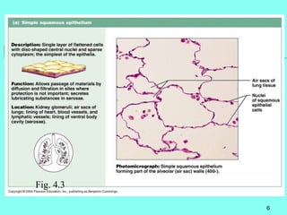
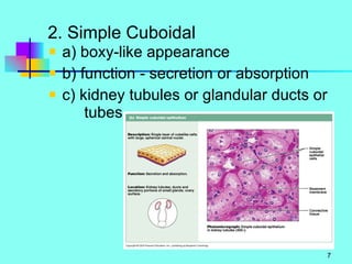
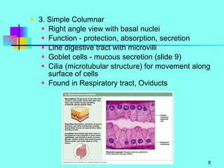
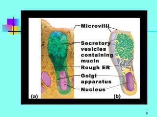
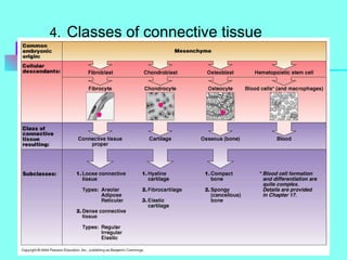
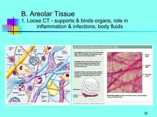
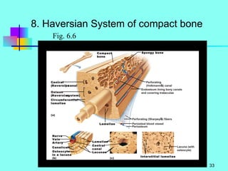
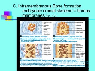
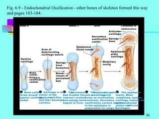
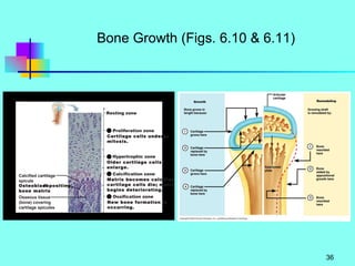
TISSUES & MEMBRANES Marieb: Chaps. 4 - 5
Introduction 1. Tissue - group of cells with related or common structure and function 2. Types of Tissues Epithelial (covering) Connective (support) Muscle (movement) Nervous (control)
I. Epithelial Tissue ( cover, line, transport ) A. Cell Types 1. Squamous - Flat or scalelike 2. Cuboidal - Cube or hexagonical 3. Columnar - Rectangular 4. Transitional - several layers with appearance of cells varying from layer to layer 5. Simple - single layer, little wear and tear, transport functions (semi-permeable) 6. Stratified - multilayer, wear & tear with replacement of surface layers 7. Pseudostratified - one lay but all cells do not reach the top
Fig. 4.2
B. Classification SIMPLE 1. Simple Squamous Epithelial - a) single layer, thin cells closely packed together b) little intercellular/extracellular space  or matrix c) non-vascular - no blood vessels d) basement membrane - epithelial layer attached to underlying connective tissue e) diffusion of gases (lungs, capillaries) f) filtration - kidney capsule g) mesothelium - squamous that lines cavities & organs
Fig. 4.3
2. Simple Cuboidal a) boxy-like appearance  b) function - secretion or absorption c) kidney tubules or glandular ducts or  tubes
3. Simple Columnar Right angle view with basal nuclei Function - protection, absorption, secretion Line digestive tract with microvilli Goblet cells - mucous secretion (slide 9) Cilia (microtubular structure) for movement along surface of cells Found in Respiratory tract, Oviducts
 
4. Pseudostratified a) variations in levels of nuclei of cells b) all touching basement membrane, but all  do not  reach the top  of the tissue c) ducts of glands d) can be ciliated with mucus goblet cells e) Respiratory tract & Female reproductive system
STRATIFIED 4. Stratified Squamous Epithelium a) multilayered - basal cells dividing appear as cuboidal or columnar cells b) Nonkeratinized Stratified Squamous -  Wet surface and wear and tear Mouth - Esophagous - Vagina
4. Stratified Squamous Epith - continued c) Keratinized Stratified Squamous Outermost cells die and contain keratin Keratin - waterproofing Barrier and protection from bacteria SKIN
5. Stratified - Transitional Epithelium a) similar to Nonkeratinized Stratified Squamous b) outer cells relaxed/nonstretched =  NOT FLAT c) outer cells stretched = flat/squamous d) urinary bladder and urethra and ureters
6. Stratified Cuboidal Epithelium a) rare - usually 2 layers b) lines ducts of larger glands - sweat glands and mammary glands 7. Stratified Columnar Epithelium a) rare - basal cells compressed b) line parts of male urethra   line parts of glandular ducts
C. Glandular Classification 1. Gland - group of cells that release a chemical into a duct or the blood a) Exocrine - gland with a duct / tube Salivary - water and chemicals Sweat - water and salts Pancreas - enzymes and buffers b) Endocrine - ductless glands Hormones released into the blood Pituitary / Thyroid
2. Exocrine - release/secretion a) Holocrine - chemical in cytoplasm - cell dies & its contents are discharged - cell replaced - SEBACEOUS b) Merocrine - secretion occurs with no other cellular contents - exocytosis - PANCREAS & SALIVARY c) Apocrine - apical portion with chemical is pinched off - MAMMARY GLANDS Fig. 4.6
3. EXOCRINE STRUCTURE a) Unicellular - goblet cells (Fig. 4.4) b) Multicellular glands and various ducts Different shapes to gland and/or duct Fig.4.5
II. Connective Tissue A. Introduction 1.loose or dense 2.diverse class 3.Fig. 4.7 Cells Fibers Matrix  extracellular
4.   Classes of connective tissue
B. Areolar Tissue 1. Loose CT - supports & binds organs, role in    inflammation & infections, body fluids
2. Cells a) Fibroblasts - immature cells Large, branching processes Formation of new tissue and repairs Produce fibers b) Fibrocyctes -mature cells Maintain tissue c) Macrophages - phagocytosis for defense d) Plasma cells - origin in wbc production of antibodies e) Mast cells - near blood vessels to initiate local inflammation Heparin - anticoaggulant Histamine - increase vessel permeability f) Pigment cells and Fat Cells (store nutrients)
3. Fibers - proteins from fibroblasts a) Collagen (white fibers) - tough, bundle arrangement, some degree flexibility b) Elastic fibers (yellow) - also protein = elastin, flexible and branching (skin, lungs, etc.) c) Reticular fibers - shorter, finer collagen fibers, more give than traditional collagen 4. Matrix - Fluid nature - nutrients and wastes Hyaluronic acid - viscous Hyaluronidase - liquefy ground substance
C. Adipose Tissue - Fat 1. Fibroblast - modified to store fats 2. Large vacuole 3. Functions Store energy raw  materials Insulation
D. Dense / Collagenous  CT  1. Collagen fibers predominate matrix (regular) 2. Fibroblasts line up 3. Tendons Muscle to bone 4. Ligaments Bone to Bone More elastic 5. Dense irregular Fig. 4.9e) Fibrous covering of organs Fig. 4.8d
E. Elastic CT 1. Elastic fibers produced by fibroblasts 2. Stretch & return to original shape 3. Walls blood vessels, vocal cords, respiratory    tract between cartilage rings F. Reticular CT 1. Reticular fibers from fibroblasts 2. Internal framework for organs - Spleen / Liver
III. Cartilage 1. Functions - tough but flexible, supportive structure, large amounts of tissue fluid 2. Chondrocyctes - cartilage cells from mesenchyme tissue Produce collagen & elastic fibers in a different matrix 3. Matrix - collagen & elastic fibers and  Chondroitin sulfate (gel like matrix) (Fig.4.7) 4. Lacunae - space in matrix around cell 5. Perichondrium - outer surface of cartilage tissue associated with fibrous CT
A. Hyaline Cartilage 1. Location a) surface of bone - joint articulating cartilage b) ribs attached to sternum c) nose cartilage d) fetal skeleton e) respiratory tract 2. Chondrocytes -  Produce collage fibers  that are NOT visible 3. Lacunae 4. Perichondium
B. Elastic Cartilage 1. Support and maintain shape 2. Larynx / Outer ear / Eustachian tube 3. Chondrocytes with visible elastic  (& collagen) fibers Lacunae Matrix - fibers and chondroitin sulfate
C. Fibrocartilage 1. Strength / Support / rigidity 2. Vertebral discs and Pubic   symphysis 3. Chondrocytes with visible collagen fibers Lacunae Matrix - fibers and chondroitin sulfate
IV. Bone (connective tissue) Chap. 6 A. Functions 1. Support 2. Protection 3. Movement 4. Mineral storage - calcium 5. Blood cell formation Blood also CT
B. Anatomy of Long Bone (Fig. 6.3) 1. Epiphysis 2. Articulating   cartilage 3. Periosteum With inner layer Osteoblasts 4. Diaphysis 5. Bone Marrow Red/Yellow 6. Osteoclasts 7. Bone Spongy Compact 1. 7. 4. 3. 2. 5. 6 7. 3.
Fig. 6.3
8. Haversian System of compact bone Fig. 6.6
C. Intramembranous Bone formation embryonic cranial skeleton = fibrous   membranes  (Fig. 6.7)
Fig. 6.9 - Endochondrial Ossification - other bones of skeleton formed this way and pages 183-184.
Bone Growth (Figs. 6.10 & 6.11)
V. Muscle Tissue - Next Unit - motion due to filament contraction - classification - location / nerve innervation / appearance -skeletal -voluntary / striated -cardiac - involuntary / striated -smooth/visceral - involuntary /  nonstriated
VI. Nervous Tissue - Neuron = Nerve Cell - Generate & Conduct Electrical signals - Cell Processes
VII. Membranes (epithelial & underlying CT) Fig. 4.11
VII. Membranes 1. Mucous Membranes  a) line body area open to the outside Digestive, respiratory, urogenital b) moist due to mucus secretion (urine) and cilia to move materials c) secretion of mucus and absorption of materials d) underlying CT binds plus flexible (lamina propria)
VII. Membranes 2. Serous Membranes a) lines areas not open to outside & their organs b) mesothelium & loose CT c) visceral portion = around organ parietal portion = around cavity d) pleural = lungs peritoneum = abdominopelvic cavity/organs 3. Synovial Membrane a) lines joint cavity b) no epithelial component
Synovial Joint ( loose CT)  Fig8.3 -.4
VIII. Skin / Cutaneous Membrane A. Epidermis - stratified keratinized sq. 1. Stratum basale - dividing columnar shape 2. Stratum spinosum - multilayers of newly formed cells 3. Stratum granulosum - flatten cells/pigment formation, 1st step in Keratin formation 4. Stratum lucidum - Dead cells, flat, keratin Formation (palm/sole) 5. Stratum corneum- 2 dozen layers dead, keratinized squamous cells that are shed
B. Dermis 1. Loose CT (dense, collagen, elastic)   Blood vessels, Nerves (sensory)   Hair follicles and Glands = Epidermal in origin 2. Dermal Ridges / Papilla 3. Subcutaneous layer  - with Adipose / Areolar CT C. Accessory Organs 1. Hair  - epidermal origin plus arrector pili muscle 2. Sebaceous Glands Holocrine Keeps hair/skim pliable, water-proof 3. Sweat Glands (merocrine) Open to surface via duct
Fig. 5.5 Fig. 5.6

4.a&p i tissues2010

  • 1.
    TISSUES & MEMBRANESMarieb: Chaps. 4 - 5
  • 2.
    Introduction 1. Tissue- group of cells with related or common structure and function 2. Types of Tissues Epithelial (covering) Connective (support) Muscle (movement) Nervous (control)
  • 3.
    I. Epithelial Tissue( cover, line, transport ) A. Cell Types 1. Squamous - Flat or scalelike 2. Cuboidal - Cube or hexagonical 3. Columnar - Rectangular 4. Transitional - several layers with appearance of cells varying from layer to layer 5. Simple - single layer, little wear and tear, transport functions (semi-permeable) 6. Stratified - multilayer, wear & tear with replacement of surface layers 7. Pseudostratified - one lay but all cells do not reach the top
  • 4.
  • 5.
    B. Classification SIMPLE1. Simple Squamous Epithelial - a) single layer, thin cells closely packed together b) little intercellular/extracellular space or matrix c) non-vascular - no blood vessels d) basement membrane - epithelial layer attached to underlying connective tissue e) diffusion of gases (lungs, capillaries) f) filtration - kidney capsule g) mesothelium - squamous that lines cavities & organs
  • 6.
  • 7.
    2. Simple Cuboidala) boxy-like appearance b) function - secretion or absorption c) kidney tubules or glandular ducts or tubes
  • 8.
    3. Simple ColumnarRight angle view with basal nuclei Function - protection, absorption, secretion Line digestive tract with microvilli Goblet cells - mucous secretion (slide 9) Cilia (microtubular structure) for movement along surface of cells Found in Respiratory tract, Oviducts
  • 9.
  • 10.
    4. Pseudostratified a)variations in levels of nuclei of cells b) all touching basement membrane, but all do not reach the top of the tissue c) ducts of glands d) can be ciliated with mucus goblet cells e) Respiratory tract & Female reproductive system
  • 11.
    STRATIFIED 4. StratifiedSquamous Epithelium a) multilayered - basal cells dividing appear as cuboidal or columnar cells b) Nonkeratinized Stratified Squamous - Wet surface and wear and tear Mouth - Esophagous - Vagina
  • 12.
    4. Stratified SquamousEpith - continued c) Keratinized Stratified Squamous Outermost cells die and contain keratin Keratin - waterproofing Barrier and protection from bacteria SKIN
  • 13.
    5. Stratified -Transitional Epithelium a) similar to Nonkeratinized Stratified Squamous b) outer cells relaxed/nonstretched = NOT FLAT c) outer cells stretched = flat/squamous d) urinary bladder and urethra and ureters
  • 14.
    6. Stratified CuboidalEpithelium a) rare - usually 2 layers b) lines ducts of larger glands - sweat glands and mammary glands 7. Stratified Columnar Epithelium a) rare - basal cells compressed b) line parts of male urethra line parts of glandular ducts
  • 15.
    C. Glandular Classification1. Gland - group of cells that release a chemical into a duct or the blood a) Exocrine - gland with a duct / tube Salivary - water and chemicals Sweat - water and salts Pancreas - enzymes and buffers b) Endocrine - ductless glands Hormones released into the blood Pituitary / Thyroid
  • 16.
    2. Exocrine -release/secretion a) Holocrine - chemical in cytoplasm - cell dies & its contents are discharged - cell replaced - SEBACEOUS b) Merocrine - secretion occurs with no other cellular contents - exocytosis - PANCREAS & SALIVARY c) Apocrine - apical portion with chemical is pinched off - MAMMARY GLANDS Fig. 4.6
  • 17.
    3. EXOCRINE STRUCTUREa) Unicellular - goblet cells (Fig. 4.4) b) Multicellular glands and various ducts Different shapes to gland and/or duct Fig.4.5
  • 18.
    II. Connective TissueA. Introduction 1.loose or dense 2.diverse class 3.Fig. 4.7 Cells Fibers Matrix extracellular
  • 19.
    4. Classes of connective tissue
  • 20.
    B. Areolar Tissue1. Loose CT - supports & binds organs, role in inflammation & infections, body fluids
  • 21.
    2. Cells a)Fibroblasts - immature cells Large, branching processes Formation of new tissue and repairs Produce fibers b) Fibrocyctes -mature cells Maintain tissue c) Macrophages - phagocytosis for defense d) Plasma cells - origin in wbc production of antibodies e) Mast cells - near blood vessels to initiate local inflammation Heparin - anticoaggulant Histamine - increase vessel permeability f) Pigment cells and Fat Cells (store nutrients)
  • 22.
    3. Fibers -proteins from fibroblasts a) Collagen (white fibers) - tough, bundle arrangement, some degree flexibility b) Elastic fibers (yellow) - also protein = elastin, flexible and branching (skin, lungs, etc.) c) Reticular fibers - shorter, finer collagen fibers, more give than traditional collagen 4. Matrix - Fluid nature - nutrients and wastes Hyaluronic acid - viscous Hyaluronidase - liquefy ground substance
  • 23.
    C. Adipose Tissue- Fat 1. Fibroblast - modified to store fats 2. Large vacuole 3. Functions Store energy raw materials Insulation
  • 24.
    D. Dense /Collagenous CT 1. Collagen fibers predominate matrix (regular) 2. Fibroblasts line up 3. Tendons Muscle to bone 4. Ligaments Bone to Bone More elastic 5. Dense irregular Fig. 4.9e) Fibrous covering of organs Fig. 4.8d
  • 25.
    E. Elastic CT1. Elastic fibers produced by fibroblasts 2. Stretch & return to original shape 3. Walls blood vessels, vocal cords, respiratory tract between cartilage rings F. Reticular CT 1. Reticular fibers from fibroblasts 2. Internal framework for organs - Spleen / Liver
  • 26.
    III. Cartilage 1.Functions - tough but flexible, supportive structure, large amounts of tissue fluid 2. Chondrocyctes - cartilage cells from mesenchyme tissue Produce collagen & elastic fibers in a different matrix 3. Matrix - collagen & elastic fibers and Chondroitin sulfate (gel like matrix) (Fig.4.7) 4. Lacunae - space in matrix around cell 5. Perichondrium - outer surface of cartilage tissue associated with fibrous CT
  • 27.
    A. Hyaline Cartilage1. Location a) surface of bone - joint articulating cartilage b) ribs attached to sternum c) nose cartilage d) fetal skeleton e) respiratory tract 2. Chondrocytes - Produce collage fibers that are NOT visible 3. Lacunae 4. Perichondium
  • 28.
    B. Elastic Cartilage1. Support and maintain shape 2. Larynx / Outer ear / Eustachian tube 3. Chondrocytes with visible elastic (& collagen) fibers Lacunae Matrix - fibers and chondroitin sulfate
  • 29.
    C. Fibrocartilage 1.Strength / Support / rigidity 2. Vertebral discs and Pubic symphysis 3. Chondrocytes with visible collagen fibers Lacunae Matrix - fibers and chondroitin sulfate
  • 30.
    IV. Bone (connectivetissue) Chap. 6 A. Functions 1. Support 2. Protection 3. Movement 4. Mineral storage - calcium 5. Blood cell formation Blood also CT
  • 31.
    B. Anatomy ofLong Bone (Fig. 6.3) 1. Epiphysis 2. Articulating cartilage 3. Periosteum With inner layer Osteoblasts 4. Diaphysis 5. Bone Marrow Red/Yellow 6. Osteoclasts 7. Bone Spongy Compact 1. 7. 4. 3. 2. 5. 6 7. 3.
  • 32.
  • 33.
    8. Haversian Systemof compact bone Fig. 6.6
  • 34.
    C. Intramembranous Boneformation embryonic cranial skeleton = fibrous membranes (Fig. 6.7)
  • 35.
    Fig. 6.9 -Endochondrial Ossification - other bones of skeleton formed this way and pages 183-184.
  • 36.
    Bone Growth (Figs.6.10 & 6.11)
  • 37.
    V. Muscle Tissue- Next Unit - motion due to filament contraction - classification - location / nerve innervation / appearance -skeletal -voluntary / striated -cardiac - involuntary / striated -smooth/visceral - involuntary / nonstriated
  • 38.
    VI. Nervous Tissue- Neuron = Nerve Cell - Generate & Conduct Electrical signals - Cell Processes
  • 39.
    VII. Membranes (epithelial& underlying CT) Fig. 4.11
  • 40.
    VII. Membranes 1.Mucous Membranes a) line body area open to the outside Digestive, respiratory, urogenital b) moist due to mucus secretion (urine) and cilia to move materials c) secretion of mucus and absorption of materials d) underlying CT binds plus flexible (lamina propria)
  • 41.
    VII. Membranes 2.Serous Membranes a) lines areas not open to outside & their organs b) mesothelium & loose CT c) visceral portion = around organ parietal portion = around cavity d) pleural = lungs peritoneum = abdominopelvic cavity/organs 3. Synovial Membrane a) lines joint cavity b) no epithelial component
  • 42.
    Synovial Joint (loose CT) Fig8.3 -.4
  • 43.
    VIII. Skin /Cutaneous Membrane A. Epidermis - stratified keratinized sq. 1. Stratum basale - dividing columnar shape 2. Stratum spinosum - multilayers of newly formed cells 3. Stratum granulosum - flatten cells/pigment formation, 1st step in Keratin formation 4. Stratum lucidum - Dead cells, flat, keratin Formation (palm/sole) 5. Stratum corneum- 2 dozen layers dead, keratinized squamous cells that are shed
  • 44.
    B. Dermis 1.Loose CT (dense, collagen, elastic) Blood vessels, Nerves (sensory) Hair follicles and Glands = Epidermal in origin 2. Dermal Ridges / Papilla 3. Subcutaneous layer - with Adipose / Areolar CT C. Accessory Organs 1. Hair - epidermal origin plus arrector pili muscle 2. Sebaceous Glands Holocrine Keeps hair/skim pliable, water-proof 3. Sweat Glands (merocrine) Open to surface via duct
  • 45.