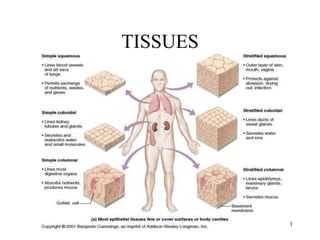
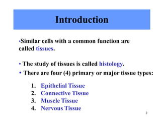
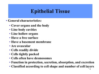
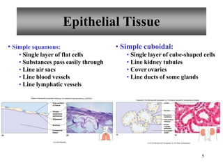
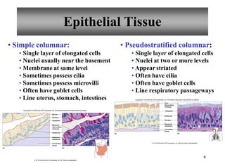
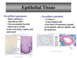
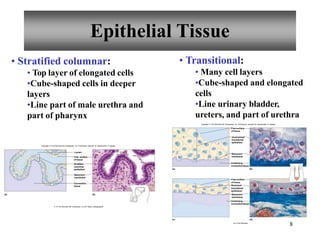
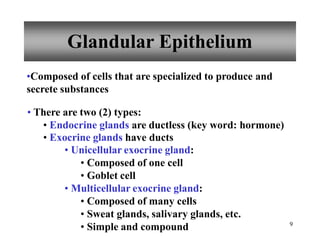
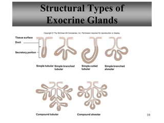
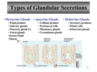
TISSUES
1
2
Introduction
•Similar cells with a common function are
called tissues.
• The study of tissues is called histology.
• There are four (4) primary or major tissue types:
1. Epithelial Tissue
2. Connective Tissue
3. Muscle Tissue
4. Nervous Tissue
Intercellular Junctions
cells
ells
ells
ells
Tight junctions
• Close space between cells
•Located among cells that
form linings
Desmosomes
• Form “spot welds” between
• Located among outer skin c
Gap junctions
• Tubular channels between c
• Located in cardiac muscle c
Tight junction
3
Cell membrane
Cell membrane
Desmosome
Cell membrane
Gap junction
Copyright © The McGraw-Hill Companies, Inc. Permission required for reproduction or display.
4
Epithelial Tissue
• General characteristics:
• Cover organs and the body
• Line body cavities
• Line hollow organs
• Have a free surface
• Have a basement membrane
• Are avascular
• Cells readily divide
• Cells tightly packed
• Cells often have desmosomes
• Function in protection, secretion, absorption, and excretion
• Classified according to cell shape and number of cell layers
Epithelial Tissue
• Simple squamous:
• Single layer of flat cells
• Substances pass easily through
• Line air sacs
• Line blood vessels
• Line lymphatic vessels
• Simple cuboidal:
• Single layer of cube-shaped cells
• Line kidney tubules
• Cover ovaries
• Line ducts of some glands
(b)
(a)
Free surface
of tissue
Simple
squamous
epithelium
Basement
Nucleus
Connective
tissue
b,d: © Ed Reschke
(a)
5
Nucleus
Basement
membrane
Free surface
of tissue
Simple
cuboidal
epithelium
Connective
tissue
(b)
Copyright © The McGraw-Hill Companies, Inc. Permission required for reproduction or display.
Lumen
b: © The McGraw-Hill Companies, Inc./Al Telser, photographer
• Simple columnar:
• Single layer of elongated cells
• Nuclei usually near the basement
• Membrane at same level
• Sometimes possess cilia
• Sometimes possess microvilli
• Often have goblet cells
• Line uterus, stomach, intestines
• Pseudostratified columnar:
• Single layer of elongated cells
• Nuclei at two or more levels
• Appear striated
• Often have cilia
• Often have goblet cells
• Line respiratory passageways
Copyright © The McGraw-Hill Companies, Inc. Permission required for reproduction or display.
Basement
membrane
Microvilli
(free surface
of tissue)
Connective
tissue
Mucus
Nucleus
Cytoplasm
Gobletcell
(a) (b)
Copyright © The McGraw-Hill Companies, Inc. Permission required for reproduction or display.
(a) (b)
Cilia
(free surface
of tissue)
Gobletcell
Basement
membrane
Nucleus
Connective
tissue
Cytoplasm
b: © The McGraw-Hill Companies, Inc./Dennis Strete, photographer
Epithelial Tissue
6
b: © The McGraw-Hill Companies, Inc./Al Telser, photographer.
7
• Stratified squamous:
• Many cell layers
• Top cells are flat
• Can accumulate keratin
• Outer layer of skin
•Line oral cavity, vagina, and
anal canal
Copyright© The McGraw-HillCompanies, Inc. Permission requiredfor reproduction or display.
• Stratified cuboidal:
• 2-3 layers
• Cube-shaped cells
•Line ducts of mammary glands,
sweat glands, salivary glands, and
the pancreas
Layer of
dividing
cells
Basement
membrane
Connective
tissue
Free surface
of tissue
Squamous
cells
(b)
(a)
b: © The McGraw-HillCompanies, Inc./Al Telser, photographer
(a) (b)
Stratified
Free surface
Lumen
of tissue
Basement
membrane
Connective
tissue
cuboidal
epithelium
Nucleus
Copyright © The McGraw-Hill Companies, Inc. Permission required for reproduction or display.
b: © The McGraw-Hill Companies, Inc./Al Telser, photographer.
Epithelial Tissue
8
• Stratified columnar:
• Top layer of elongated cells
•Cube-shaped cells in deeper
layers
•Line part of male urethra and
part of pharynx
• Transitional:
• Many cell layers
•Cube-shaped and elongated
cells
•Line urinary bladder,
ureters, and part of urethra
(b)
(a)
Lumen
Connective
tissue
Basement
membrane
Free surface
of tissue
Stratified
columnar
epithelium
Copyright © The McGraw-Hill Companies, Inc. Permission required for reproduction or display.
b: © The McGraw-Hill Companies, Inc./Al Telser, photographer
Basement
membrane
Underlying
connectivetissue
connectivetissue
(b)
(a)
Free surface
of tissue
Stretched
transitional
epithelium
Basement
membrane
Underlying
(d)
(c)
Copyright © The McGraw-Hill Companies, Inc. Permission required for reproduction or display.
Free surface
of tissue
Unstretched
transitional
epithelium
b,d: © Ed Reschke
Epithelial Tissue
9
Glandular Epithelium
•Composed of cells that are specialized to produce and
secrete substances
• There are two (2) types:
• Endocrine glands are ductless (key word: hormone)
• Exocrine glands have ducts
• Unicellular exocrine gland:
• Composed of one cell
• Goblet cell
• Multicellular exocrine gland:
• Composed of many cells
• Sweat glands, salivary glands, etc.
• Simple and compound
10
Structural Types of
Exocrine Glands
Tissue surface
Duct
Secretory portion
Simple tubular Simple branched
tubular
Simple branched
alveolar
Simple coiled
tubular
Compound tubular Compound alveolar
Copyright © The McGraw-Hill Companies, Inc. Permission required for reproduction or display.
11
Types of Glandular Secretions
• Merocrine Glands
• Fluid product
• Salivary glands
• Pancreas gland (?)
• Sweat glands
•Serous Fluid
•Mucus
• Apocrine Glands
• Cellular product
• Portions of cells
• Mammary glands
• Ceruminous glands
• Holocrine Glands
• Secretory products
• Whole cells
• Sebaceous glands
(a) Merocrine gland (b) Apocrine gland (c) Holocrine gland
Secretion
Pinched off
portion of cell
(secretion)
Intact
cell
Disintegrating cell
and its contents
(secretion)
New cell
forming by
mitosis and
cytokinesis
12
Connective Tissues
• General characteristics:
• Most abundant tissue type
• Many functions:
• Bind structures
• Provide support and protection
• Serve as frameworks
• Fill spaces
• Store fat
• Produce blood cells
• Protect against infections
• Help repair tissue damage
• Have an extracellular matrix
• Have varying degrees of vascularity
• Have cells that usually divide
Connective Tissue
Major Cell Types Present
13
• Fibroblasts
• Fixed cell
• Most common cell
• Large, star-shaped
• Produce fibers
• Mast cells
• Fixed cell
• Release heparin
• Release histamine
• Macrophages
• Wandering cell
• Phagocytic
•Important in injury or
infection
Connective Tissue
Fiber Types Present
14
• Collagenous fibers
• Thick
• Composed of collagen
• Great tensile strength
• Abundant in dense CT
• Hold structures together
• Tendons, ligaments
• Reticular fibers
• Elastic fibers
•Bundles of microfibrils
embedded in elastin
• Fibers branch
• Elastic
• Vocal cords, air passages
• Very thin collagenous fibers
• Highly branched
• Form supportive networks
Connective Tissues
15
• Connective Tissue Proper:
• Loose connective tissue
• Adipose tissue
• Reticular connective tissue
• Dense connective tissue
• Elastic connective tissue
• Specialized Connective Tissue:
• Cartilage
• Bone
• Blood
Connective Tissue Types
• Loose Connective Tissue
• Mainly fibroblasts
• Fluid to gel-like matrix
• Collagenous fibers
• Elastic fibers
• Bind skin to structures
• Beneath most epithelia
•Blood vessels nourish
nearby epithelial cells
• Between muscles
Copyright © The McGraw-Hill Companies, Inc. Permission required for reproduction or display.
• Adipose Tissue
• Adipocytes
• Cushions
• Insulates
• Store fats
• Beneath skin
• Behind eyeballs
• Around kidneys and heart
(a) (b)
Collagenous
fiber
Fibroblast
Ground
substance
Elastic
fiber
(a) (b)
Cell
membrane
Nucleus
Cytsol
Fat droplet
Copyright © The McGraw-Hill Companies, Inc. Permission required for reproduction or display.
16
b: © The McGraw-Hill Companies, Inc./Dennis Strete, photographer
Connective Tissue Types
• Reticular Connective Tissue
• Composed of reticular fibers
• Supports internal organ walls
•Walls of liver, spleen,
lymphatic organs
• Dense Connective Tissue
• Packed collagenous fibers
• Elastic fibers
• Few fibroblasts
• Bind body parts together
• Tendons, ligaments, dermis
• Poor blood supply
Copyright © The McGraw-Hill Companies, Inc. Permission required for reproduction or display.
Collagenous
fibers
White blood
cell
Fibroblast
(a) (b)
Copyright © The McGraw-Hill Companies, Inc. Permission required for reproduction or display.
b: © The McGraw-Hill Companies, Inc./Al Telser, photographer
Fibroblasts
Collagenous
fibers
(a) (b)
b: © The McGraw-Hill Companies, Inc./Dennis Strete, photographer
17
18
Connective Tissue Types
• Elastic Connective Tissue
• Abundant in elastic fibers
• Some collagenous fibers
• Fibroblasts
• Attachments between bones
• Walls of large arteries, airways, heart
• Bone (Osseous Tissue)
• Solid matrix
• Supports
• Protects
• Forms blood cells
• Attachment for muscles
• Skeleton
• Osteocytes in lacunae
Copyright © The McGraw-Hill Companies, Inc. Permission required for reproductionor display
Elastic fibers
Collagenous
fibers
Fibroblast
(a) (b)
Copyright © The McGraw-Hill Companies, Inc. Permission required for reproduction or display.
b: © The McGraw-Hill Companies, Inc./Al Telser, photographer
21
Canaliculi
Osteocyte
in lacuna
Central
canal
Lamella
Osteocyte
Nucleus
Cell process in
canaliculus
(a) (b)
Osteon
b: © The McGraw-Hill Companies, Inc./Dennis Strete, photographer
• Hyaline cartilage
• Most abundant
• Ends of bones
• Nose, respiratory passages
• Embryonic skeleton
• Elastic cartilage
• Flexible
• External ear, larynx
• Fibrocartilage
• Very tough
• Shock absorber
• Intervertebral discs
• Pads of knee and pelvic girdle
19
Connective Tissue Types
• Cartilage
• Rigid matrix
• Chondrocytes in lacunae
• Poor blood supply
• Three (3) types:
• Hyaline Cartilage
• Elastic Cartilage
• Fibrocartilage
20
Connective Tissue Types
Three (3) types of cartilage:
Copyright © The McGraw-Hill Companies, Inc. Permission required for reproduction or display. Copyright © The McGraw-Hill Companies, Inc. Permission required for reproduction or display.
Hyaline Cartilage Elastic Cartilage
Fibrocartilage
Chondrocyte
Nucleus
Extracellular
matrix
(a) (b)
Lacuna
b: © The McGraw-Hill Companies, Inc./Al Telser, photographer
Chondrocyte
Elastic fibers
Nucleus
Extracellular
matrix
(a) (b)
Lacuna
b: © The McGraw-Hill Companies, Inc./Al Telser, photographer
Nucleus
Collagenous
fiber
Extracellular
matrix
(a) (b)
Lacuna
Chondrocyte
Copyright © The McGraw-Hill Companies, Inc. Permission required for reproduction or display.
b: © The McGraw-Hill Companies, Inc./Al Telser, photographer
Connective Tissue Types
• Blood
• Fluid matrix called plasma
• Red blood cells
• White blood cells
• Platelets
• Transports
• Defends
• Involved in clotting
•Throughout body in blood
vessels
• Heart
Red blood
cells
Plasma
(extracellular
matrix of blood)
Platelets
White blood
cell
(a)
21
(b)
Copyright © The McGraw-Hill Companies, Inc. Permission required for reproduction or display.
b: © The McGraw-Hill Companies, Inc./Al Telser, photographer
22
Types of Membranes
1. Serous Membranes
•Line body cavities that
do not open to the
outside
• Reduce friction
•Inner lining of thorax
and abdomen
•Cover organs of thorax
and abdomen
• Secrete serous fluid
2. Mucous Membranes
• Line tubes and organs
that open to outside world
•Lining of mouth, nose,
throat, etc.
• Secrete mucus
2. Cutaneous Membranes
• Covers body
• Skin
3. Synovial Membranes
•Composed entirely of
connective tissue
• Lines joints
• There are four (4) types of epithelial membranes:
23
Muscle Tissues
• General characteristics:
•Muscle cells also called
muscle fibers
• Contractile
• Three (3) types:
• Skeletal muscle
• Smooth muscle
• Cardiac muscle
• Skeletal muscle
• Attached to bones
• Striated
• Voluntary
• Smooth muscle
• Walls of organs
• Skin
• Walls of blood vessels
• Involuntary
• Non-striated
• Cardiac muscle
• Heart wall
• Involuntary
• Striated
• Intercalated discs
Muscle Tissue
Skeletal Muscle Smooth Muscle
Striations
Portion of a
muscle fiber
Nuclei
(a) (b)
Copyright © The McGraw-Hill Companies, Inc. Permission required for reproduction or display.
b: © The McGraw-Hill Companies, Inc./Al Telser, photographer
Nucleus
Cytoplasm
(a) (b)
Copyright © The McGraw-Hill Companies, Inc. Permission required for reproduction or display.
b: © The McGraw-Hill Companies, Inc./Dennis Strete, photographer
Intercalated
disc
Striations
Nucleus
(a)
24
Cardiac Muscle
(b)
Copyright © The McGraw-Hill Companies, Inc. Permission required for reproduction or display.
b: © The McGraw-Hill Companies, Inc./Al Telser, photographer
Nervous Tissue
•Found in brain, spinal cord, and
peripheral nerves
• Functional cells are neurons
•Neuroglial cells support and
bind nervous tissue components
• Sensory reception
• Conduction of nerve impulses
Cell
membrane
Neuroglial
cells
Cytoplasm
Cellular
process
Nucleus
(a)
25
(b)
Copyright © The McGraw-Hill Companies, Inc. Permission required for reproduction or display.
b: © Ed Reschke.

Cell theory documents for the kids presentation

  • 1.
  • 2.
    2 Introduction •Similar cells witha common function are called tissues. • The study of tissues is called histology. • There are four (4) primary or major tissue types: 1. Epithelial Tissue 2. Connective Tissue 3. Muscle Tissue 4. Nervous Tissue
  • 3.
    Intercellular Junctions cells ells ells ells Tight junctions •Close space between cells •Located among cells that form linings Desmosomes • Form “spot welds” between • Located among outer skin c Gap junctions • Tubular channels between c • Located in cardiac muscle c Tight junction 3 Cell membrane Cell membrane Desmosome Cell membrane Gap junction Copyright © The McGraw-Hill Companies, Inc. Permission required for reproduction or display.
  • 4.
    4 Epithelial Tissue • Generalcharacteristics: • Cover organs and the body • Line body cavities • Line hollow organs • Have a free surface • Have a basement membrane • Are avascular • Cells readily divide • Cells tightly packed • Cells often have desmosomes • Function in protection, secretion, absorption, and excretion • Classified according to cell shape and number of cell layers
  • 5.
    Epithelial Tissue • Simplesquamous: • Single layer of flat cells • Substances pass easily through • Line air sacs • Line blood vessels • Line lymphatic vessels • Simple cuboidal: • Single layer of cube-shaped cells • Line kidney tubules • Cover ovaries • Line ducts of some glands (b) (a) Free surface of tissue Simple squamous epithelium Basement Nucleus Connective tissue b,d: © Ed Reschke (a) 5 Nucleus Basement membrane Free surface of tissue Simple cuboidal epithelium Connective tissue (b) Copyright © The McGraw-Hill Companies, Inc. Permission required for reproduction or display. Lumen b: © The McGraw-Hill Companies, Inc./Al Telser, photographer
  • 6.
    • Simple columnar: •Single layer of elongated cells • Nuclei usually near the basement • Membrane at same level • Sometimes possess cilia • Sometimes possess microvilli • Often have goblet cells • Line uterus, stomach, intestines • Pseudostratified columnar: • Single layer of elongated cells • Nuclei at two or more levels • Appear striated • Often have cilia • Often have goblet cells • Line respiratory passageways Copyright © The McGraw-Hill Companies, Inc. Permission required for reproduction or display. Basement membrane Microvilli (free surface of tissue) Connective tissue Mucus Nucleus Cytoplasm Gobletcell (a) (b) Copyright © The McGraw-Hill Companies, Inc. Permission required for reproduction or display. (a) (b) Cilia (free surface of tissue) Gobletcell Basement membrane Nucleus Connective tissue Cytoplasm b: © The McGraw-Hill Companies, Inc./Dennis Strete, photographer Epithelial Tissue 6 b: © The McGraw-Hill Companies, Inc./Al Telser, photographer.
  • 7.
    7 • Stratified squamous: •Many cell layers • Top cells are flat • Can accumulate keratin • Outer layer of skin •Line oral cavity, vagina, and anal canal Copyright© The McGraw-HillCompanies, Inc. Permission requiredfor reproduction or display. • Stratified cuboidal: • 2-3 layers • Cube-shaped cells •Line ducts of mammary glands, sweat glands, salivary glands, and the pancreas Layer of dividing cells Basement membrane Connective tissue Free surface of tissue Squamous cells (b) (a) b: © The McGraw-HillCompanies, Inc./Al Telser, photographer (a) (b) Stratified Free surface Lumen of tissue Basement membrane Connective tissue cuboidal epithelium Nucleus Copyright © The McGraw-Hill Companies, Inc. Permission required for reproduction or display. b: © The McGraw-Hill Companies, Inc./Al Telser, photographer. Epithelial Tissue
  • 8.
    8 • Stratified columnar: •Top layer of elongated cells •Cube-shaped cells in deeper layers •Line part of male urethra and part of pharynx • Transitional: • Many cell layers •Cube-shaped and elongated cells •Line urinary bladder, ureters, and part of urethra (b) (a) Lumen Connective tissue Basement membrane Free surface of tissue Stratified columnar epithelium Copyright © The McGraw-Hill Companies, Inc. Permission required for reproduction or display. b: © The McGraw-Hill Companies, Inc./Al Telser, photographer Basement membrane Underlying connectivetissue connectivetissue (b) (a) Free surface of tissue Stretched transitional epithelium Basement membrane Underlying (d) (c) Copyright © The McGraw-Hill Companies, Inc. Permission required for reproduction or display. Free surface of tissue Unstretched transitional epithelium b,d: © Ed Reschke Epithelial Tissue
  • 9.
    9 Glandular Epithelium •Composed ofcells that are specialized to produce and secrete substances • There are two (2) types: • Endocrine glands are ductless (key word: hormone) • Exocrine glands have ducts • Unicellular exocrine gland: • Composed of one cell • Goblet cell • Multicellular exocrine gland: • Composed of many cells • Sweat glands, salivary glands, etc. • Simple and compound
  • 10.
    10 Structural Types of ExocrineGlands Tissue surface Duct Secretory portion Simple tubular Simple branched tubular Simple branched alveolar Simple coiled tubular Compound tubular Compound alveolar Copyright © The McGraw-Hill Companies, Inc. Permission required for reproduction or display.
  • 11.
    11 Types of GlandularSecretions • Merocrine Glands • Fluid product • Salivary glands • Pancreas gland (?) • Sweat glands •Serous Fluid •Mucus • Apocrine Glands • Cellular product • Portions of cells • Mammary glands • Ceruminous glands • Holocrine Glands • Secretory products • Whole cells • Sebaceous glands (a) Merocrine gland (b) Apocrine gland (c) Holocrine gland Secretion Pinched off portion of cell (secretion) Intact cell Disintegrating cell and its contents (secretion) New cell forming by mitosis and cytokinesis
  • 12.
    12 Connective Tissues • Generalcharacteristics: • Most abundant tissue type • Many functions: • Bind structures • Provide support and protection • Serve as frameworks • Fill spaces • Store fat • Produce blood cells • Protect against infections • Help repair tissue damage • Have an extracellular matrix • Have varying degrees of vascularity • Have cells that usually divide
  • 13.
    Connective Tissue Major CellTypes Present 13 • Fibroblasts • Fixed cell • Most common cell • Large, star-shaped • Produce fibers • Mast cells • Fixed cell • Release heparin • Release histamine • Macrophages • Wandering cell • Phagocytic •Important in injury or infection
  • 14.
    Connective Tissue Fiber TypesPresent 14 • Collagenous fibers • Thick • Composed of collagen • Great tensile strength • Abundant in dense CT • Hold structures together • Tendons, ligaments • Reticular fibers • Elastic fibers •Bundles of microfibrils embedded in elastin • Fibers branch • Elastic • Vocal cords, air passages • Very thin collagenous fibers • Highly branched • Form supportive networks
  • 15.
    Connective Tissues 15 • ConnectiveTissue Proper: • Loose connective tissue • Adipose tissue • Reticular connective tissue • Dense connective tissue • Elastic connective tissue • Specialized Connective Tissue: • Cartilage • Bone • Blood
  • 16.
    Connective Tissue Types •Loose Connective Tissue • Mainly fibroblasts • Fluid to gel-like matrix • Collagenous fibers • Elastic fibers • Bind skin to structures • Beneath most epithelia •Blood vessels nourish nearby epithelial cells • Between muscles Copyright © The McGraw-Hill Companies, Inc. Permission required for reproduction or display. • Adipose Tissue • Adipocytes • Cushions • Insulates • Store fats • Beneath skin • Behind eyeballs • Around kidneys and heart (a) (b) Collagenous fiber Fibroblast Ground substance Elastic fiber (a) (b) Cell membrane Nucleus Cytsol Fat droplet Copyright © The McGraw-Hill Companies, Inc. Permission required for reproduction or display. 16 b: © The McGraw-Hill Companies, Inc./Dennis Strete, photographer b: © The McGraw-Hill Companies, Inc./Dennis Strete, photographer
  • 17.
    Connective Tissue Types •Reticular Connective Tissue • Composed of reticular fibers • Supports internal organ walls •Walls of liver, spleen, lymphatic organs • Dense Connective Tissue • Packed collagenous fibers • Elastic fibers • Few fibroblasts • Bind body parts together • Tendons, ligaments, dermis • Poor blood supply Copyright © The McGraw-Hill Companies, Inc. Permission required for reproduction or display. Collagenous fibers White blood cell Fibroblast (a) (b) Copyright © The McGraw-Hill Companies, Inc. Permission required for reproduction or display. b: © The McGraw-Hill Companies, Inc./Al Telser, photographer Fibroblasts Collagenous fibers (a) (b) b: © The McGraw-Hill Companies, Inc./Dennis Strete, photographer 17
  • 18.
    18 Connective Tissue Types •Elastic Connective Tissue • Abundant in elastic fibers • Some collagenous fibers • Fibroblasts • Attachments between bones • Walls of large arteries, airways, heart • Bone (Osseous Tissue) • Solid matrix • Supports • Protects • Forms blood cells • Attachment for muscles • Skeleton • Osteocytes in lacunae Copyright © The McGraw-Hill Companies, Inc. Permission required for reproductionor display Elastic fibers Collagenous fibers Fibroblast (a) (b) Copyright © The McGraw-Hill Companies, Inc. Permission required for reproduction or display. b: © The McGraw-Hill Companies, Inc./Al Telser, photographer 21 Canaliculi Osteocyte in lacuna Central canal Lamella Osteocyte Nucleus Cell process in canaliculus (a) (b) Osteon b: © The McGraw-Hill Companies, Inc./Dennis Strete, photographer
  • 19.
    • Hyaline cartilage •Most abundant • Ends of bones • Nose, respiratory passages • Embryonic skeleton • Elastic cartilage • Flexible • External ear, larynx • Fibrocartilage • Very tough • Shock absorber • Intervertebral discs • Pads of knee and pelvic girdle 19 Connective Tissue Types • Cartilage • Rigid matrix • Chondrocytes in lacunae • Poor blood supply • Three (3) types: • Hyaline Cartilage • Elastic Cartilage • Fibrocartilage
  • 20.
    20 Connective Tissue Types Three(3) types of cartilage: Copyright © The McGraw-Hill Companies, Inc. Permission required for reproduction or display. Copyright © The McGraw-Hill Companies, Inc. Permission required for reproduction or display. Hyaline Cartilage Elastic Cartilage Fibrocartilage Chondrocyte Nucleus Extracellular matrix (a) (b) Lacuna b: © The McGraw-Hill Companies, Inc./Al Telser, photographer Chondrocyte Elastic fibers Nucleus Extracellular matrix (a) (b) Lacuna b: © The McGraw-Hill Companies, Inc./Al Telser, photographer Nucleus Collagenous fiber Extracellular matrix (a) (b) Lacuna Chondrocyte Copyright © The McGraw-Hill Companies, Inc. Permission required for reproduction or display. b: © The McGraw-Hill Companies, Inc./Al Telser, photographer
  • 21.
    Connective Tissue Types •Blood • Fluid matrix called plasma • Red blood cells • White blood cells • Platelets • Transports • Defends • Involved in clotting •Throughout body in blood vessels • Heart Red blood cells Plasma (extracellular matrix of blood) Platelets White blood cell (a) 21 (b) Copyright © The McGraw-Hill Companies, Inc. Permission required for reproduction or display. b: © The McGraw-Hill Companies, Inc./Al Telser, photographer
  • 22.
    22 Types of Membranes 1.Serous Membranes •Line body cavities that do not open to the outside • Reduce friction •Inner lining of thorax and abdomen •Cover organs of thorax and abdomen • Secrete serous fluid 2. Mucous Membranes • Line tubes and organs that open to outside world •Lining of mouth, nose, throat, etc. • Secrete mucus 2. Cutaneous Membranes • Covers body • Skin 3. Synovial Membranes •Composed entirely of connective tissue • Lines joints • There are four (4) types of epithelial membranes:
  • 23.
    23 Muscle Tissues • Generalcharacteristics: •Muscle cells also called muscle fibers • Contractile • Three (3) types: • Skeletal muscle • Smooth muscle • Cardiac muscle • Skeletal muscle • Attached to bones • Striated • Voluntary • Smooth muscle • Walls of organs • Skin • Walls of blood vessels • Involuntary • Non-striated • Cardiac muscle • Heart wall • Involuntary • Striated • Intercalated discs
  • 24.
    Muscle Tissue Skeletal MuscleSmooth Muscle Striations Portion of a muscle fiber Nuclei (a) (b) Copyright © The McGraw-Hill Companies, Inc. Permission required for reproduction or display. b: © The McGraw-Hill Companies, Inc./Al Telser, photographer Nucleus Cytoplasm (a) (b) Copyright © The McGraw-Hill Companies, Inc. Permission required for reproduction or display. b: © The McGraw-Hill Companies, Inc./Dennis Strete, photographer Intercalated disc Striations Nucleus (a) 24 Cardiac Muscle (b) Copyright © The McGraw-Hill Companies, Inc. Permission required for reproduction or display. b: © The McGraw-Hill Companies, Inc./Al Telser, photographer
  • 25.
    Nervous Tissue •Found inbrain, spinal cord, and peripheral nerves • Functional cells are neurons •Neuroglial cells support and bind nervous tissue components • Sensory reception • Conduction of nerve impulses Cell membrane Neuroglial cells Cytoplasm Cellular process Nucleus (a) 25 (b) Copyright © The McGraw-Hill Companies, Inc. Permission required for reproduction or display. b: © Ed Reschke.