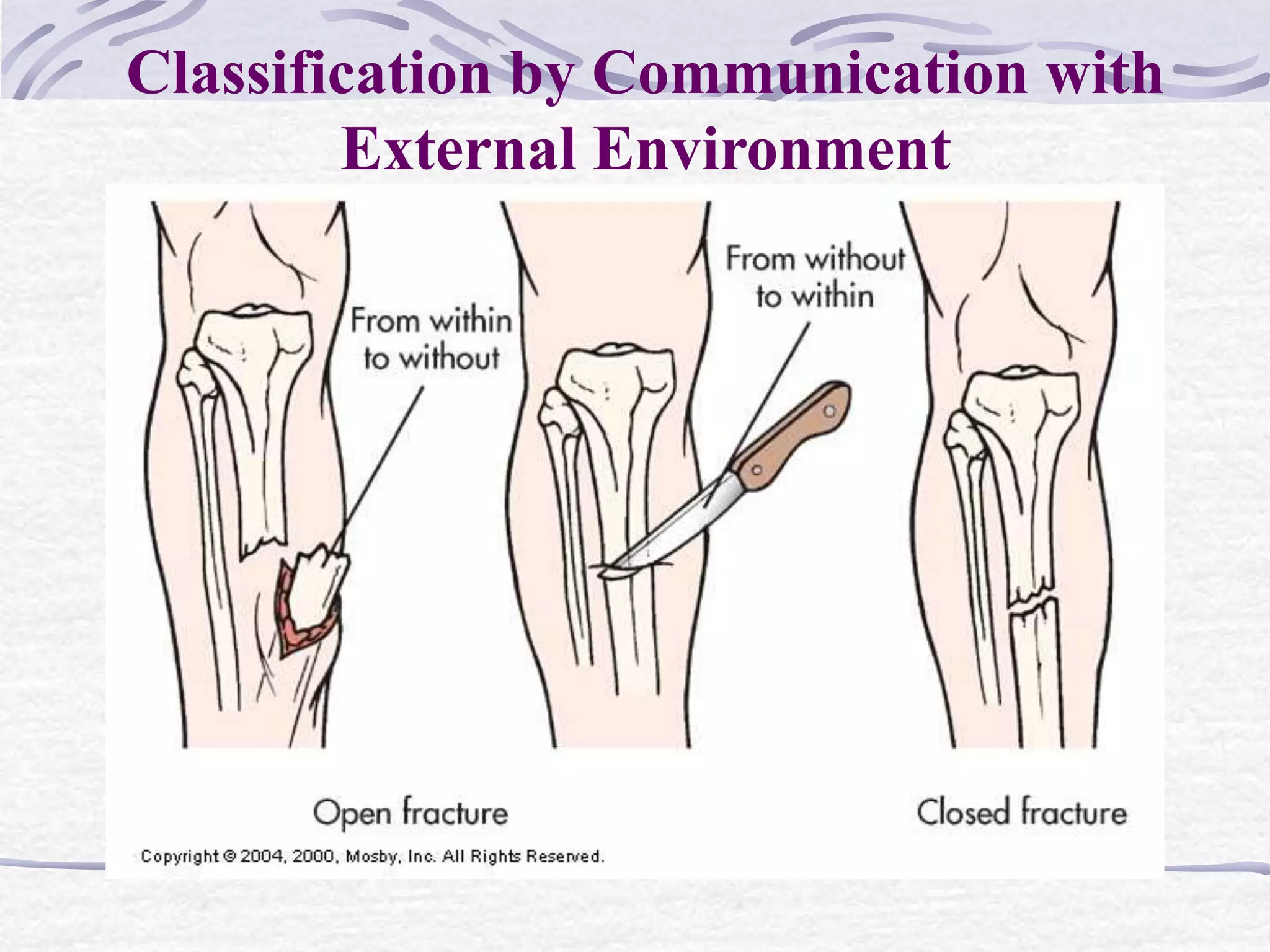
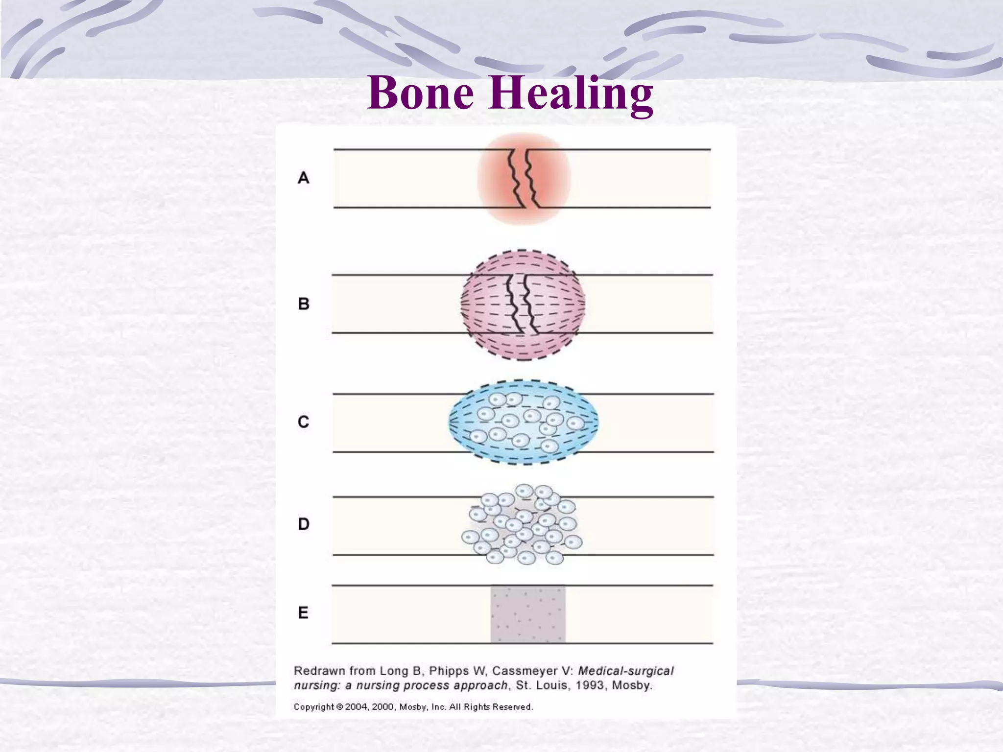
12. Description
Stable fractures
Occur when a piece of the periosteum is
intact across the fracture
External or internal fixation has rendered
the fragments stationary
15. Fracture Healing
Reparative process of self-healing (union)
occurs in the following stages:
1. Fracture hematoma (d/t bleeding, edema)
2. Granulation tissue → osteoid (3 – 14 days
post injury)
3. Callus formation (minerals deposited in osteoid)
16. Fracture Healing
Reparative process of self-healing (union)
occurs in the following stages:
4. Ossification (3 wks – 6 mos)
5. Consolidation (distance between fragments
decreases → closes).
6. Remodeling (union completed; remodels to
original shape, strength)
18. Collaborative Care
Overall goals of treatment:
Anatomic realignment of bone fragments
(reduction)
Immobilization to maintain alignment
(fixation)
Restoration of normal function
19. Collaborative Care
Fracture Reduction
Closed reduction
Nonsurgical, manual realignment
Open reduction
Correction of bone alignment through a
surgical incision
20. Collaborative Care
Fracture Reduction
Traction (with simultaneous counter-traction)
Application of pulling force to attain
realignment
Skin traction (short-term: 48-72 hrs)
Skeletal traction (longer periods)
28. Complications of Fractures
Infection
Collaborative Care
Open fractures require aggressive surgical
debridement
Post-op IV antibiotics for 3 to 7 days
(prophylactic)
29. Complications of Fractures
Compartment Syndrome
Condition in which elevated intracompartmental
pressure within a confined myofascial
compartment compromises the neurovascular
function of tissues within that space
Causes capillary perfusion to be reduced below a
level necessary for tissue viability
34. Complications of Fractures
Compartment Syndrome
Clinical Manifestations
Absence of peripheral pulse = ominous late
sign
Myoglobinuria
Dark reddish-brown urine
35. Complications of Fractures
Compartment Syndrome
Collaborative Care
Prompt, accurate diagnosis is critical
Early recognition is the key
Do not apply ice or elevate above heart level
36. Complications of Fractures
Compartment Syndrome
Collaborative Care
Remove/loosen the bandage and bivalve the
cast
Reduce traction weight
Surgical decompression (fasciotomy)
37. Complications of Fractures
Venous Thrombosis
Veins of the lower extremities and pelvis are
highly susceptible to thrombus formation after
fracture, especially hip fracture
38. Complications of Fractures
Venous Thrombosis
Precipitating factors:
Venous stasis caused by incorrectly applied
casts or traction
Local pressure on a vein
Immobility
Prevent with anticoagulant medications
39. Complications of Fractures
Fat Embolism Syndrome (FES)
Characterized by the presence of fat globules in
tissues and organs after a traumatic skeletal
injury
40. Complications of Fractures
Fat Embolism Syndrome (FES)
Fractures that most often cause FES:
Long bones
Ribs
Tibia
Pelvis
41. Complications of Fractures
Fat Embolism Syndrome (FES)
Clinical Manifestations
Usually occur 24-48 hours after injury
Interstitial pneumonitis
Produce symptoms of ARDS
42. Complications of Fractures
Fat Embolism Syndrome (FES)
Clinical Manifestations
Symptoms of ARDS:
Chest pain
Tachypnea
Cyanosis
PaO2
43. Complications of Fractures
Fat Embolism Syndrome (FES)
Clinical Manifestations
Symptoms of ARDS:
Dyspnea
Apprehension
Tachycardia
44. Complications of Fractures
Fat Embolism Syndrome (FES)
Clinical Manifestations
Rapid and acute course
Feeling of impending disaster
Patient may become comatose in a short time
45. Complications of Fractures
Fat Embolism Syndrome (FES)
Clinical Manifestations
Rapid and acute course
Feeling of impending disaster
Patient may become comatose in a short time