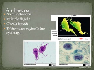
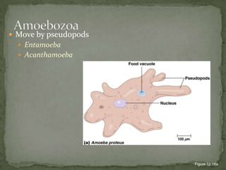
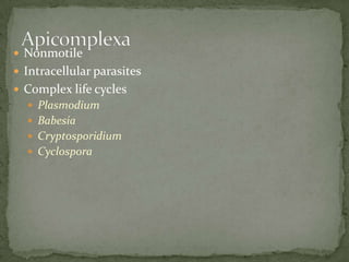
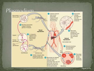
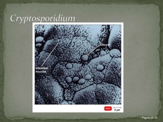
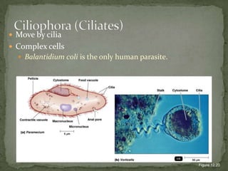
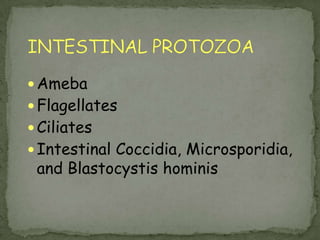
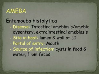
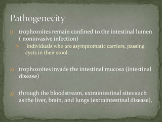
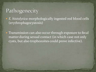
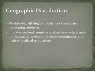
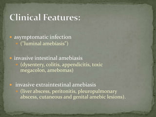
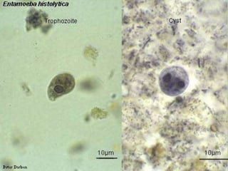
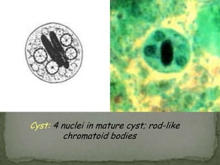
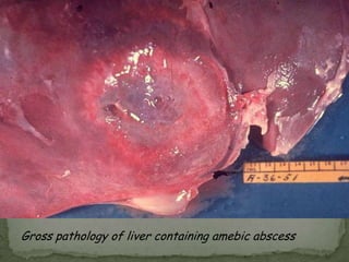
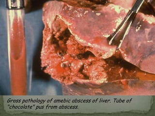
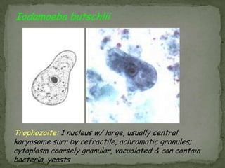
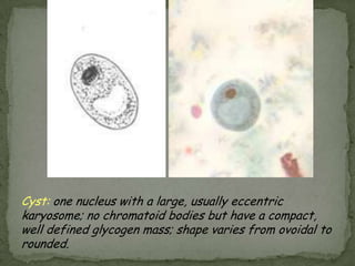
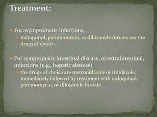
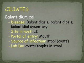
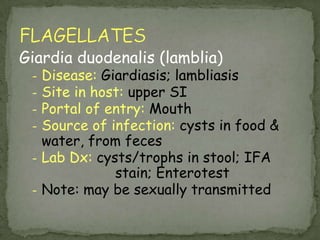
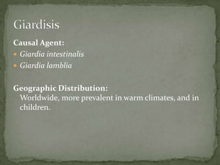
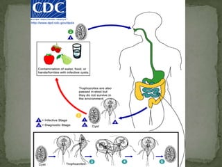
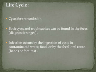
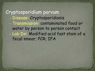
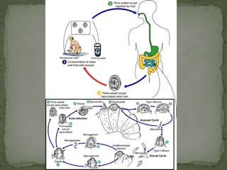
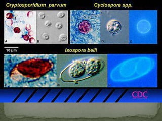
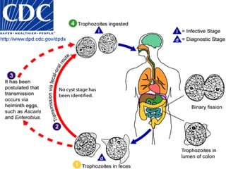
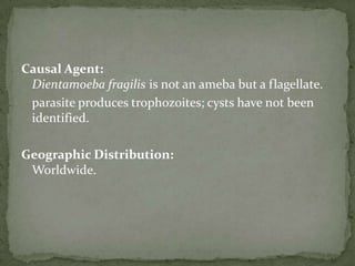
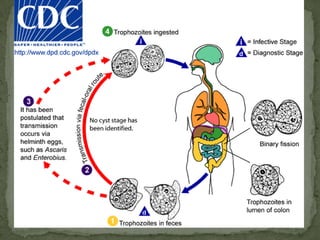
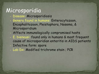
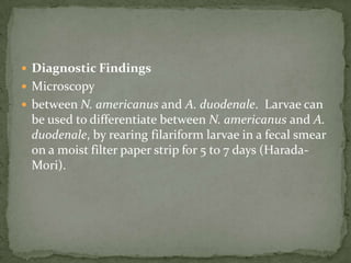
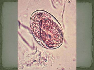
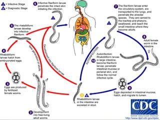
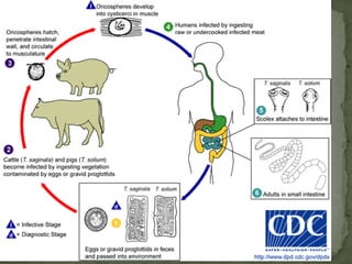
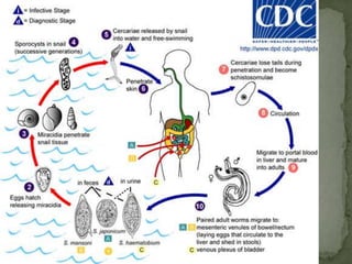
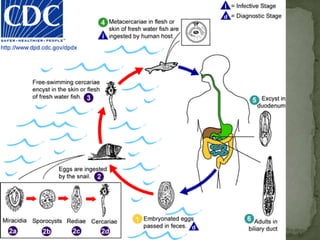
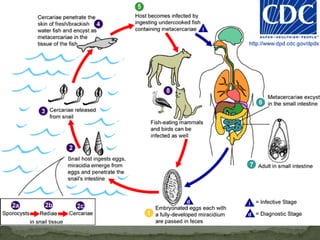
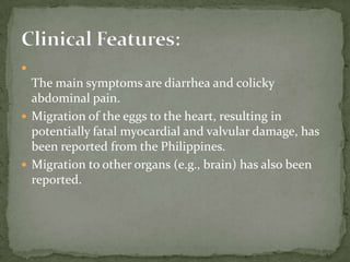
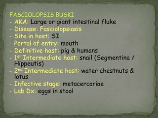
The Prokaryotes:Domains Bacteria and Archaea11
The ProtozoaTable 12.1
EukaryoticUnicellularChemoheterotrophsVegetative form is a trophozoite.Asexual reproduction is by fission,budding, or schizogony.Sexual reproduction by conjugation.Some produce cysts.ProtozoaFigure 12.16
No mitochondriaMultiple flagellaGiardia lambliaTrichomonas vaginalis (no cyst stage)ArchaezoaFigure 12.17b–d
No mitochondriaNonmotileIntracellular parasitesNosemaMicrospora
Move by pseudopodsEntamoebaAcanthamoebaAmoebozoaFigure 12.18a
NonmotileIntracellular parasitesComplex life cyclesPlasmodiumBabesiaCryptosporidiumCyclosporaApicomplexa
23876PlasmodiumFigure 12.19
CryptosporidiumFigure 25.19
Move by ciliaComplex cellsBalantidium coli is the only human parasite.Figure 12.20Ciliophora (Ciliates)
Move by flagellaPhotoautotrophsEuglenoidsChemoheterotrophsNaegleria: Flagellated and amoeboid forms; causes meningoencephalitis.Trypanosoma: Undulating membrane, transmitted by vectors.Leishmania: Flagellated form in sand fly vector, ovoid form in vertebrate host.Euglenozoa
EuglenozoaFigure 12.21
Why are these studied with algae and protozoa?DinoflagellatesFigure 12.14
INTESTINAL PROTOZOAAmebaFlagellatesCiliatesIntestinal Coccidia, Microsporidia, and Blastocystis hominis
EntamoebahistolyticaDisease:Intestinal amebiasis/amebic dysentery, extraintestinalamebiasis
Site in host:lumen & wall of LI
Portal of entry:Mouth
Source of infection:cysts in food & water, from fecesAMEBA
Causal Agent:Entamoebahistolyticapathogenic amebaassociated with intestinal and extraintestinal infections. Amoebiasis
Cysts and trophozoites are passed in feces .  ingestion of mature cysts in fecally contaminated food, water, or hands. Excystation occurs in the small intestine -  trophozoitesare released, which migrate to the large intestine.  The trophozoites multiply by binary fission - produce cysts , and both stages are passed in the feces .  Pathogenecity
Cysts and trophozoites are passed in feces .  ingestion of mature cysts in fecally contaminated food, water, or hands. Excystation occurs in the small intestine  and trophozoites are released, which migrate to the large intestine.  The trophozoites multiply by binary fission and produce cysts , and both stages are passed in the feces .  protection by their walls, the cysts can survive days to weeks in the external environmentPathogenecity
trophozoitesremain confined to the intestinal lumen ( noninvasive infection)individuals who are asymptomatic carriers, passing cysts in their stool.  trophozoitesinvade the intestinal mucosa (intestinal disease)through the bloodstream, extraintestinal sites such as the liver, brain, and lungs (extraintestinal disease), Pathogenecity
E. histolytica morphologically ingested red blood cells (erythrophagocystosis)  Transmission can also occur through exposure to fecal matter during sexual contact (in which case not only cysts, but also trophozoites could prove infective).Pathogenecity
Worldwide, with higher incidence of amebiasis in developing countries.  In industrialized countries, risk groups include male homosexuals, travelers and recent immigrants, and institutionalized populations.Geographic Distribution:
asymptomatic infection ("luminal amebiasis")invasive intestinal amebiasis(dysentery, colitis, appendicitis, toxic megacolon, amebomas) invasive extraintestinalamebiasis(liver abscess, peritonitis, pleuropulmonary abscess, cutaneous and genital amebic lesions).Clinical Features:
Fresh stool: wet mounts and permanently stained preparations (e.g., trichrome)Concentrates from fresh stool: wet mounts, with or without iodine stain, and permanently stained preparations E. histolyticatrophozoites can also be identified in aspirates or biopsy samples obtained during colonoscopy or surgery.Laboratory Diagnosis:
MicroscopyImmunodiagnosisMolecular methods for discriminating between E. histolytica and E. disparMorphologic comparison with other intestinal parasitesBench aid for E. histolyticaDiagnostic findings:
Trophozoite:small, usually central karyosome;			 finely granular chromatin
Trophozoite with ingested RBCs
Cyst:4 nuclei in mature cyst; rod-like			chromatoid bodies
Gross pathology of liver containing amebic abscess 
Gross pathology of amebic abscess of liver. Tube of "chocolate" pus from abscess.
Entamoeba hartmanniTrophozoite: small, usually eccentric karyosome;			finely granular chromatin
Cyst:4 nuclei in mature cyst; rod-like			chromatoid bodies (6-8 um, smaller		than E. histolytica cysts)
Entamoeba coliTrophozoite: 1 nucleus with large eccentric		 karyosome; coarse, irregular peripheral	 chromatin
Cyst:8 nuclei in mature cyst; splinter-like 		chromatoid bodies w/ pointed ends	large, eccentric karyosome
Endolimax nanaTrophozoites:1 nucleus w/ large, irregularly shaped, blot-like karyosome; has no peripheral chromatin; cytoplasm is granular and vacuolated
Cyst: mature cyst w/ 4 nuclei with large,		 blot-like karyosomes; no have			 chromatoid bodies
Iodamoeba butschliiTrophozoite: 1 nucleus w/ large, usually central karyosome surr by refractile, achromatic granules;  cytoplasm coarsely granular, vacuolated & can contain bacteria, yeasts  
Cyst: one nucleus with a large, usually eccentric karyosome; no chromatoid bodies but have a compact, well defined glycogen mass; shape varies from ovoidal to rounded.
For asymptomatic infections, iodoquinol, paromomycin, or diloxanidefuroate are the drugs of choice.  For symptomatic intestinal disease, or extraintestinal, infections (e.g., hepatic abscess) the drugs of choice are metronidazole or tinidazole, immediately followed by treatment with iodoquinol, paromomycin, or diloxanidefuroate. Treatment:
Balantidium coliDisease:Balantidiasis;balantidiosis; balantidial dysentery
Site in host:LI
Portal of entry:Mouth
Source of infection:stool (cysts)
Lab Dx: cysts/trophs in stoolCILIATES
Causal AgentBalantidium coli, a large ciliated protozoan parasite.Geographic Distribution:Worldwide.  Because pigs are an animal reservoir.  Other reservoirs include rodents and nonhuman primates.Balantidiasis
cysts – infective stageingestion of contaminated food or water Life cycle
excystation occurs in the small intestine- trophozoitescolonize the large intestine Trophozoites undergo encystation to produce infective cysts .  Some trophozoites invade the wall of the colon and multiply. Mature cysts are passed with feces .Life cycle
Most cases are asymptomatic.  Clinical manifestations, when present, include persistent diarrheaoccasionally dysenteryabdominal painweight loss.  Symptoms can be severe in debilitated persons.Clinical Features:
trophozoites - stool specimens or in tissueCysts are less frequently encountered.  Laboratory Diagnosis:
Trophozoites: large size (50-70 µm); rows of cilia on the cell surface; a cytostome; a bean shaped macronucleus and a smaller, less conspicuous micronucleus
Cyst: spherical to oval; cilia present in the young cyst but are absent in older forms; large, kidney-shaped macronucleus & contractile vacuoles in cytoplasm
The drug of choice is tetracycline*, with metronidazole* and  iodoquinol* as alternatives.  Tetracycline is contraindicated in pregnancy and in children less than 8 years old. Treatment:
May be pathogenic
Currently classified as an amoebaBlastocystis hominis
BlastocystishominisGeographic Distribution:Worldwide.Causal Agent:
thick-walled cyst present in the stools (fecal-oral route)cysts infect epithelial cells of the digestive tract and multiply asexually Vacuolar forms - give origin to multi vacuolar and ameboidforms multi-vacuolar -- pre-cyst -- thin-walled cyst (autoinfection)ameboid-- pre-cyst --thick-walled cystLife Cycle:
can cause both asymptomatic and symptomaticsymptoms of illness including watery diarrhea, abdominal pain, perianalpruritus, and excessive flatulence.Clinical Features:
Cyst-like forms appear round with large, central vacuole-like body. The nuclei in the peripheral cytoplasmic rim are clearly visible, staining purple.
metronidazoleor iodoquinolTreatment:
Giardiaduodenalis (lamblia)Disease:Giardiasis;lambliasis
Site in host:upper SI
Portal of entry:Mouth
Source of infection:cysts in food & water, from feces
Lab Dx: cysts/trophs in stool; IFA				stain; Enterotest
Note: may be sexually transmittedFLAGELLATES
Causal Agent:GiardiaintestinalisGiardialambliaGeographic Distribution:Worldwide, more prevalent in warm climates, and in children.Giardisis
Cysts for transmissionBoth cysts and trophozoites can be found in the feces (diagnostic stages) .  Infection occurs by the ingestion of cysts in contaminated water, food, or by the fecal-oral route (hands or fomites) . Life Cycle:
(small intestine) excystationTrophozoites multiply (lumen of the proximal small bowel )-- free or attached to the mucosa by a ventral sucking disk .  Encystation(colon).  cyst (nondiarrhealfeces )Life Cycle:
The spectrum varies from asymptomatic carriage to severe diarrhea and malabsorption  Acute giardiasis develops after an incubation period of 1 to 14 days (average of 7 days) and usually lasts 1 to 3 weeksSymptoms include diarrhea, abdominal pain, bloating, nausea, and vomiting.  In chronic giardiasis the symptoms are recurrent and malabsorption and debilitation may occur.Clinical Features:
Trophozoite: pyriform shape w/ 2 nuclei & a large, central karyosome; large ventral sucking disc, 4 pairs of flagella, 2 curved median bodies
Cysts: ellipsoid shape w/ 2 nuclei each (more mature ones will have four); lengthwise running central fibrils; short fibers laterally or obliquely across fibrils in lower half of cyst
metronidazoleand tinidazole.  Nitazoxanide(giardiasisin children)Treatment:
INTESTINAL COCCIDIA, MICROSPORIDIA, and BLASTOCYSTIS HOMINIS
Cryptosporidium parvumDisease: Cryptosporidiosis
Transmission: contaminated food or water by person to person contact
Lab Dx: Modified acid fast stain of a fecal smear; PCR; IFA
Causal Agent:Cryptosporidium parvum and Cryptosporidium hominisare (most prevalent species)Geographic Distribution:first reports of human cases in 1976, worldwide.  Waterborne outbreak in Milwaukee (Wisconsin) in 1993, that affected more than 400,000 people.Crytosporidiosis
Sporulatedoocysts, containing 4 sporozoites-- excreted by the infected host through feces and possibly other routes such as respiratory secretions .  Transmission occurs mainly through contact with contaminated water (e.g., drinking or recreational water). food sourcesoutbreaks U S -- waterparks, community swimming pools, and day care centers.  Zoonotic and anthroponotictransmissionLife cycle
Life cycle Following ingestion (and possibly inhalation) Excystation-- sporozoitesare released --parasitize gastrointestinal AND respiratory tract.  asexual multiplication (schizogony or merogony)  -- sexual multiplication (gametogony) -- producing microgamonts (male) and macrogamonts (female)
Life cycle Upon fertilization -- oocysts that sporulate in the infected hostTwo different types of oocysts are produced thick-walled, which is commonly excreted from the host thin-walled oocyst , which is primarily involved in autoinfection. 
asymptomatic infections  severe, life-threatening illnessincubation period is an average of 7 days (2 to 10 days).  Watery diarrhea is the most frequent symptom accompanied by dehydration, wt loss, abd. pain, fever, n/vimmunocompetent persons, symptoms are usually short lived (1 to 2 weeks-- can be chronic and more severe in immunocompromised patients, especially those with CD4 counts <200/µl.  Clinical Features:
asymptomatic infections severe, life-threatening illnessincubation period is an average of 7 days (but can range from 2 to 10 days).  Watery diarrhea is the most frequent symptom, and can be accompanied by dehydration, weight loss, abdominal pain, fever, nausea and vomiting. Clinical Features:
Clinical Features: In immunocompetent persons, symptoms are usually short lived (1 to 2 weeks); they can be chronic more severe in immunocompromised patients, especially those with CD4 counts <200/µl.  also found in other digestive tract, lungs, and conjunctiva.
Treatment:Rapid loss of fluids -- fluid and electrolyte replacement.  healthy, immunocompetent persons (self-limited)-- Nitazoxanide  Immunocompromisedand high risk pt.-- nitazoxanide is unclear.  For persons with AIDS, anti-retroviral therapyis encourage
Laboratory Diagnosis:Acid-fast staining methodsimmunofluorescencemicroscopy method of choice (followed closely by enzyme immunoassays)
Oocysts are rounded, 4.2 µm - 5.4 µm in diameter.  Sporozoites are visible inside the oocysts, indicating that sporulation has occurred. 
Oocysts stained by the modified acid-fast method: against a blue-green background, the oocysts stand out in a bright red stain.  Sporozoites are visible inside the two oocysts to the right.
Oocysts of C. parvum (upper left) and cysts of Giardia intestinalis (lower right) labeled with immunofluorescent antibodies.
CyclosporacayetanensisDisease:Cyclosporiasis
Transmission: contaminated water
Lab Dx: Modified acid fast stain of a fecal smearCyclospora oocysts from fresh stool fixed in 10% formalin and stained with modified acid-fast stain.  Compared to wet mount preparations, the oocysts are less perfectly round and have a wrinkled appearance.  Most importantly, the staining is variable among the four oocysts.
Sporulation of Cyclospora oocysts.  The sequence shows, as observed by DIC microscopy of wet mounts: an oocyst passed in fresh stool (Day 0); sporulated oocysts at days 5 (Day 5) and 10 (Day 10), which both contain 2 sporocysts; and a ruptured oocyst (Rupture), with a sporocyst still inside the oocyst and the other sporocyst just outside ­ the coiled sporozoites are barely visible inside the sporocysts. 
Causal Agent:unicellular coccidian parasite- CyclosporacayetanensisGeographic Distribution:most common in tropical and subtropical areas1990, foodborneoutbreaks of cyclosporiasis, 3600 persons, in the United States and Canada.
sporulation -- sporont -- two sporocysts (contains 2 sporozoites)  sporulatedoocysts are ingested (in contaminated food or water) oocystsexcyst in the gastrointestinal tract-- sporozoites invade the small intestine asexual multiplication and sexual development -- oocystsLife Cycle:
Clinical Features:incubation period of 1 week—severe watery diarrheas/sx- anorexia, wt loss, abd. pain, N/V,  myalgias, low-grade fever, and fatigue.  Untreated infections typically last for 10-12 weeks -- follow a relapsing course.  In disease-endemic settings -- asymptomatic.
identification of oocysts in stool specimensLaboratory Diagnosis:
combination of two antibiotics, trimethoprim-sulfamethoxazole*, also known as Bactrim, Septra, or Cotrim.  Supportive measures include management of fluid and electrolyte balance, and rest.   Treatment:
Isospora belliDisease:Isosporiasis; intestinal coccidiosis
Transmission: hand to mouth or through		 contaminated food or water
Lab Dx: wet mount of formalin-ethyl acetate conc. of fecal sample; modified acid fast stainOocysts: large (25 to 30 µm); typical ellipsoidal shape; when excreted, they are immature and contain one sporoblast; oocyst matures after excretion: the single sporoblast divides in two sporoblasts which develop cyst walls, becoming sporocysts, which eventually contain four sporozoites each
Causal Agent:coccidian parasite, Cystoisospora belli, is the least common of the three intestinal coccidiaGeographic Distribution:Worldwide, especially in tropical and subtropical areas.  Infection occurs in immunodepressedpt. and outbreaks in institutionalized groups in US
infection occurs by ingestion of sporocysts-containing oocystssporocystsexcyst in the small intestine --  release their sporozoites, which invade the epithelial cells -- initiate schizogony .  Life Cycle:
acute, nonbloody diarrhea with crampyabdpain (weeks)--malabsorption& wt loss. immuno-depressed patients , infants & children—severe diarrhea.  EosinophiliaClinical Features:
Microscopic wet mounts by bright-field, differential interference contrast (DIC), and epifluorescencemodified acid-fast stain.Laboratory Diagnosis:
Trimethoprim-sulfamethoxazole is the drug of choice. Treatment:
Dientamoebafragilis- NO cyst stage not an ameba, but a flagellate!
 may possess some pathogenicity
Site in host: LI
Portal of entry:mout
Source of infection: stool (trophs)
Causal Agent:Dientamoebafragilis is not an ameba but a flagellate.  parasite produces trophozoites; cysts have not been identified. Geographic Distribution:Worldwide.
the trophozoite is the only stage in stoolsTrophozoites have characteristically one or two nuclei Life Cycle:
children –intermittent diarrhea, abd pain, n/v, anorexia, fatigue, malaise, poor  wt gainClinical Features:
detection of trophozoites in permanently stained fecal smears (e.g., trichrome). Laboratory Diagnosis:
Nucleus: cluster of granules, with no peripheral chromatin; size range 5-15 µm.
  The drug of choice is iodoquinolParomomycin*, tetracycline*, (contraindicated in children under age 8, pregnant and lactating women) or metronidazole can also be used.  Treatment:
MicrosporidiaDisease:Microsporidiosis
Genera found in humans:Enterocytozoon, Encephalitozoon, Pleistophora, Nosema, & Microsporidium
Affects immunologically compromised hosts
E. bieneusi: found only in humans & most frequent cause of microsporidian enteritis in AIDS patients
Infective form: spore
Lab Dx: Modified trichrome stain ; PCR
Stool smear stained with Chromotrope 2R containing Enterocytozoon bieneusi spores. Black arrows indicate E. bieneusi spores with their belt-like stripe visible.  Red arrow indicates an unidentified yeast.  The yellow arrow indicates a vacuolated spore.
Stool smear stained with Quick-Hot Gram Chromotrope stain containing Enterocytozoon bieneusi spores. Black arrows indicate E. bieneusi spores with their belt-like stripe visible.  The red arrow indicates an unidentified yeast.  The yellow arrow indicates a vacuolated spore.
BLOOD and TISSUE PROTOZOA
OTHER PROTOZOABLOOD and TISSUE PROTOZOAPlasmodiumBabesiaTrypanosomabruceiTrypanosomacruziToxoplasmagondiiLeishmania
PROTOZOA FROM OTHER BODY SITESFree-living AmebaeNaegleriaAcanthamoebaTrichomonasvaginalis
PLASMODIUMDisease: MalariaP. vivax: Benign tertian malariaP. malariae: Quartan malariaP. falciparum: Malignant tertian malariaP. ovale: Ovale tertian malariaLab Dx: Giemsa stained thick and thin blood smears; IFA; PCR
Infected RBC:P. vivax and P. ovale: reticulocytesP. malariae: senescent erythrocytesP. falciparum: erythrocytes of all agesCyclic paroxysm of fever:P. vivax and P. ovale: every 48 hoursP. malariae: every 72 hoursP. falciparum: every 36-48 hours
P. falciparum: Blood Stage ParasitesThin Blood SmearsFig. 1: Normal red cell; Figs. 2-18: Trophozoites (among these, Figs. 2-10 correspond to ring-stage trophozoites); Figs. 19-26:Schizonts (Fig. 26 is a ruptured schizont); Figs. 27, 28: Mature macrogametocytes (female); Figs. 29, 30:  Mature microgametocytes (male).
Gametocytes of P. falciparum in thin blood smears.  Note the presence of a “Laveran’s bib”, which is not always visible.
P. falciparum rings have delicate cytoplasm and 1 or 2 small chromatin dots.  Red blood cells (RBCs) that are infected are not enlarged; multiple infection of RBCs more common in P. falciparum than in other species.  Occasional appliqué forms (rings appearing on the periphery of the RBC) can be present.
P. falciparum schizonts: seldom seen in peripheral blood.  Mature schizonts have 8 to 24 small merozoites; dark pigment, clumped in one mass.
P. malariae schizonts: have 6 to 12 merozoites with large nuclei, clustered around a mass of coarse, dark-brown pigment.  Merozoites can occasionally be arranged as a rosette pattern.
P. malariae trophozoites: have compact cytoplasm and a large chromatin dot.  Occasional band forms and/or "basket" forms with coarse, dark-brown pigment can be seen.
P. vivax gametocytes: round to oval with scattered brown pigment and may almost fill the red blood cell (RBC).  RBCs are enlarged 1 1/2 to 2 × and may be distorted.  Under optimal conditions, Schüffner's dots may appear more fine than those seen in P. ovale.
Rex Karl S. Teoxon, R.N, M.D133Vector: (night biting)anopheles mosquito minimus flavire
134SIGNS AND SYMPTOMSFever, chills, profuse sweating, convulsion, Anemia and fluid and electrolytes imbalance, hepatomegaly, splenomegalyDx: blood extraction (extract blood at the height of fever) thin and thick smear. Fluorescently labeled Ab
over 1 million deaths/year - mainly Africa
Incubation period - 8-30 days
influenza-like symptoms; paroxysms (fever, chills, rigors) every 36 to 48 hours, depending on species
sickle cell trait and P. falicparum136COMPLICATIONSP. ovale and vivax – relapse after 10 years of first exposureP. falciparum – Cerebral malaria – severe headache, drowsiness, seizure,  delirium, comaBlack water fever – jaundice, ARF, hemoglobinuria, black colored urine
137MANAGEMENTP. Vivax and P. Ovale – Primaquine (relapse) P. falciparum - Chloroquine For chloroquine resistant plasmodium – quinine* Prophylaxis – chloroquine or mefloquine, pyrimethamine/ sulfadoxine (fansidar)
Disease:BabesiosisLab Dx:Giemsa stained thick and thin blood smearsBABESIA
Babesia microti infection, Giemsa stained thin smear.  The organisms resemble P. falciparum; however Babesia parasites present several distinguishing features: they vary more in shape and in size; and they do not produce pigment. 
Infection with Babesia.  Giemsa stained thin smears showing the tetrad, a dividing form pathognomonic for Babesia.  Note also the variation in size and shape of the ring stage parasites and the absence of pigment. 
TRYPANOSOMA BRUCEIDisease: African trypanosomiasisT. b. gambiense: Gambian trypanosomiasis, West & Mid-African sleeping sicknessT. b. rhodesiense: Rhodesian trypanosomiasis, East African sleeping sicknessLab Dx:Giemsa stained thick and thin blood smears or lymph exudate (early stage); Giemsa stained smears of CSF (late stage)
Site in host: lymph glands, blood stream, brainPortal of entry: skinSource of infection: tsetse flyWinterbottom’s sign: enlargement of posterior cervical LNs
Trypomastigote: slender to fat and stumpy forms; in Giemsa stained films – C or U shaped forms NOT seen; small, oval kinetoplast located posterior to the nucleus; a centrally located nucleus, an undulating membrane, and an anterior flagellum. The trypanosomes length range is 14-33 µm
A dividing parasite is seen at the right. Dividing forms are seen in African trypanosomiasis, but not in American trypanosomiasis (Chagas' disease)
Tsetse fly. The vector of African trypanosomiasis
Winterbottoms sign
TRYPANOSOMA CRUZIDisease: American trypanosomiasis, Chaga’sdiseaseLab Dx:Giemsa stained thick and thin blood smears for the trypomastigote; histopath exam for the amastigoteSite in host: Tissues – heart; bloodPortal of entry: skinSource of infection: Kissing bug Triatomidae
Trypomastigote: shape is short & stubby to long & slender; in Giemsa stained blood films – C or U shaped; kinetoplast is large, oval & located posterior to the nucleus; anterior long free flagellum
Trypanosoma cruzi crithidia
Trypanosoma cruzi: Leishmanial form
Riduviid bug: the vector of American trypanosomiasis
Ramana's sign: unilateral conjunctivitis and orbital edema 
Chaga'sDiseaseasymptomatic- mostly
acute- rash, edema on face (site of bite), flu-like symptomschronic– rare but serious
g.i. tract nerve damage leading to megacolon
heart- conductive problems and cardiomyopathy, sudden death
"kissing bug" reduviid bug vector --(epimastigotereplicative form)Entry, Spread, Multiplicationbug defecates on wound releasing trypomastigotes that get rubbed into wound and vasculature
convert to amastigotes that invade and replicate in host cells
Diagnosis– clinical, serology, blood smear microscopyTreatment- not very good, especially for late complicationsPrevention– clear houses of bugs, use netting for sleeping
TOXOPLASMA GONDIIDisease: ToxoplasmosisSite in host: All organsPortal of entry:Ingestion of oocyst contaminated waterAerosolization of oocyst contaminated dust or litterConsumption of raw or undercooked cyst infected meatTransplacental passage of the tachyzoite
- Definitive host: domestic cats    - Intermediate host:  infected rodentsAccidental intermediate host: humansLab Dx: IFAT and ELISA; Giemsa-stained smears of exudates, aspirates or tissues
Toxoplasma gondii, parasiteAffects birds, mammals i.e. catsInfected person may carry the organism for life (reactivation is possible)161TOXOPLASMOSIS
162PATHOGENESISingestion of cyst from uncooked meat / fecal oral route from infected cats (feces)Quickly multiply in the GITDistributed to CNS, lymphatic tissue, skeletal muscle, myocardium, retina and placenta
T. gondiitachyzoites:crescentic to pyriform shaped with a prominent, centrally placed nucleus.
Toxoplasma gondii cyst in brain tissue stained with hematoxylin and eosin (100×).
166SIGNS AND SYMPTOMSMalaise, fever,  myalgia, headache, fatigue, sore throat, lymphadenopathy or asymptomaticFULMINANT = vomiting, cough and dyspnea, hyperpyrexia, delirium and seizures, encephalopathy, meningitisINFANTS = hydrocephalus or microcephalus, seizure, jaundice later strabismus, blindness, epilepsy, mental retardation
167DIAGNOSISSerology – high IgM or rising IgMCT scanMgmt:4-6 weeks of sulfadiazine +  pyrimethamine (take folic acid to counteract drug’s adverse effects)
LEISHMANIADisease:
L. tropica complex: Old Word Cutaneousleishmaniasis (oriental sore, Aleppo boil, Delhi ulcer, Baghdad boil)
L. mexicana complex: New Word Cutaneousleishmaniasis (chiclero ulcer, bay sore)
L. braziliensis complex: Mucocutaneusleishmaniasis (espundia, uta)
L. donovani: Visceral leishmaniasis (kala-azar or black disease, Dumdum fever)Lab Dx: Giemsa stained tissue sections or impression smears
Site in host: Monocytes/macrophages of skin & mucosa
Portal of entry: Skin
Source of infection: Phlebotomus or Lutzomiya fly
L. tropica amastigotes: ovoid in shape; large & eccentric nucleus; small, rodlike kinetoplast positioned opposite the nucleus; rodlike axoneme perpendicular to the kinetoplast
FREE LIVING AMEBAEDisease:
Naegleria:Primary Amebic Meningoencephalitis (PAM)
Acanthamoeba:Chronic Granulomatous Amebic Encephalitis and keratitis
Lab Dx: Direct microscopic exam (Wheatley’s trichromestain)PROTOZOA FROM OTHER BODY SITES
FREE LIVING AMEBAEPortal of Entry:
Naegleria:nose
Acanthamoeba:respiratory tract or ulcers in skin or mucosa / direct invasion of eye
Source of infection:
Naegleria: warm lakes, streams, ponds or inadequately chlorinated swimming pools
Acanthamoeba: immunocompromised or debilitated host
N. fowleri trophozoites cultured from cerebrospinal fluid: cells have characteristically large nuclei, with a large, dark staining karyosome.  The amebae are very active and extend and retract broad pseudopods. Trichrome stain. 
Acanthamoeba spp.:the cysts are spherical, 15-20 µm in diameter, having a thick double wall. The outer wall may be spherical or wrinkled, the inner wall appear stellate or polyhedral
Acanthamoeba spp.: cysts stained with Heidenhain’s iron alum-haematoxylin method.
TRICHOMONAS VAGINALISDisease: Trichomonadvaginitis
Site in host: vagina & prostate
Portal of entry: genitalia
Sources of infection:trophs in vaginal & prostatic secretions
NO cyst stage
Lab Dx:trophs in vaginal & prostatic fluids
Trophozoites of T. vaginalis: large, pyriform flagellate exhibiting rapid & jerky motility. The wavelike motion of the undulating membrane is often apparent
Trichomonas vaginalis:flagellates are 10-30 µm in lenght and 6-20 µm in breadth. Flagella, nucleus, axostyle and undullating membrane are visible. Filamentous form of Lactobacillus Döderleini is present. Giemsa-Romanowski stain.
Trichomonas vaginalisparasite182TRICHOMONIASIS
183SIGNS AND SYMPTOMSFemales: itching, burning on urination, yellow gray frothy malodorous vaginal discharge, foul smellingMales: usually asymptomaticDx: microscopic exam of vaginal discharge
184MANAGEMENTMetronidazole (Flagyl)include partnersCX: PROM
The HelminthsTable 12.1
EukaryoticMulticellular animalsChemoheterotrophicKingdom: AnimaliaPhylum: Platyhelminthes (flatworms)Class: Trematodes (flukes)Class: Cestodes (tapeworms)Phylum: Nematodes (roundworms)Helminths (Parasitic Worms)
TrematodesFigure 12.25
Humans as Definitive HostFigure 12.26
CestodesFigure 12.27
Humans as Intermediate Host Figure 12.28
Nematodes: Eggs Infective for HumansFigure 12.29
Nematodes: Larvae Infective for HumansFigure 25.26
Kingdom: AnimaliaPhylum: Arthropoda (exoskeleton, jointed legs)Class: Insecta (6 legs)Lice, fleas, mosquitoesClass: Arachnida (8 legs)Mites and ticksMay transmit diseases (vectors)Arthropods as VectorsFigures 12.31a, 12.32
HELMINTHS INTESTINAL NEMATODESEnterobius vermicularis
Ascaris lumbricoides
Necator americanus
Ancylostoma duodenale
Trichuris trichiura
Strongyloides stercoralisINTESTINAL CESTODES
Taenia saginata
Taenia solium
Hymenolepis diminuta
Diphyllobothrium latum
Dipylidium caninumINTESTINAL TREMATODES
Clonorchis sinensis
Fasciola hepatica
Paragonimus westermani
Fasciolopsis buski
Heterophyes heterophyes
Metagonimus yokogawai
Schistosoma mansoni
Schistosoma haematobium
Schistosoma japonicumTISSUE NEMATODES & CESTODES
Trichinella spiralis
Echinococcus granulosus
Echinococcus multilocularis
Spirometra speciesCausal Agents:The nematode (roundworm) Capillariaphilippinensis causes human intestinal capillariasisC. hepatica-- humans hepatic capillariasisC. aerophila-- humans pulmonary capillariasis.
Geographic Distribution:Capillariaphilippinensis is endemic in the Philippines and also occurs in Thailand. 
Life Cycle:unembryonated eggs are passed in the human stool and become embryonatedafter ingestion by freshwater fish-- larvae hatch & penetrate the intestine-- migrate to the tissues -Ingestion of raw or undercooked fishAdults worm -small intestinefemales deposit unembryonatedeggs (autoinfection)  -- hyperinfection(a massive number of adult worms) . 
Life Cycle:Capillaria hepatica adult worms reside in the liver of various animals, especially rats. Capillariaaerophila adult worms reside in the epithelium of the tracheo-bronchial tract of various animals. 
Clinical Features: Intestinal capillariasis-- pain and diarrhea autoinfection.  protein-losing enteropathy-- cachexiaand deathHepatic capillariasis (C. hepatica) -- acute or subacute hepatitis with eosinophilia-- dissemination --  fatalPulmonary capillariasis (C. aerophila) -- fever, cough, asthma, and pneumonia-- fatal.
Diagnostic findingsMicroscopyTreatment:The drug of choice is mebendazole*, and albendazole* is an alternative. 
ASCARIS LUMBRICOIDESDisease: Ascariasis; roundworm infection
Site in host: SI
Portal of entry: Mouth
Infective stage: ova containing second stage larva
Sources of infection: eggs from soil or vegetables
Lab Dx: eggs in stoolFertilized Ascaris egg (A) still at the unicellular stage. Unfertilized and fertilized eggs, (B and C, respectively).
Adult Ascaris worm: tapered ends; length 15 to 35 cm (the females tend to be the larger ones).  This worm is a female, as evidenced by the size and genital girdle (the dark circular groove at bottom area of image).
Causal Agent:Ascarislumbricoidesis the largest nematode (Adult females: 20 to 35 cm; adult male: 15 to 30 cm.)Geographic Distribution:most common human helminthic infection.  Worldwide distribution.  Highest prevalence in tropical and subtropical regions, and areas with inadequate sanitation. 
Life Cycle:Adult worms live in the lumen of the small intestine--produce 200,000 eggs/day  Fertile eggs embryonate-  infectiveeggs swallowed -- the larvae hatch &, invade the intestinal mucosa--  portal-- systemic --  lungs .  lungs -- alveolar walls-- bronchial tree – throat swallowed-- small intestine-- adult worms .  
Clinical Features:adult worms usually cause no acute symptoms.  High worm burdens –abd pain &  obstruction.  Migrating worms – occlusion of biltract or oral expulsion.  lung phase of larval migration, pulmonary symptoms can occur (cough, dyspnea, hemoptysis, eosinophilicpneumonitis - Loeffler’s syndrome).
Diagnostic findingsMicroscopyTreatment:The drugs of choice for treatment of ascariasis are albendazole* with mebendazole, ivermectin*, and nitazoxanide as alternatives.     In the United States, ascariasis is generally treated for 1-3 days with medication prescribed by a health care provider.  The drugs are effective and appear to have few side effects. 
Ascaris lumbricoidesIn GI tract, few symptoms in light infectionsNauseaVomitingObstruction of small bowel or common bile duct.Pulmonary: symptoms due to migrationAlveoli (verminous pneumonia)—cough, fever wheeze, dyspnea, X-ray changes, eosinophilia
Effects of Adult Ascaris WormsDepends on worm loadEffectsMechanical: obstruction, volvulus, intussusception, appendicitis, obstructive jaundice, liver abscesses, pancreatitis, asphyxiaToxic and MetabolicMalnutrition (complex)
Ascaris lumbricoidesDiagnosisCharacteristic eggs on direct smear examinationIf treating mixed infections, treat Ascaris firstMebendazolePyrantel Control: Periodic mass treatment of children, health education, environmental sanitation
Causal Agent:The nematode (roundworm) Trichuristrichiura, also called the human whipworm.Geographic Distribution:The third most common round worm of humans.  Worldwide, with infections more frequent in areas with tropical weather and poor sanitation practices, and among children.  It is estimated that 800 million people are infected worldwide.  Trichuriasis occurs in the southern United States.
Life Cycle:The unembryonated eggs are passed with the stool .  In the soil, the eggs develop into a 2-cell stage embryonate eggs After ingestion (soil-contaminated hands or food), the eggs hatch in the small intestine-larvae - adults in the cecum and ascending colon.  Female worms in the cecum shed between 3,000 and 20,000 eggs per day. 
Clinical Features:Most frequently asymptomatic.  Heavy infections, especially in small children, can cause gastrointestinal problems (abdominal pain, diarrhea, rectal prolapse) and possibly growth retardation.
Diagnostic findingsmicroscopyExamination of the rectal mucosa by proctoscopy (or directly in case of prolapses) can occasionally demonstrate adult worms.Treatment:Mebendazole is the drug of choice, with albendazole as an alternative. 
Case 138-yr-old schoolgirl visiting the U.S. from Malaysia1 week history of epigastric pain, flatulence, anorexia, bloody diarrheaNo eosinophilia notedClinical diagnosis of amoebic dysentery made However, microscopy of stool prep…
Diagnosis?
Trichuris trichiura (Whipworm)Common in Southeast U.S. Frequently coexists with ascarisEntirely intraluminal life cycle—eggs are ingestedFrequently asymptomaticSevere infections: diarrhea, abdominal pain and tenesmusRectal prolapse in childrenDS-eggs in stoolMebendazole 100 mg bid x 3 days
ENTEROBIUS VERMICULARISDisease: Enterobiasis; pinworm infection; seatworm infection; oxyuriasis
Site in host: LI, appendix
Portal of entry: Mouth
Infective stage: ova containing rhabditiform larva
Sources of infection: oral-fecal route; through contaminated fomites/food; inhalation ff by ingestion of airborne ova; retroinfection
Lab Dx: eggs in perianal region; Scotch tape swabEnterobius eggs: oval, asymmetric w/ one side noticeably flattened; smooth, thin-shelled, maycontain embryo 
Anterior end of Enterobius vermicularis adult worm.
Causal Agent:The nematode (roundworm) Enterobiusvermicularis (previously Oxyurisvermicularis) also called human pinworm.  (Adult females: 8 to 13 mm, adult male: 2 to 5 mm.)  Humans are considered to be the only hosts of E. vermicularis. Geographic Distribution:Worldwide, with infections more frequent in school- or preschool-children and in crowded conditions.  Enterobiasis appears to be more common in temperate than tropical countries.  The most common helminthic infection in the United States (an estimated 40 million persons infected).
Life Cycle:Eggs are deposited on perianal folds .  Self-infection occurs by transferring infective eggs to the mouth with hands that have scratched the perianal area .  Person-to-person transmission can also occur through handling of contaminated clothes or bed linens.  Enterobiasis may also be acquired through surfaces in the environment that are contaminated with pinworm eggs (e.g., curtains, carpeting).  Some small number of eggs may become airborne and inhaled.  .
Life Cycle:These would be swallowed and follow the same development as ingested eggs.  -larvae hatch in the small intestine -adults in the colon .Gravid females migrate nocturnally outside the anus and oviposit while crawling on the skin of the perianal area .  The larvae contained inside the eggs develop (the eggs become infective) in 4 to 6 hours under optimal conditions .  Retroinfection, or the migration of newly hatched larvae from the anal skin back into the rectum
Clinical FeaturesEnterobiasis is frequently asymptomatic.  The most typical symptom is perianalpruritus, especially at night, which may lead to excoriations and bacterial superinfection.  Occasionally, invasion of the female genital tract with vulvovaginitis and pelvic or peritoneal granulomas can occur.  Other symptoms include anorexia, irritability, and abdominal pain.
Diagnostic findingsMicroscopyTreatment:The drug of choice is pyrantelpamoate.  Measures to prevent reinfection, such as personal hygiene and laundering of bedding, should be discussed and implemented in cases where infection affects other household members. 
Case 1011-year-old femaleDoing poorly in schoolNot sleeping wellAnorecticComplains of itching in rectal region throughout the dayA Scotch-tape test reveals…
Diagnosis?
Enterobius (Pinworm)18 million infections in U.S.Incidence higher in whitesPreschool and elementary school most oftenMostly asymptomaticNocturnal anal pruritis cardinal feature due to migration and eggsMay have insomnia, possible emotional symptomsDS-eggs or adults on perineum {scotch tape}Mebendazole 100 mg.  Repeat in 2 weeks.  Pyrantel pamoate 11 mg/kg; repeat 2 weeks
NECATOR AMERICANUSDisease: New World Hookworm Disease
Site in host: SI, attached
Portal of entry: Skin
Infective stage: filariform larva
Sources of infection: infective filariform larvae in soil
Lab Dx: eggs in stoolHookworm egg: oval or ellipsoid; thin shell; usually  embryo at four cell stage
The embryo has begun cellular division and is at an early (gastrula) developmental stage.
Hookworm rhabditiform larva (wet preparation).
Hookworm filariform larva (wet preparation).
Anterior end of Necator americanus:oral opening of this species contains cutting "plates" . The muscular esophagus is labeled in this image (*).
ANCYLOSTOMA DUODENALEDisease: Old World Hookworm Disease
Site in host: SI, attached
Portal of entry: skin, usually feet
Infective stage: filariform larva
Sources of infection: infective filariform larvae in soil
Lab Dx: eggs in stoolOral opening of Ancylostoma duodenale:presence of four cutting "teeth," two on each side.
BAA: Adult worm of Ancylostoma duodenale.  Anterior end is depicted showing cutting teeth.B: Adult worm of Necator americanus.  Anterior end showing mouth parts with cutting plates.
Causal Agents:The human hookworms include two nematode (roundworm) species, Ancylostomaduodenale and Necatoramericanus.  A smaller group of hookworms infecting animals can invade and parasitize humans (A. ceylanicum) or can penetrate the human skin (causing cutaneous larva migrans), but do not develop any further (A. braziliense, A. caninum, Uncinariastenocephala).  Occasionally A. caninum larva may migrate to the human intestine causing eosinophilic enteritis; this may happen when larva is ingested rather than through skin invasion.
Geographic Distribution:The second most common human helminthic infection (after ascariasis).  Worldwide distribution, mostly in areas with moist, warm climate.  Both N. americanus and A. duodenale are found in Africa, Asia and the Americas.  Necatoramericanus predominates in the Americas and Australia, while only A. duodenale is found in the Middle East, North Africa and southern Europe.
Life Cycle:Eggs are passed in the stool-released rhabditiform larvae grow in the feces and/or the soil , and after 5 to 10 days (and two molts) they become filariform (third-stage) larvae that are infective .    On contact with the human host, the larvae penetrate the skin and are carried through the veins to the heart and then to the lungs.  They penetrate into the pulmonary alveoli, ascend the bronchial tree to the pharynx, and are swallowed The larvae reach the small intestine- adults.  Adult worms live in the lumen of the small intestine, where they attach to the intestinal wall with resultant blood loss by the host .  In addition, infection by A. duodenale may probably also occur by the oral and transmammary route.  N. americanus, however, requires a transpulmonary migration phase
Clinical Features:Iron deficiency anemia is the most common symptom of hookworm infection, and can be accompanied by cardiac complications.  Gastrointestinal and nutritional/metabolic symptoms can also occur.  local skin manifestations ("ground itch") can occur during penetration by the filariform (L3) larvae, and respiratory symptoms can be observed during pulmonary migration of the larvae.
Diagnostic FindingsMicroscopybetween N. americanus and A. duodenale.  Larvae can be used to differentiate between N. americanus and A. duodenale, by rearing filariform larvae in a fecal smear on a moist filter paper strip for 5 to 7 days (Harada-Mori). 
Treatment:In countries where hookworm is common and reinfection is likely, light infections are often not treated.  In the United States, hookworm infections are generally treated with albendazole*  Mebendazole* or pyrantelpamoate* can also be used.  Eosinophilic enteritis caused by A. caninum and for cutaneous larva migrans(creeping eruption) caused by canine and feline hookworms.
Case 1257 year old farmer from Dixie CountyPresents with profound SOBPhysical examination: anemic otherwise  unremarkableLaboratory examination reveals a profound anemia (hct 24) with aniso and poikilocytosisRemainder of laboratory examination normal.
Diagnosis?
Hookworm	Hookworm responsible for development of USPHSCaused by two different species (North American and Old World)Very similar to strongyloides in life cycleAttaches to duodenum, feeds on bloodElaborates anticoagulant, attaches and reattaches many timesLoss of around 0.1 ml/d of blood per worm
Case 1418-year-old trailer park handyman seen in ERWorked under trailers wearing shorts and no shirtDeveloped intensely pruritic skin rashUnable to sleepWBC 18,00065% eosinophils.
Case 15An 8 year old boyPresents with skin lesions and itching after spending the summer at a beach condo in St. Augustine with his family (mother, father, younger sister, dog and cat).Legs show several raised, reddened, serpiginous lesions that are intensely pruritic.
Diagnosis ?
Cutaneous Larva MigransCaused by filariform larvae of dog or cat hookworm (Ancylostoma braziliense or Ancylostoma duodenaleCommon in Southeast U.S.Red papule at entry with serpiginous tunnelIntense pruritisSelf limiting conditionDiagnosis clinicalTopical or oral thiabendazole 25 mg/kg bid for 3-5 daysMay use ethyl chloride topically
Cutaneous larva migrans (creeping eruption)More common in childrenLarvae penetrate skin and cause tingling followed by intense itching.Eggs shed from dog and cat bowels develop into infectious larvae outside the body in places protected from desiccation and extremes of temperatureShady, sandy areas under houses, at beach, etc.
Cutaneous larva migrans (creeping eruption)Usually not associated with systemic symptoms
Cutaneous larva migrans (creeping eruption)Diagnosis and treatmentSkin lesions are readily recognizedUsually diagnosed clinicallyGenerally do not require biopsyReveal eosinophilia inflammatory infiltrateMigrating parasite is generally not seenStool smear will reveal eggs
Visceral Larva Migrans Infection with dog or cat round wormsToxocara canis; Toxocara catisUnderdiagnosed based on seroprevalence surveysHeavy infections associated with fever, cough, nausea, vomiting, hepatomegaly, and eosinophiliaUncommon in adultsOcular type more common in adultsDiagnosis-ELISAThiabendazole: 25 mg/kg bid X 5 days
Case 17A 34 yr-old woman from Saudi ArabiaRadiation and cyclophosphamide, adriamycin, vincristine and prednisone for diffuse large B cell lymphoma of the neck.Mild eosinophilia (AEC=500) at the time of diagnosis4 months after initiation of chemo, c/o intermittent diffuse abdominal pain, bloating, constipation and occasional rectal bleeding.Absolute eosinophil count: 1000
Case 17No evidence of lymphoma found on re-stagingCompleted chemo, was deemed to be in complete remission, but had persistence of GI complaints.Upper endoscopy was unrevealing.Colonoscopy and biopsy revealed granulomatous inflammation, prominent eosinophilic infiltrate, surrounding a collection of eggs.
STRONGYLOIDES STERCORALISDisease: Strongyloidiasis, Cochin-China diarrhea or Vietnam diarrhea
Site in host: wall of SI
Portal of entry: skin
Infective stage: filariform larva
Sources of infection: larvae in soil; autoinfection
Lab Dx: larvae in stoolStrongyloides stercoralis first-stage larva: The rhabditoid esophagus is clearly visible in this larva; it consists of a club-shaped anterior portion, a postmedian constriction, and a posterior bulb.
Causal Agent:The nematode (roundworm) Strongyloidesstercoralis.  Other Strongyloides include S. fülleborni, which infects chimpanzees and baboons and may produce limited infections in humans..Geographic Distribution:Tropical and subtropical areas, but cases also occur in temperate areas (including the South of the United States).  More frequently found in rural areas, institutional settings, and lower socioeconomic groups
Life Cycle:The Strongyloides life cycle is more complex than that of most nematodes with its alternation between free-living and parasitic cycles, and its potential for autoinfection and multiplication within the host.  Two types of cycles exist:
Life Cycle:Free-living cycle: The rhabditiform larvae passed in the stool (see "Parasitic cycle" below) can either molt twice and become infective filariform larvae (direct development) or molt four times and become free living adult males and females that mate and produce eggs from which rhabditiform larvae hatch .  The latter in turn can either develop into a new generation of free-living adults (as represented in ), or into infective filariform larvae .  The filariform larvae penetrate the human host skin to initiate the parasitic cycle (see below) .
Life Cycle:Parasitic cycle:Filariform larvae in contaminated soil penetrate the human skin , and are transported to the lungs where they penetrate the alveolar spaces; they are carried through the bronchial tree to the pharynx, are swallowed and then reach the small intestine .  In the small intestine become adult female worms .  The females live threaded in the epithelium of the small intestine and by parthenogenesis produce eggs , which yield rhabditiform larvae.  The rhabditiform larvae can either be passed in the stool (see "Free-living cycle" above), or can cause autoinfection . 
Life Cycle:Parasitic cycle:   In autoinfection, the rhabditiform larvae become infective filariform larvae, which can penetrate either the intestinal mucosa (internal autoinfection) or the skin of the perianal area (external autoinfection);  To date, occurrence of autoinfection in humans with helminthic infections is recognized only in Strongyloidesstercoralis and Capillariaphilippinensis infections. 
Clinical FeaturesFrequently asymptomatic.  Gastrointestinal symptoms include abdominal pain and diarrhea.  Pulmonary symptoms (including Loeffler’s syndrome) can occur during pulmonary migration of the filariform larvae.  Dermatologic manifestations include urticarial rashes in the buttocks and waist areas.  Disseminated strongyloidiasis occurs in immunosuppressed patients, can present with abdominal pain, distension, shock, pulmonary and neurologic complications and septicemia, and is potentially fatal.  Blood eosinophilia is generally present during the acute and chronic stages, but may be absent with dissemination.
Diagnostic findingsMicroscopyTreatment:The drug of choice for the treatment of uncomplicated strongyloidiasis is ivermectin with albendazole* as the alternative.  All patients who are at risk of disseminated strongyloidiasis should be treated. 
On the day of admission…Fever, confusion, and not able to get out of bed---transported to the hospitalInitial blood work:Elevated WBCRaised eosinophil count 4 times normalUnderwent UGI endoscopyDuodenal biopsy obtained
Strongyloides: Crucial Aspects of Life CycleInfection acquired through penetration of intact skinInfection may persist for many years via autoinfectionIn immunocompromised patients, there is risk of dissemination or hyperinfectionHyperinfection syndrome
Disseminated StrongyloidiasisHigh mortality75%Penetration of gut wall by infective larvaeGut organisms carried on the surface of larvae results in polymicrobial sepsis, meningitisLarvae disseminate into all parts of body: CNS, lungs, bladder, peritoneum
Summary—Clinical FindingsDefective cell-meditated immunity: steroids, burns, lymphomas, AIDS (?)Gl symptoms in about two-thirds:Abdominal painBloatingDiarrheaConstipationWheezing, SOB, hemoptysis
Summary—Clinical FindingsSkin rash or pruritis in ~ one-thirdLarva currens (racing larva)Intensely pruriticLinear or serpiginous urticaria with flare that moves 5-15 cm/hrUsually buttocks, groin, and trunkIn dissemination, diffuse petechiae and purpura
Summary-Clinical FindingsEosinophilia 60-95%Less if on steroids
DIPHYLLOBOTHRIUM LATUMDisease: Diphyllobothriasis; fish tapeworm infection; broad tapeworm infection
Site in host: SI
Portal of entry: mouth
Definitive host: human, dogs, cats
1st Intermediate host: crustaceans (Cyclops or Diaptomus)
2nd Intermediate host: freshwater fish
Infective stage: plerocercoid larvae
Sources of infection: plerocercoid in freshwater fish
Lab Dx: eggs in stool
Eggs of D. latum:oval or ellipsoidal, with at one end an operculum that can be inconspicuous.  At the opposite (abopercular) end is a small knob that can be barely discernible. 
Eggs of Diphyllobothrium latum:are oval or ellipsoidal, with at one end an operculum (arrows) that can be inconspicuous.  The eggs are passed in the stool unembryonated. 
D. latum scolex and gravid proglottids
Proglottids of Diphyllobothrium latum.  These proglottids tend to be passed in strands of variable length in the stool.  The proglottids tend to be broader than long. 
Proglottids of D. latum:broader than it is long; size 2 to 4 mm long by 10 to 12 mm wide; uterus coiled in rosette appearance; genital pore at the center of the proglottid.
Causal Agents:The cestodeDiphyllobothriumlatum (the fish or broad tapeworm), the largest human tapeworm. Geographic Distribution:Diphyllobothriasis occurs in the Northern Hemisphere Freshwater fish infected with Diphyllobothrium sp. larva may be transported to and consumed in geographic areas where active transmission does not occur, resulting in human diphyllobothriasis. 
Life Cycle:Immature eggs are passed in feces -oncospheres -develop into a coracidia .  After ingestion by a suitable freshwater crustacean (the copepod first intermediate host) the coracidia develop into procercoid larvae .  second intermediate host, typically minnows and other small freshwater fish, the procercoid larvae are released from the crustacean and migrate into the fish flesh where they develop into a plerocercoid larvae (sparganum)  plerocercoid larvae are the infective stage for humans.  Because humans do not generally eat undercooked minnows and similar small freshwater fish, these do not represent an important source of infection.   
Life Cycle:  After ingestion of the infected fish, the plerocercoid develop into immature adults and then into mature adult tapeworms which will reside in the small intestine.  The adults of D. latum attach to the intestinal mucosa by means of the two bilateral groves (bothria) of their scolex .  The adults can reach more than 10 m in length, with more than 3,000 proglottids.  Immature eggs are discharged from the proglottids (up to 1,000,000 eggs per day per worm) and are passed in the feces . 
Clinical Features:Diphyllobothriasis can be a long-lasting infection (decades).  Most infections are asymptomatic.  Manifestations may include abdominal discomfort, diarrhea, vomiting, and weight loss.  Vitamin B12 deficiency with pernicious anemia may occur.  Massive infections may result in intestinal obstruction.  Migration of proglottids can cause cholecystitis or cholangitis.
Diagnostic findingsMicroscopyTreatment:Praziquantel* is the drug of choice.  Alternatively, Niclosamide can also be used to treat diphyllobothriasis.  
DIPYLIDIUM CANINUMDisease: Dipylidiasis; dog tapeworm infection
Site in host: SI
Portal of entry: mouth
Definitive host: dog & cat (or humans)
Intermediate host: larval flea
Infective stage: eggs
Sources of infection: flea & louse
Lab Dx: eggs in stool or egg sacks in stoolEgg of Dipylidium caninum:round to oval (average size 35 to 40 µm) and contain an oncosphere that has 6 hooklets. Ovum contains hexacanth wmbryo (8-15 ova are usually enclosed within sac-like membrane) 
Egg packets of Dipylidium caninum:Proglottids of Dipylidium caninum contain characteristic egg packets that are round to ovoid and contain 5 to 15 (sometimes more) eggs each. 
Proglottids of D. caninum: barrel-shaped proglottids (average mature size 12 mm × 3 mm) have two genital pores, one in the middle of each lateral margin.  Proglottids may be passed singly or in chains, and occasionally may be seen dangling from the anus. Proglottids are much longer than broad.
Adult tapeworm of Dipylidium caninum.  The scolex of the worm is very narrow and the proglottids, as they mature, get larger. 
Causal Agent:Dipylidiumcaninum(the double-pored dog tapeworm) mainly infects dogs and cats, but is occasionally found in humans.Geographic Distribution:Worldwide.  Human infections have been reported in Europe, the Philippines, China, Japan, Argentina, and the United States
Life Cycle:Gravid proglottids are passed intact in the feces or emerge from the perianal region of the host .  Subsequently they release typical egg packets . ingestion of an egg by the intermediate host (larval stages of the dog or cat flea Ctenocephalides spp.), an oncosphere is released into the flea's intestine.  The oncosphere penetrates the intestinal wall, invades the insect's hemocoel (body cavity), and develops into a cysticercoid larva .  The larva develops into an adult, and the adult flea harbours the infective cysticercoid .  The vertebrate host becomes infected by ingesting the adult flea containing the cysticercoid .  The dog is the principal definitive host for Dipylidiumcaninum.  Other potential hosts include cats, foxes, and humans (mostly children) , . 
Life Cycle:Humans acquire infection by ingesting the cysticercoid contaminated flea.  This can be promulgated by close contact between children and their infected pets.  In the small intestine of the vertebrate host the cysticercoid develops into the adult tapeworm (measuring up to 60 cm in length and 3 mm in width) reside in the small intestine of the host, where they each attach by their scolex.  They produce proglottids (or segments) which have two genital pores (hence the name "double-pored" tapeworm).  The proglottids mature, become gravid, detach from the tapeworm, and migrate to the anus or are passed in the stool .
Clinical Features:Most infections with Dipylidiumcaninum are asymptomatic.  Pets may exhibit behavior to relieve anal pruritis (such as scraping anal region across grass or carpeting).  Mild gastrointestinal disturbances may occur.  The most striking feature in animals and children consists of the passage of proglottids.  These can be found in the perianal region, in the feces, on diapers, and occasionally on floor covering and furniture.  The proglottids are motile when freshly passed and may be mistaken for maggots or fly larvae.
Diagnostic findingsMicroscopyTreatment:Treatment for both animals and humans is simple and very effective.  Praziquantel is given either orally or by injection (pets only).  The medication causes the tapeworm to dissolve within the intestines.  Since the worm is usually digested before it passes, it may not be visible in the dog's stool.  These drugs are generally well tolerated. 
HYMENOLEPIS NANADisease: Hymenolepiasis; dwarf tapeworm infection
Site in host: adults & cysts in SI
Portal of entry: mouth
Definitive host: human, mice & rats
Intermediate host: DO NOT require an IH
Infective stage: eggs
Sources of infection: eggs fr feces in soil; autoinfection
Lab Dx: eggs in stoolEgg of Hymenolepis nana: oval or subspherical and smaller than those of H. diminuta, their size being 40 - 60 µm x 30 - 50 µm.  On the inner membrane are two poles, from which 4-8 polar filaments spread out between the two membranes. The oncosphere has six hooks (seen as dark lines at 8 o'clock). 
Three adult Hymenolepis nana tapeworms. Each tapeworm (length: 15-40 mm) has a small, rounded scolex at the anterior end, and proglottids can be distinguished at the posterior, wider end.
HYMENOLEPIS DIMINUTADisease: Hymenolepiasis; rat tapeworm infection
Site in host: SI
Portal of entry: mouth
Definitive host: human, mice & rats
Intermediate host: insects (rat & mouse flea, the flour moth and flour beetle)
Infective stage: eggs
Sources of infection: cysts from insects
Lab Dx: eggs in stoolEgg of Hymenolepis diminuta:round or slightly oval, size 70 - 86 µm X 60 - 80 µm, with a striated outer membrane and a thin inner membrane.  The space between the membranes is smooth or faintly granular.  The oncosphere has six hooks.
Mature proglottids of Hymenolepis diminuta.
Causal Agents:Hymenolepiasis is caused by two cestodes (tapeworm) species, Hymenolepis nana (the dwarf tapeworm,) and Hymenolepisdimnuta (rat tapeworm).  Hymenolepisdiminuta is a cestode of rodents infrequently seen in humans and frequently found in rodents.Geographic Distribution:Hymenolepis nana is the most common cause of all cestode infections, and is encountered worldwide.  In temperate areas its incidence is higher in children and institutionalized groups.  Hymenolepisdiminuta, while less frequent, has been reported from various areas of the world.
Life Cycle:Eggs of Hymenolepis nana eggs are ingested by an arthropod intermediate host (various species of beetles and fleas may serve as intermediate hosts), they develop into cysticercoids, which can infect humans or rodents upon ingestion and develop into adults in the small intestine.  When eggs are ingested (in contaminated food or water or from hands contaminated with feces- oncospheres (hexacanth larvae) penetrate the intestinal villus and develop into cysticercoid larvae .  Upon rupture of the villus, the cysticercoids return to the intestinal lumen, evaginate their scoleces , attach to the intestinal mucosa and develop into adults that reside in the ileal portion of the small intestine producing gravid proglottids .  Eggs are passed in the stool when released from proglottids through its genital atrium or when proglottids disintegrate in the small intestine internal autoinfection, where the eggs release their hexacanth embryo, which penetrates the villus continuing the infective cycle without passage through the external environment . 
Life Cycle:.Eggs of Hymenolepisdiminuta are passed out in the feces of the infected definitive host (rodents, man) .  The mature eggs are ingested by an intermediate host (various arthropod adults or larvae) , and oncospheres are released from the eggs and penetrate the intestinal wall of the host , which develop into cysticercoid larvae.  Species from the genus Tribolium are common intermediate hosts for H. diminuta.  The cysticercoid larvae persist through the arthropod's morphogenesis to adulthood.  H. diminuta infection is acquired by the mammalian host after ingestion of an intermediate host carrying the cysticercoid larvae .  Humans can be accidentally infected through the ingestion of insects in precooked cereals, or other food items, and directly from the environment (e.g., oral exploration of the environment by children).  After ingestion, the tissue of the infected arthropod is digested releasing the cysticercoid larvae in the stomach and small intestine.  Eversion of the scoleces occurs shortly after the cysticercoid larvae are released.  Using the four suckers on the scolex, the parasite attaches to the small intestine wall.  Maturation of the parasites occurs within 20 days and the adult worms can reach an average of 30 cm in length .  Eggs are released in the small intestine from gravid proglottids that disintegrate after breaking off from the adult worms.  The eggs are expelled to the environment in the mammalian host's feces .
Clinical Features:Hymenolepis nana and H. diminuta infections are most often asymptomatic.  Heavy infections with H. nana can cause weakness, headaches, anorexia, abdominal pain, and diarrhea.
Treatment:Diagnostic findingsMicroscopyTreatment:Praziquantel* is the drug of choice. 
TAENIA SAGINATADisease: Taeniasis; beef tapeworm infection
Site in host: SI
Portal of entry: mouth
Definitive host: human
Intermediate host: grazing cattle
Infective stage: eggs
Sources of infection: cysts in beef
Lab Dx: segments and eggs in stool; Scotch tape swab
Taeniid eggs: rounded or subspherical, diameter 31 to 43 µm, with a thick radially striated brown shell.  Inside each shell is an embryonated oncosphere with 6 hooks (hexacanth embryo). 
Taenia egg.  Note the thick, "striated" shell and several of the larval hooks; approximate size = 40 µm. 
T. Saginata gravid proglottid:has 15 to 30 main uterine branches on each side of central stem; proglottids are much longer than wide
TAENIA SOLIUMDisease: Taeniasis; pork tapeworm infection
Site in host: SI
Portal of entry: mouth
Definitive host: human
Intermediate host: pig
Infective stage: eggs
Sources of infection: cysts in pork; autoinfection
Lab Dx: segments and eggs in stool; Scotch tape swabT. saginata gravid proglottid:has 15 to 30 main uterine branches on each side of central stem; proglottids are much longer than wide
Scoleces of Taenia saginata and Taenia solium: Scolex of T. saginata has 4 suckers and no hooks.  T. solium has 4 suckers in addition to a double row of hooks.
Scolex of Taenia solium:measures approximately 1 mm across.  The four suckers are numbered.  Note the presence of an armed (hooked) rostellum (*); the scolex of Taenia saginata, the beef tapeworm, does not have an armed rostellum.
A cysticercus of Taenia in muscle.  Note the fibrous capsule (*) around the cysticercus.
Causal Agents:The cestodes (tapeworms) Taeniasaginata (beef tapeworm) and T. solium (pork tapeworm).  Taeniasolium can also cause cysticercosis.Geographic Distribution:Both species are worldwide in distribution.  Taeniasolium is more prevalent in poorer communities where humans live in close contact with pigs and eat undercooked pork and is very rare in Muslim countries
Life Cycle:Taeniasis is the infection of humans with the adult tapeworm of Taeniasaginata or Taeniasolium.  Humans are the only definitive hosts for T. saginata and T. solium.  Eggs or gravid proglottids are passed with feces ; Cattle (T. saginata) and pigs (T. solium) become infected by ingesting vegetation contaminated with eggs or gravid proglottids .  Humans become infected by ingesting raw or undercooked infected meat .  In the human intestine, the cysticercus develops into an adult tapeworm  The adult tapeworms attach to the small intestine by their scolex and reside in the small intestine . 
Life Cycle:Length of adult worms is usually 5 m or less for T. saginata (however it may reach up to 25 m) and 2 to 7 m for T. solium.  The adults produce proglottids which mature, become gravid, detach from the tapeworm, and migrate to the anus or are passed in the stool (approximately 6 per day).  T. saginata adults usually have 1,000 to 2,000 proglottids, while T. solium adults have an average of 1,000 proglottids.  The eggs contained in the gravid proglottids are released after the proglottids are passed with the feces.  T. saginata may produce up to 100,000 and T. solium may produce 50,000 eggs per proglottid respectively.
Clinical Features:Taeniasaginatataeniasis produces only mild abdominal symptoms.  The most striking feature consists of the passage (active and passive) of proglottids.  Occasionally, appendicitis or cholangitis can result from migrating proglottids.  Taeniasoliumtaeniasis is less frequently symptomatic than Taeniasaginatataeniasis.  The main symptom is often the passage (passive) of proglottids.  The most important feature of Taeniasoliumtaeniasis is the risk of development of cysticercosis.
Diagnostic findingsTAKE EXTREME CARE IN PROCESSING THE SAMPLES!  INGESTION OF EGGS CAN RESULT IN CYSTICERCOSIS!Microscopy Antibody detection may prove useful especially in the early invasive stages, when the eggs and proglottids are not yet apparent in the stools.Treatment:Treatment is simple and very effective.  Praziquantel* is the drug of choice. 
Taenia saginataIngestion of raw or poorly cooked beefCows infected via the ingestion of human waste containing the eggs of the parasiteCows contain viable cysticercus larvae in the muscleHumans act as the host only to the adult tapewormsUp to 25 meters in the lumen of intestineFound all over the world, including the U.S.
Beef Tapeworm
TreatmentPraziquantelAlbendazoleNiclosamide
Tapeworms (Cestodes)Adult worms inhabit GI tract of definitive vertebrate hostLarvae inhabit tissues of intermediate hostHumansDefinitive for T. saginataIntermediate for Echinococcus granulosus (hydatid)Both definitive and intermediate for T. soliumAdult worms shed egg-containing segments in stool    ingested by intermediate host       larval form in tissues
CystercercosisSymptoms depend on location of cysts, but frequently include motor spasms, seizures, confusion, irritability, and personality changeIn the eye, often subretinal or in vitreous.  Movement may be seen by the patient.  Pain, amaurosis, and loss of vision may occur.
CysticercosisClinical manifestationsAdult worms rarely cause sxsLarvae penetrate intestine, enter blood, and eventually encyst in the brain.Cerebral ventircles  hydrocephalusSpinal cord  compression, paraplegiaSubarachnoid space  chronic meningitisCerebral cortex  seizuresCysts may remain asymptomatic for years, and become clinically apparent when larvae dieLarvae may encyst in other organs, but are rarely symptomatic
CysticercosisDiagnosisCT and MRI preferred studiesDiscrete cysts that may enhanceUsually multiple lesionsSingle lesions especially common in cases from IndiaOlder lesions may calcifyCSFLymphs or eos, low glucose, elevated proteinSerologyEspecially in cases with multiple cysts
CysticercosisTreatmentComplex and controversialPraziquantel and albendazole may kill cysts, but death of larvae can increase inflammation, edema and exacerbate sxsWhen possible, surgical resection of symptomatic cyst is preferredCorticosteroids vs. edema and inflammation; antiseizure meds
Causal Agents:Schistosomiasis is caused by digenetic blood trematodes.  The three main species infecting humans are Schistosomahaematobium, S. japonicum, and S. mansoni.  In addition, other species of schistosomes, which parasitize birds and mammals, can cause cercarial dermatitis in humans.Geographic Distribution: Schistosomamansoni is found in parts of South America and the Caribbean, Africa, and the Middle East; S. haematobium in Africa and the Middle East; and S. japonicum in the Far East. 
Life Cycle:Eggs are eliminated with feces or urine .- eggs hatch and release miracidia , which swim and penetrate specific snail intermediate hosts .  The stages in the snail include 2 generations of sporocysts and the production of cercariae .  Upon release from the snail, the infective cercariae swim, penetrate the skin of the human host , and shed their forked tail, becoming schistosomulae .  The schistosomulae migrate through several tissues and stages to their residence in the veins (, ).  Adult worms in humans reside in the mesenteric venules in various locations, which at times seem to be specific for each species . S. japonicum is more frequently found in the superior mesenteric veins draining the small intestine , andS. mansoni occurs more often in the superior mesenteric veins draining the large intestine .    S. haematobium most often occurs in the venous plexus of bladder , but it can also be found in the rectal venules. 
Life Cycle:.  Pathology of S. mansoni and S. japonicumschistosomiasis includes: Katayama fever, hepatic perisinusoidal egg granulomas, Symmers’ pipe stem periportal fibrosis, portal hypertension, and occasional embolic egg granulomas in brain or spinal cord.  Pathology of S. haematobiumschistosomiasis includes: hematuria, scarring, calcification, squamous cell carcinoma, and occasional embolic egg granulomas in brain or spinal cord.Human contact with water is thus necessary for infection by schistosomes.  Various animals, such as dogs, cats, rodents, pigs, hourse and goats, serve as reservoirs for S. japonicum, and dogs for S. mekongi.
Clinical FeaturesMany infections are asymptomatic.  Acute schistosomiasis (Katayama's fever) may occur weeks after the initial infection, especially by S. mansoni and S. japonicum.  Manifestations include fever, cough, abdominal pain, diarrhea, hepatospenomegaly, and eosinophilia.  Occasionally central nervous system lesions occur: cerebral granulomatous disease may be caused by ectopic S. japonicum eggs in the brain, and granulomatous lesions around ectopic eggs in the spinal cord from S. mansoni and S. haematobium infections may result in a transverse myelitis with flaccid paraplegia.  .
Clinical FeaturesContinuing infection may cause granulomatous reactions and fibrosis in the affected organs, which may result in manifestations that include: colonic polyposis with bloody diarrhea (Schistosomamansoni mostly); portal hypertension with hematemesis and splenomegaly (S. mansoni, S. japonicum, S. mansoni); cystitis and ureteritis (S. haematobium) with hematuria, which can progress to bladder cancer; pulmonary hypertension (S. mansoni, S. japonicum, more rarely S. haematobium); glomerulonephritis; and central nervous system lesions.
Diagnostic findingsmicroscopyAntobodydetrectioncan be useful in both in clinical management (e.g., recent infections) and for epidemiologic surveys.Treatment:Safe and effective drugs are available for the treatment of schistosomiasis.  The drug of choice is praziquantel for infections caused by all Schistosoma species.  Oxamniquine has been effective in treating infections caused by S. mansoni in some areas in which praziquantel is less effective. 
FASCIOLA HEPATICAAKA: Sheep Liver Fluke
Disease: Fascioliasis, “liver rot”
Site in host: Bile ducts
Portal of entry: mouth
Definitive host: sheep, cattle & other mammals, including humans
Intermediate host: snail (Lymnaea)
Source of infection: eating watercress, lettuce or radishes or drinking water infested with metacercariae
Infective stage: metacercariae
Lab Dx: eggs in stool Fasciola hepatica eggs: eggs are ellipsoidal, with small, barely distinct operculum. The operculum can be opened. The eggs have a thin shell which is slightly thicker at the abopercular end.  They are passed unembryonated. 
Causal Agents:The trematodesFasciola hepatica (the sheep liver fluke) and Fasciolagigantica, parasites of herbivores that can infect humans accidentally.Geographic Distribution:Fascioliasis occurs worldwide.  Human infections with F. hepatica are found in areas where sheep and cattle are raised, and where humans consume raw watercress, including Europe, the Middle East, and Asia.  Infections with F. gigantica have been reported, more rarely, in Asia, Africa, and Hawaii.

Paralect

  • 1.
  • 2.
  • 3.
    EukaryoticUnicellularChemoheterotrophsVegetative form isa trophozoite.Asexual reproduction is by fission,budding, or schizogony.Sexual reproduction by conjugation.Some produce cysts.ProtozoaFigure 12.16
  • 4.
    No mitochondriaMultiple flagellaGiardialambliaTrichomonas vaginalis (no cyst stage)ArchaezoaFigure 12.17b–d
  • 5.
  • 6.
  • 7.
    NonmotileIntracellular parasitesComplex lifecyclesPlasmodiumBabesiaCryptosporidiumCyclosporaApicomplexa
  • 8.
  • 9.
  • 10.
    Move by ciliaComplexcellsBalantidium coli is the only human parasite.Figure 12.20Ciliophora (Ciliates)
  • 11.
    Move by flagellaPhotoautotrophsEuglenoidsChemoheterotrophsNaegleria:Flagellated and amoeboid forms; causes meningoencephalitis.Trypanosoma: Undulating membrane, transmitted by vectors.Leishmania: Flagellated form in sand fly vector, ovoid form in vertebrate host.Euglenozoa
  • 12.
  • 13.
    Why are thesestudied with algae and protozoa?DinoflagellatesFigure 12.14
  • 15.
  • 16.
  • 17.
    Site in host:lumen& wall of LI
  • 18.
  • 19.
    Source of infection:cystsin food & water, from fecesAMEBA
  • 20.
    Causal Agent:Entamoebahistolyticapathogenic amebaassociatedwith intestinal and extraintestinal infections. Amoebiasis
  • 22.
    Cysts and trophozoitesare passed in feces .  ingestion of mature cysts in fecally contaminated food, water, or hands. Excystation occurs in the small intestine - trophozoitesare released, which migrate to the large intestine.  The trophozoites multiply by binary fission - produce cysts , and both stages are passed in the feces .  Pathogenecity
  • 23.
    Cysts and trophozoitesare passed in feces .  ingestion of mature cysts in fecally contaminated food, water, or hands. Excystation occurs in the small intestine and trophozoites are released, which migrate to the large intestine.  The trophozoites multiply by binary fission and produce cysts , and both stages are passed in the feces .  protection by their walls, the cysts can survive days to weeks in the external environmentPathogenecity
  • 24.
    trophozoitesremain confined tothe intestinal lumen ( noninvasive infection)individuals who are asymptomatic carriers, passing cysts in their stool.  trophozoitesinvade the intestinal mucosa (intestinal disease)through the bloodstream, extraintestinal sites such as the liver, brain, and lungs (extraintestinal disease), Pathogenecity
  • 25.
    E. histolytica morphologicallyingested red blood cells (erythrophagocystosis)  Transmission can also occur through exposure to fecal matter during sexual contact (in which case not only cysts, but also trophozoites could prove infective).Pathogenecity
  • 26.
    Worldwide, with higherincidence of amebiasis in developing countries.  In industrialized countries, risk groups include male homosexuals, travelers and recent immigrants, and institutionalized populations.Geographic Distribution:
  • 27.
    asymptomatic infection ("luminalamebiasis")invasive intestinal amebiasis(dysentery, colitis, appendicitis, toxic megacolon, amebomas) invasive extraintestinalamebiasis(liver abscess, peritonitis, pleuropulmonary abscess, cutaneous and genital amebic lesions).Clinical Features:
  • 28.
    Fresh stool: wetmounts and permanently stained preparations (e.g., trichrome)Concentrates from fresh stool: wet mounts, with or without iodine stain, and permanently stained preparations E. histolyticatrophozoites can also be identified in aspirates or biopsy samples obtained during colonoscopy or surgery.Laboratory Diagnosis:
  • 29.
    MicroscopyImmunodiagnosisMolecular methods fordiscriminating between E. histolytica and E. disparMorphologic comparison with other intestinal parasitesBench aid for E. histolyticaDiagnostic findings:
  • 30.
    Trophozoite:small, usually centralkaryosome; finely granular chromatin
  • 31.
  • 33.
    Cyst:4 nuclei inmature cyst; rod-like chromatoid bodies
  • 34.
    Gross pathology ofliver containing amebic abscess 
  • 35.
    Gross pathology ofamebic abscess of liver. Tube of "chocolate" pus from abscess.
  • 37.
    Entamoeba hartmanniTrophozoite: small,usually eccentric karyosome; finely granular chromatin
  • 38.
    Cyst:4 nuclei inmature cyst; rod-like chromatoid bodies (6-8 um, smaller than E. histolytica cysts)
  • 39.
    Entamoeba coliTrophozoite: 1nucleus with large eccentric karyosome; coarse, irregular peripheral chromatin
  • 40.
    Cyst:8 nuclei inmature cyst; splinter-like chromatoid bodies w/ pointed ends large, eccentric karyosome
  • 42.
    Endolimax nanaTrophozoites:1 nucleusw/ large, irregularly shaped, blot-like karyosome; has no peripheral chromatin; cytoplasm is granular and vacuolated
  • 43.
    Cyst: mature cystw/ 4 nuclei with large, blot-like karyosomes; no have chromatoid bodies
  • 44.
    Iodamoeba butschliiTrophozoite: 1nucleus w/ large, usually central karyosome surr by refractile, achromatic granules; cytoplasm coarsely granular, vacuolated & can contain bacteria, yeasts  
  • 45.
    Cyst: one nucleuswith a large, usually eccentric karyosome; no chromatoid bodies but have a compact, well defined glycogen mass; shape varies from ovoidal to rounded.
  • 46.
    For asymptomatic infections,iodoquinol, paromomycin, or diloxanidefuroate are the drugs of choice.  For symptomatic intestinal disease, or extraintestinal, infections (e.g., hepatic abscess) the drugs of choice are metronidazole or tinidazole, immediately followed by treatment with iodoquinol, paromomycin, or diloxanidefuroate. Treatment:
  • 47.
  • 48.
  • 49.
  • 50.
  • 51.
    Lab Dx: cysts/trophsin stoolCILIATES
  • 53.
    Causal AgentBalantidium coli,a large ciliated protozoan parasite.Geographic Distribution:Worldwide.  Because pigs are an animal reservoir.  Other reservoirs include rodents and nonhuman primates.Balantidiasis
  • 54.
    cysts – infectivestageingestion of contaminated food or water Life cycle
  • 55.
    excystation occurs inthe small intestine- trophozoitescolonize the large intestine Trophozoites undergo encystation to produce infective cysts .  Some trophozoites invade the wall of the colon and multiply. Mature cysts are passed with feces .Life cycle
  • 56.
    Most cases areasymptomatic.  Clinical manifestations, when present, include persistent diarrheaoccasionally dysenteryabdominal painweight loss.  Symptoms can be severe in debilitated persons.Clinical Features:
  • 57.
    trophozoites - stoolspecimens or in tissueCysts are less frequently encountered.  Laboratory Diagnosis:
  • 58.
    Trophozoites: large size(50-70 µm); rows of cilia on the cell surface; a cytostome; a bean shaped macronucleus and a smaller, less conspicuous micronucleus
  • 59.
    Cyst: spherical tooval; cilia present in the young cyst but are absent in older forms; large, kidney-shaped macronucleus & contractile vacuoles in cytoplasm
  • 60.
    The drug ofchoice is tetracycline*, with metronidazole* and  iodoquinol* as alternatives.  Tetracycline is contraindicated in pregnancy and in children less than 8 years old. Treatment:
  • 61.
  • 62.
    Currently classified asan amoebaBlastocystis hominis
  • 63.
  • 64.
    thick-walled cyst presentin the stools (fecal-oral route)cysts infect epithelial cells of the digestive tract and multiply asexually Vacuolar forms - give origin to multi vacuolar and ameboidforms multi-vacuolar -- pre-cyst -- thin-walled cyst (autoinfection)ameboid-- pre-cyst --thick-walled cystLife Cycle:
  • 66.
    can cause bothasymptomatic and symptomaticsymptoms of illness including watery diarrhea, abdominal pain, perianalpruritus, and excessive flatulence.Clinical Features:
  • 67.
    Cyst-like forms appearround with large, central vacuole-like body. The nuclei in the peripheral cytoplasmic rim are clearly visible, staining purple.
  • 69.
  • 70.
  • 71.
  • 72.
  • 73.
    Source of infection:cystsin food & water, from feces
  • 74.
    Lab Dx: cysts/trophsin stool; IFA stain; Enterotest
  • 75.
    Note: may besexually transmittedFLAGELLATES
  • 76.
    Causal Agent:GiardiaintestinalisGiardialambliaGeographic Distribution:Worldwide,more prevalent in warm climates, and in children.Giardisis
  • 78.
    Cysts for transmissionBothcysts and trophozoites can be found in the feces (diagnostic stages) .  Infection occurs by the ingestion of cysts in contaminated water, food, or by the fecal-oral route (hands or fomites) . Life Cycle:
  • 79.
    (small intestine) excystationTrophozoitesmultiply (lumen of the proximal small bowel )-- free or attached to the mucosa by a ventral sucking disk .  Encystation(colon).  cyst (nondiarrhealfeces )Life Cycle:
  • 80.
    The spectrum variesfrom asymptomatic carriage to severe diarrhea and malabsorption  Acute giardiasis develops after an incubation period of 1 to 14 days (average of 7 days) and usually lasts 1 to 3 weeksSymptoms include diarrhea, abdominal pain, bloating, nausea, and vomiting.  In chronic giardiasis the symptoms are recurrent and malabsorption and debilitation may occur.Clinical Features:
  • 81.
    Trophozoite: pyriform shapew/ 2 nuclei & a large, central karyosome; large ventral sucking disc, 4 pairs of flagella, 2 curved median bodies
  • 82.
    Cysts: ellipsoid shapew/ 2 nuclei each (more mature ones will have four); lengthwise running central fibrils; short fibers laterally or obliquely across fibrils in lower half of cyst
  • 83.
  • 84.
    INTESTINAL COCCIDIA, MICROSPORIDIA,and BLASTOCYSTIS HOMINIS
  • 85.
  • 86.
    Transmission: contaminated foodor water by person to person contact
  • 87.
    Lab Dx: Modifiedacid fast stain of a fecal smear; PCR; IFA
  • 88.
    Causal Agent:Cryptosporidium parvumand Cryptosporidium hominisare (most prevalent species)Geographic Distribution:first reports of human cases in 1976, worldwide.  Waterborne outbreak in Milwaukee (Wisconsin) in 1993, that affected more than 400,000 people.Crytosporidiosis
  • 90.
    Sporulatedoocysts, containing 4sporozoites-- excreted by the infected host through feces and possibly other routes such as respiratory secretions .  Transmission occurs mainly through contact with contaminated water (e.g., drinking or recreational water). food sourcesoutbreaks U S -- waterparks, community swimming pools, and day care centers.  Zoonotic and anthroponotictransmissionLife cycle
  • 91.
    Life cycle Followingingestion (and possibly inhalation) Excystation-- sporozoitesare released --parasitize gastrointestinal AND respiratory tract.  asexual multiplication (schizogony or merogony) -- sexual multiplication (gametogony) -- producing microgamonts (male) and macrogamonts (female)
  • 92.
    Life cycle Uponfertilization -- oocysts that sporulate in the infected hostTwo different types of oocysts are produced thick-walled, which is commonly excreted from the host thin-walled oocyst , which is primarily involved in autoinfection. 
  • 93.
    asymptomatic infections severe, life-threatening illnessincubation period is an average of 7 days (2 to 10 days).  Watery diarrhea is the most frequent symptom accompanied by dehydration, wt loss, abd. pain, fever, n/vimmunocompetent persons, symptoms are usually short lived (1 to 2 weeks-- can be chronic and more severe in immunocompromised patients, especially those with CD4 counts <200/µl.  Clinical Features:
  • 94.
    asymptomatic infections severe,life-threatening illnessincubation period is an average of 7 days (but can range from 2 to 10 days).  Watery diarrhea is the most frequent symptom, and can be accompanied by dehydration, weight loss, abdominal pain, fever, nausea and vomiting. Clinical Features:
  • 95.
    Clinical Features: Inimmunocompetent persons, symptoms are usually short lived (1 to 2 weeks); they can be chronic more severe in immunocompromised patients, especially those with CD4 counts <200/µl.  also found in other digestive tract, lungs, and conjunctiva.
  • 96.
    Treatment:Rapid loss offluids -- fluid and electrolyte replacement.  healthy, immunocompetent persons (self-limited)-- Nitazoxanide  Immunocompromisedand high risk pt.-- nitazoxanide is unclear.  For persons with AIDS, anti-retroviral therapyis encourage
  • 97.
    Laboratory Diagnosis:Acid-fast stainingmethodsimmunofluorescencemicroscopy method of choice (followed closely by enzyme immunoassays)
  • 98.
    Oocysts are rounded,4.2 µm - 5.4 µm in diameter.  Sporozoites are visible inside the oocysts, indicating that sporulation has occurred. 
  • 99.
    Oocysts stained bythe modified acid-fast method: against a blue-green background, the oocysts stand out in a bright red stain.  Sporozoites are visible inside the two oocysts to the right.
  • 100.
    Oocysts of C.parvum (upper left) and cysts of Giardia intestinalis (lower right) labeled with immunofluorescent antibodies.
  • 101.
  • 102.
  • 103.
    Lab Dx: Modifiedacid fast stain of a fecal smearCyclospora oocysts from fresh stool fixed in 10% formalin and stained with modified acid-fast stain.  Compared to wet mount preparations, the oocysts are less perfectly round and have a wrinkled appearance.  Most importantly, the staining is variable among the four oocysts.
  • 104.
    Sporulation of Cyclosporaoocysts.  The sequence shows, as observed by DIC microscopy of wet mounts: an oocyst passed in fresh stool (Day 0); sporulated oocysts at days 5 (Day 5) and 10 (Day 10), which both contain 2 sporocysts; and a ruptured oocyst (Rupture), with a sporocyst still inside the oocyst and the other sporocyst just outside ­ the coiled sporozoites are barely visible inside the sporocysts. 
  • 106.
    Causal Agent:unicellular coccidianparasite- CyclosporacayetanensisGeographic Distribution:most common in tropical and subtropical areas1990, foodborneoutbreaks of cyclosporiasis, 3600 persons, in the United States and Canada.
  • 108.
    sporulation -- sporont-- two sporocysts (contains 2 sporozoites)  sporulatedoocysts are ingested (in contaminated food or water) oocystsexcyst in the gastrointestinal tract-- sporozoites invade the small intestine asexual multiplication and sexual development -- oocystsLife Cycle:
  • 109.
    Clinical Features:incubation periodof 1 week—severe watery diarrheas/sx- anorexia, wt loss, abd. pain, N/V, myalgias, low-grade fever, and fatigue.  Untreated infections typically last for 10-12 weeks -- follow a relapsing course.  In disease-endemic settings -- asymptomatic.
  • 110.
    identification of oocystsin stool specimensLaboratory Diagnosis:
  • 111.
    combination of twoantibiotics, trimethoprim-sulfamethoxazole*, also known as Bactrim, Septra, or Cotrim.  Supportive measures include management of fluid and electrolyte balance, and rest.   Treatment:
  • 112.
  • 113.
    Transmission: hand tomouth or through contaminated food or water
  • 114.
    Lab Dx: wetmount of formalin-ethyl acetate conc. of fecal sample; modified acid fast stainOocysts: large (25 to 30 µm); typical ellipsoidal shape; when excreted, they are immature and contain one sporoblast; oocyst matures after excretion: the single sporoblast divides in two sporoblasts which develop cyst walls, becoming sporocysts, which eventually contain four sporozoites each
  • 115.
    Causal Agent:coccidian parasite,Cystoisospora belli, is the least common of the three intestinal coccidiaGeographic Distribution:Worldwide, especially in tropical and subtropical areas.  Infection occurs in immunodepressedpt. and outbreaks in institutionalized groups in US
  • 117.
    infection occurs byingestion of sporocysts-containing oocystssporocystsexcyst in the small intestine -- release their sporozoites, which invade the epithelial cells -- initiate schizogony .  Life Cycle:
  • 118.
    acute, nonbloody diarrheawith crampyabdpain (weeks)--malabsorption& wt loss. immuno-depressed patients , infants & children—severe diarrhea.  EosinophiliaClinical Features:
  • 119.
    Microscopic wet mountsby bright-field, differential interference contrast (DIC), and epifluorescencemodified acid-fast stain.Laboratory Diagnosis:
  • 120.
    Trimethoprim-sulfamethoxazole is thedrug of choice. Treatment:
  • 121.
    Dientamoebafragilis- NO cyststage not an ameba, but a flagellate!
  • 122.
    may possesssome pathogenicity
  • 123.
  • 124.
  • 125.
    Source of infection:stool (trophs)
  • 126.
    Causal Agent:Dientamoebafragilis isnot an ameba but a flagellate.  parasite produces trophozoites; cysts have not been identified. Geographic Distribution:Worldwide.
  • 127.
    the trophozoite isthe only stage in stoolsTrophozoites have characteristically one or two nuclei Life Cycle:
  • 129.
    children –intermittent diarrhea,abd pain, n/v, anorexia, fatigue, malaise, poor wt gainClinical Features:
  • 130.
    detection of trophozoitesin permanently stained fecal smears (e.g., trichrome). Laboratory Diagnosis:
  • 131.
    Nucleus: cluster ofgranules, with no peripheral chromatin; size range 5-15 µm.
  • 133.
      The drugof choice is iodoquinolParomomycin*, tetracycline*, (contraindicated in children under age 8, pregnant and lactating women) or metronidazole can also be used.  Treatment:
  • 135.
  • 136.
    Genera found inhumans:Enterocytozoon, Encephalitozoon, Pleistophora, Nosema, & Microsporidium
  • 137.
  • 138.
    E. bieneusi: foundonly in humans & most frequent cause of microsporidian enteritis in AIDS patients
  • 139.
  • 140.
    Lab Dx: Modifiedtrichrome stain ; PCR
  • 141.
    Stool smear stainedwith Chromotrope 2R containing Enterocytozoon bieneusi spores. Black arrows indicate E. bieneusi spores with their belt-like stripe visible.  Red arrow indicates an unidentified yeast.  The yellow arrow indicates a vacuolated spore.
  • 142.
    Stool smear stainedwith Quick-Hot Gram Chromotrope stain containing Enterocytozoon bieneusi spores. Black arrows indicate E. bieneusi spores with their belt-like stripe visible.  The red arrow indicates an unidentified yeast.  The yellow arrow indicates a vacuolated spore.
  • 143.
  • 144.
    OTHER PROTOZOABLOOD andTISSUE PROTOZOAPlasmodiumBabesiaTrypanosomabruceiTrypanosomacruziToxoplasmagondiiLeishmania
  • 145.
    PROTOZOA FROM OTHERBODY SITESFree-living AmebaeNaegleriaAcanthamoebaTrichomonasvaginalis
  • 146.
    PLASMODIUMDisease: MalariaP. vivax:Benign tertian malariaP. malariae: Quartan malariaP. falciparum: Malignant tertian malariaP. ovale: Ovale tertian malariaLab Dx: Giemsa stained thick and thin blood smears; IFA; PCR
  • 147.
    Infected RBC:P. vivaxand P. ovale: reticulocytesP. malariae: senescent erythrocytesP. falciparum: erythrocytes of all agesCyclic paroxysm of fever:P. vivax and P. ovale: every 48 hoursP. malariae: every 72 hoursP. falciparum: every 36-48 hours
  • 149.
    P. falciparum: Blood StageParasitesThin Blood SmearsFig. 1: Normal red cell; Figs. 2-18: Trophozoites (among these, Figs. 2-10 correspond to ring-stage trophozoites); Figs. 19-26:Schizonts (Fig. 26 is a ruptured schizont); Figs. 27, 28: Mature macrogametocytes (female); Figs. 29, 30:  Mature microgametocytes (male).
  • 150.
    Gametocytes of P.falciparum in thin blood smears.  Note the presence of a “Laveran’s bib”, which is not always visible.
  • 151.
    P. falciparum ringshave delicate cytoplasm and 1 or 2 small chromatin dots.  Red blood cells (RBCs) that are infected are not enlarged; multiple infection of RBCs more common in P. falciparum than in other species.  Occasional appliqué forms (rings appearing on the periphery of the RBC) can be present.
  • 152.
    P. falciparum schizonts: seldomseen in peripheral blood.  Mature schizonts have 8 to 24 small merozoites; dark pigment, clumped in one mass.
  • 153.
    P. malariae schizonts:have 6 to 12 merozoites with large nuclei, clustered around a mass of coarse, dark-brown pigment.  Merozoites can occasionally be arranged as a rosette pattern.
  • 154.
    P. malariae trophozoites:have compact cytoplasm and a large chromatin dot.  Occasional band forms and/or "basket" forms with coarse, dark-brown pigment can be seen.
  • 155.
    P. vivax gametocytes:round to oval with scattered brown pigment and may almost fill the red blood cell (RBC).  RBCs are enlarged 1 1/2 to 2 × and may be distorted.  Under optimal conditions, Schüffner's dots may appear more fine than those seen in P. ovale.
  • 156.
    Rex Karl S.Teoxon, R.N, M.D133Vector: (night biting)anopheles mosquito minimus flavire
  • 157.
    134SIGNS AND SYMPTOMSFever,chills, profuse sweating, convulsion, Anemia and fluid and electrolytes imbalance, hepatomegaly, splenomegalyDx: blood extraction (extract blood at the height of fever) thin and thick smear. Fluorescently labeled Ab
  • 158.
    over 1 milliondeaths/year - mainly Africa
  • 159.
  • 160.
    influenza-like symptoms; paroxysms(fever, chills, rigors) every 36 to 48 hours, depending on species
  • 161.
    sickle cell traitand P. falicparum136COMPLICATIONSP. ovale and vivax – relapse after 10 years of first exposureP. falciparum – Cerebral malaria – severe headache, drowsiness, seizure, delirium, comaBlack water fever – jaundice, ARF, hemoglobinuria, black colored urine
  • 162.
    137MANAGEMENTP. Vivax andP. Ovale – Primaquine (relapse) P. falciparum - Chloroquine For chloroquine resistant plasmodium – quinine* Prophylaxis – chloroquine or mefloquine, pyrimethamine/ sulfadoxine (fansidar)
  • 163.
    Disease:BabesiosisLab Dx:Giemsa stainedthick and thin blood smearsBABESIA
  • 165.
    Babesia microti infection,Giemsa stained thin smear.  The organisms resemble P. falciparum; however Babesia parasites present several distinguishing features: they vary more in shape and in size; and they do not produce pigment. 
  • 166.
    Infection with Babesia. Giemsa stained thin smears showing the tetrad, a dividing form pathognomonic for Babesia.  Note also the variation in size and shape of the ring stage parasites and the absence of pigment. 
  • 167.
    TRYPANOSOMA BRUCEIDisease: AfricantrypanosomiasisT. b. gambiense: Gambian trypanosomiasis, West & Mid-African sleeping sicknessT. b. rhodesiense: Rhodesian trypanosomiasis, East African sleeping sicknessLab Dx:Giemsa stained thick and thin blood smears or lymph exudate (early stage); Giemsa stained smears of CSF (late stage)
  • 168.
    Site in host:lymph glands, blood stream, brainPortal of entry: skinSource of infection: tsetse flyWinterbottom’s sign: enlargement of posterior cervical LNs
  • 170.
    Trypomastigote: slender tofat and stumpy forms; in Giemsa stained films – C or U shaped forms NOT seen; small, oval kinetoplast located posterior to the nucleus; a centrally located nucleus, an undulating membrane, and an anterior flagellum. The trypanosomes length range is 14-33 µm
  • 171.
    A dividing parasiteis seen at the right. Dividing forms are seen in African trypanosomiasis, but not in American trypanosomiasis (Chagas' disease)
  • 172.
    Tsetse fly. Thevector of African trypanosomiasis
  • 173.
  • 174.
    TRYPANOSOMA CRUZIDisease: Americantrypanosomiasis, Chaga’sdiseaseLab Dx:Giemsa stained thick and thin blood smears for the trypomastigote; histopath exam for the amastigoteSite in host: Tissues – heart; bloodPortal of entry: skinSource of infection: Kissing bug Triatomidae
  • 175.
    Trypomastigote: shape isshort & stubby to long & slender; in Giemsa stained blood films – C or U shaped; kinetoplast is large, oval & located posterior to the nucleus; anterior long free flagellum
  • 176.
  • 177.
  • 178.
    Riduviid bug: thevector of American trypanosomiasis
  • 179.
    Ramana's sign: unilateralconjunctivitis and orbital edema 
  • 180.
  • 181.
    acute- rash, edemaon face (site of bite), flu-like symptomschronic– rare but serious
  • 182.
    g.i. tract nervedamage leading to megacolon
  • 183.
    heart- conductive problemsand cardiomyopathy, sudden death
  • 184.
    "kissing bug" reduviidbug vector --(epimastigotereplicative form)Entry, Spread, Multiplicationbug defecates on wound releasing trypomastigotes that get rubbed into wound and vasculature
  • 185.
    convert to amastigotesthat invade and replicate in host cells
  • 186.
    Diagnosis– clinical, serology,blood smear microscopyTreatment- not very good, especially for late complicationsPrevention– clear houses of bugs, use netting for sleeping
  • 187.
    TOXOPLASMA GONDIIDisease: ToxoplasmosisSitein host: All organsPortal of entry:Ingestion of oocyst contaminated waterAerosolization of oocyst contaminated dust or litterConsumption of raw or undercooked cyst infected meatTransplacental passage of the tachyzoite
  • 188.
    - Definitive host:domestic cats - Intermediate host: infected rodentsAccidental intermediate host: humansLab Dx: IFAT and ELISA; Giemsa-stained smears of exudates, aspirates or tissues
  • 189.
    Toxoplasma gondii, parasiteAffectsbirds, mammals i.e. catsInfected person may carry the organism for life (reactivation is possible)161TOXOPLASMOSIS
  • 190.
    162PATHOGENESISingestion of cystfrom uncooked meat / fecal oral route from infected cats (feces)Quickly multiply in the GITDistributed to CNS, lymphatic tissue, skeletal muscle, myocardium, retina and placenta
  • 192.
    T. gondiitachyzoites:crescentic topyriform shaped with a prominent, centrally placed nucleus.
  • 193.
    Toxoplasma gondii cystin brain tissue stained with hematoxylin and eosin (100×).
  • 194.
    166SIGNS AND SYMPTOMSMalaise,fever, myalgia, headache, fatigue, sore throat, lymphadenopathy or asymptomaticFULMINANT = vomiting, cough and dyspnea, hyperpyrexia, delirium and seizures, encephalopathy, meningitisINFANTS = hydrocephalus or microcephalus, seizure, jaundice later strabismus, blindness, epilepsy, mental retardation
  • 195.
    167DIAGNOSISSerology – highIgM or rising IgMCT scanMgmt:4-6 weeks of sulfadiazine + pyrimethamine (take folic acid to counteract drug’s adverse effects)
  • 196.
  • 197.
    L. tropica complex:Old Word Cutaneousleishmaniasis (oriental sore, Aleppo boil, Delhi ulcer, Baghdad boil)
  • 198.
    L. mexicana complex:New Word Cutaneousleishmaniasis (chiclero ulcer, bay sore)
  • 199.
    L. braziliensis complex:Mucocutaneusleishmaniasis (espundia, uta)
  • 200.
    L. donovani: Visceralleishmaniasis (kala-azar or black disease, Dumdum fever)Lab Dx: Giemsa stained tissue sections or impression smears
  • 201.
    Site in host:Monocytes/macrophages of skin & mucosa
  • 202.
  • 203.
    Source of infection:Phlebotomus or Lutzomiya fly
  • 204.
    L. tropica amastigotes:ovoid in shape; large & eccentric nucleus; small, rodlike kinetoplast positioned opposite the nucleus; rodlike axoneme perpendicular to the kinetoplast
  • 205.
  • 206.
  • 207.
  • 208.
    Lab Dx: Directmicroscopic exam (Wheatley’s trichromestain)PROTOZOA FROM OTHER BODY SITES
  • 209.
  • 210.
  • 211.
    Acanthamoeba:respiratory tract orulcers in skin or mucosa / direct invasion of eye
  • 212.
  • 213.
    Naegleria: warm lakes,streams, ponds or inadequately chlorinated swimming pools
  • 214.
  • 215.
    N. fowleri trophozoitescultured from cerebrospinal fluid: cells have characteristically large nuclei, with a large, dark staining karyosome.  The amebae are very active and extend and retract broad pseudopods. Trichrome stain. 
  • 216.
    Acanthamoeba spp.:the cystsare spherical, 15-20 µm in diameter, having a thick double wall. The outer wall may be spherical or wrinkled, the inner wall appear stellate or polyhedral
  • 217.
    Acanthamoeba spp.: cystsstained with Heidenhain’s iron alum-haematoxylin method.
  • 218.
  • 219.
    Site in host:vagina & prostate
  • 220.
  • 221.
    Sources of infection:trophsin vaginal & prostatic secretions
  • 222.
  • 223.
    Lab Dx:trophs invaginal & prostatic fluids
  • 224.
    Trophozoites of T.vaginalis: large, pyriform flagellate exhibiting rapid & jerky motility. The wavelike motion of the undulating membrane is often apparent
  • 225.
    Trichomonas vaginalis:flagellates are10-30 µm in lenght and 6-20 µm in breadth. Flagella, nucleus, axostyle and undullating membrane are visible. Filamentous form of Lactobacillus Döderleini is present. Giemsa-Romanowski stain.
  • 226.
  • 227.
    183SIGNS AND SYMPTOMSFemales:itching, burning on urination, yellow gray frothy malodorous vaginal discharge, foul smellingMales: usually asymptomaticDx: microscopic exam of vaginal discharge
  • 228.
  • 230.
  • 231.
    EukaryoticMulticellular animalsChemoheterotrophicKingdom: AnimaliaPhylum:Platyhelminthes (flatworms)Class: Trematodes (flukes)Class: Cestodes (tapeworms)Phylum: Nematodes (roundworms)Helminths (Parasitic Worms)
  • 232.
  • 233.
    Humans as DefinitiveHostFigure 12.26
  • 234.
  • 235.
    Humans as IntermediateHost Figure 12.28
  • 236.
    Nematodes: Eggs Infectivefor HumansFigure 12.29
  • 237.
    Nematodes: Larvae Infectivefor HumansFigure 25.26
  • 238.
    Kingdom: AnimaliaPhylum: Arthropoda(exoskeleton, jointed legs)Class: Insecta (6 legs)Lice, fleas, mosquitoesClass: Arachnida (8 legs)Mites and ticksMay transmit diseases (vectors)Arthropods as VectorsFigures 12.31a, 12.32
  • 239.
  • 240.
  • 241.
  • 242.
  • 243.
  • 244.
  • 245.
  • 246.
  • 247.
  • 248.
  • 249.
  • 250.
  • 251.
  • 252.
  • 253.
  • 254.
  • 255.
  • 256.
  • 257.
  • 258.
  • 259.
  • 260.
  • 261.
  • 262.
    Spirometra speciesCausal Agents:Thenematode (roundworm) Capillariaphilippinensis causes human intestinal capillariasisC. hepatica-- humans hepatic capillariasisC. aerophila-- humans pulmonary capillariasis.
  • 263.
    Geographic Distribution:Capillariaphilippinensis isendemic in the Philippines and also occurs in Thailand. 
  • 265.
    Life Cycle:unembryonated eggsare passed in the human stool and become embryonatedafter ingestion by freshwater fish-- larvae hatch & penetrate the intestine-- migrate to the tissues -Ingestion of raw or undercooked fishAdults worm -small intestinefemales deposit unembryonatedeggs (autoinfection)  -- hyperinfection(a massive number of adult worms) . 
  • 266.
    Life Cycle:Capillaria hepaticaadult worms reside in the liver of various animals, especially rats. Capillariaaerophila adult worms reside in the epithelium of the tracheo-bronchial tract of various animals. 
  • 267.
    Clinical Features: Intestinalcapillariasis-- pain and diarrhea autoinfection.  protein-losing enteropathy-- cachexiaand deathHepatic capillariasis (C. hepatica) -- acute or subacute hepatitis with eosinophilia-- dissemination -- fatalPulmonary capillariasis (C. aerophila) -- fever, cough, asthma, and pneumonia-- fatal.
  • 268.
    Diagnostic findingsMicroscopyTreatment:The drugof choice is mebendazole*, and albendazole* is an alternative. 
  • 269.
  • 270.
  • 271.
  • 272.
    Infective stage: ovacontaining second stage larva
  • 273.
    Sources of infection:eggs from soil or vegetables
  • 274.
    Lab Dx: eggsin stoolFertilized Ascaris egg (A) still at the unicellular stage. Unfertilized and fertilized eggs, (B and C, respectively).
  • 275.
    Adult Ascaris worm:tapered ends; length 15 to 35 cm (the females tend to be the larger ones).  This worm is a female, as evidenced by the size and genital girdle (the dark circular groove at bottom area of image).
  • 277.
    Causal Agent:Ascarislumbricoidesis thelargest nematode (Adult females: 20 to 35 cm; adult male: 15 to 30 cm.)Geographic Distribution:most common human helminthic infection.  Worldwide distribution.  Highest prevalence in tropical and subtropical regions, and areas with inadequate sanitation. 
  • 279.
    Life Cycle:Adult wormslive in the lumen of the small intestine--produce 200,000 eggs/day  Fertile eggs embryonate- infectiveeggs swallowed -- the larvae hatch &, invade the intestinal mucosa-- portal-- systemic -- lungs .  lungs -- alveolar walls-- bronchial tree – throat swallowed-- small intestine-- adult worms .  
  • 280.
    Clinical Features:adult wormsusually cause no acute symptoms.  High worm burdens –abd pain & obstruction.  Migrating worms – occlusion of biltract or oral expulsion.  lung phase of larval migration, pulmonary symptoms can occur (cough, dyspnea, hemoptysis, eosinophilicpneumonitis - Loeffler’s syndrome).
  • 281.
    Diagnostic findingsMicroscopyTreatment:The drugsof choice for treatment of ascariasis are albendazole* with mebendazole, ivermectin*, and nitazoxanide as alternatives.  In the United States, ascariasis is generally treated for 1-3 days with medication prescribed by a health care provider.  The drugs are effective and appear to have few side effects. 
  • 282.
    Ascaris lumbricoidesIn GItract, few symptoms in light infectionsNauseaVomitingObstruction of small bowel or common bile duct.Pulmonary: symptoms due to migrationAlveoli (verminous pneumonia)—cough, fever wheeze, dyspnea, X-ray changes, eosinophilia
  • 283.
    Effects of AdultAscaris WormsDepends on worm loadEffectsMechanical: obstruction, volvulus, intussusception, appendicitis, obstructive jaundice, liver abscesses, pancreatitis, asphyxiaToxic and MetabolicMalnutrition (complex)
  • 284.
    Ascaris lumbricoidesDiagnosisCharacteristic eggson direct smear examinationIf treating mixed infections, treat Ascaris firstMebendazolePyrantel Control: Periodic mass treatment of children, health education, environmental sanitation
  • 286.
    Causal Agent:The nematode(roundworm) Trichuristrichiura, also called the human whipworm.Geographic Distribution:The third most common round worm of humans.  Worldwide, with infections more frequent in areas with tropical weather and poor sanitation practices, and among children.  It is estimated that 800 million people are infected worldwide.  Trichuriasis occurs in the southern United States.
  • 288.
    Life Cycle:The unembryonatedeggs are passed with the stool .  In the soil, the eggs develop into a 2-cell stage embryonate eggs After ingestion (soil-contaminated hands or food), the eggs hatch in the small intestine-larvae - adults in the cecum and ascending colon.  Female worms in the cecum shed between 3,000 and 20,000 eggs per day. 
  • 289.
    Clinical Features:Most frequentlyasymptomatic.  Heavy infections, especially in small children, can cause gastrointestinal problems (abdominal pain, diarrhea, rectal prolapse) and possibly growth retardation.
  • 290.
    Diagnostic findingsmicroscopyExamination ofthe rectal mucosa by proctoscopy (or directly in case of prolapses) can occasionally demonstrate adult worms.Treatment:Mebendazole is the drug of choice, with albendazole as an alternative. 
  • 291.
    Case 138-yr-old schoolgirlvisiting the U.S. from Malaysia1 week history of epigastric pain, flatulence, anorexia, bloody diarrheaNo eosinophilia notedClinical diagnosis of amoebic dysentery made However, microscopy of stool prep…
  • 293.
  • 294.
    Trichuris trichiura (Whipworm)Commonin Southeast U.S. Frequently coexists with ascarisEntirely intraluminal life cycle—eggs are ingestedFrequently asymptomaticSevere infections: diarrhea, abdominal pain and tenesmusRectal prolapse in childrenDS-eggs in stoolMebendazole 100 mg bid x 3 days
  • 298.
    ENTEROBIUS VERMICULARISDisease: Enterobiasis;pinworm infection; seatworm infection; oxyuriasis
  • 299.
    Site in host:LI, appendix
  • 300.
  • 301.
    Infective stage: ovacontaining rhabditiform larva
  • 302.
    Sources of infection:oral-fecal route; through contaminated fomites/food; inhalation ff by ingestion of airborne ova; retroinfection
  • 303.
    Lab Dx: eggsin perianal region; Scotch tape swabEnterobius eggs: oval, asymmetric w/ one side noticeably flattened; smooth, thin-shelled, maycontain embryo 
  • 304.
    Anterior end ofEnterobius vermicularis adult worm.
  • 306.
    Causal Agent:The nematode(roundworm) Enterobiusvermicularis (previously Oxyurisvermicularis) also called human pinworm.  (Adult females: 8 to 13 mm, adult male: 2 to 5 mm.)  Humans are considered to be the only hosts of E. vermicularis. Geographic Distribution:Worldwide, with infections more frequent in school- or preschool-children and in crowded conditions.  Enterobiasis appears to be more common in temperate than tropical countries.  The most common helminthic infection in the United States (an estimated 40 million persons infected).
  • 308.
    Life Cycle:Eggs aredeposited on perianal folds .  Self-infection occurs by transferring infective eggs to the mouth with hands that have scratched the perianal area .  Person-to-person transmission can also occur through handling of contaminated clothes or bed linens.  Enterobiasis may also be acquired through surfaces in the environment that are contaminated with pinworm eggs (e.g., curtains, carpeting).  Some small number of eggs may become airborne and inhaled.  .
  • 309.
    Life Cycle:These wouldbe swallowed and follow the same development as ingested eggs.  -larvae hatch in the small intestine -adults in the colon .Gravid females migrate nocturnally outside the anus and oviposit while crawling on the skin of the perianal area .  The larvae contained inside the eggs develop (the eggs become infective) in 4 to 6 hours under optimal conditions .  Retroinfection, or the migration of newly hatched larvae from the anal skin back into the rectum
  • 310.
    Clinical FeaturesEnterobiasis isfrequently asymptomatic.  The most typical symptom is perianalpruritus, especially at night, which may lead to excoriations and bacterial superinfection.  Occasionally, invasion of the female genital tract with vulvovaginitis and pelvic or peritoneal granulomas can occur.  Other symptoms include anorexia, irritability, and abdominal pain.
  • 311.
    Diagnostic findingsMicroscopyTreatment:The drugof choice is pyrantelpamoate.  Measures to prevent reinfection, such as personal hygiene and laundering of bedding, should be discussed and implemented in cases where infection affects other household members. 
  • 312.
    Case 1011-year-old femaleDoingpoorly in schoolNot sleeping wellAnorecticComplains of itching in rectal region throughout the dayA Scotch-tape test reveals…
  • 316.
  • 317.
    Enterobius (Pinworm)18 millioninfections in U.S.Incidence higher in whitesPreschool and elementary school most oftenMostly asymptomaticNocturnal anal pruritis cardinal feature due to migration and eggsMay have insomnia, possible emotional symptomsDS-eggs or adults on perineum {scotch tape}Mebendazole 100 mg. Repeat in 2 weeks. Pyrantel pamoate 11 mg/kg; repeat 2 weeks
  • 318.
    NECATOR AMERICANUSDisease: NewWorld Hookworm Disease
  • 319.
    Site in host:SI, attached
  • 320.
  • 321.
  • 322.
    Sources of infection:infective filariform larvae in soil
  • 323.
    Lab Dx: eggsin stoolHookworm egg: oval or ellipsoid; thin shell; usually embryo at four cell stage
  • 324.
    The embryo hasbegun cellular division and is at an early (gastrula) developmental stage.
  • 325.
    Hookworm rhabditiform larva(wet preparation).
  • 326.
    Hookworm filariform larva(wet preparation).
  • 329.
    Anterior end ofNecator americanus:oral opening of this species contains cutting "plates" . The muscular esophagus is labeled in this image (*).
  • 330.
    ANCYLOSTOMA DUODENALEDisease: OldWorld Hookworm Disease
  • 331.
    Site in host:SI, attached
  • 332.
    Portal of entry:skin, usually feet
  • 333.
  • 334.
    Sources of infection:infective filariform larvae in soil
  • 335.
    Lab Dx: eggsin stoolOral opening of Ancylostoma duodenale:presence of four cutting "teeth," two on each side.
  • 336.
    BAA: Adult wormof Ancylostoma duodenale.  Anterior end is depicted showing cutting teeth.B: Adult worm of Necator americanus.  Anterior end showing mouth parts with cutting plates.
  • 339.
    Causal Agents:The humanhookworms include two nematode (roundworm) species, Ancylostomaduodenale and Necatoramericanus.  A smaller group of hookworms infecting animals can invade and parasitize humans (A. ceylanicum) or can penetrate the human skin (causing cutaneous larva migrans), but do not develop any further (A. braziliense, A. caninum, Uncinariastenocephala).  Occasionally A. caninum larva may migrate to the human intestine causing eosinophilic enteritis; this may happen when larva is ingested rather than through skin invasion.
  • 340.
    Geographic Distribution:The secondmost common human helminthic infection (after ascariasis).  Worldwide distribution, mostly in areas with moist, warm climate.  Both N. americanus and A. duodenale are found in Africa, Asia and the Americas.  Necatoramericanus predominates in the Americas and Australia, while only A. duodenale is found in the Middle East, North Africa and southern Europe.
  • 342.
    Life Cycle:Eggs arepassed in the stool-released rhabditiform larvae grow in the feces and/or the soil , and after 5 to 10 days (and two molts) they become filariform (third-stage) larvae that are infective .    On contact with the human host, the larvae penetrate the skin and are carried through the veins to the heart and then to the lungs.  They penetrate into the pulmonary alveoli, ascend the bronchial tree to the pharynx, and are swallowed The larvae reach the small intestine- adults.  Adult worms live in the lumen of the small intestine, where they attach to the intestinal wall with resultant blood loss by the host .  In addition, infection by A. duodenale may probably also occur by the oral and transmammary route.  N. americanus, however, requires a transpulmonary migration phase
  • 343.
    Clinical Features:Iron deficiencyanemia is the most common symptom of hookworm infection, and can be accompanied by cardiac complications.  Gastrointestinal and nutritional/metabolic symptoms can also occur.  local skin manifestations ("ground itch") can occur during penetration by the filariform (L3) larvae, and respiratory symptoms can be observed during pulmonary migration of the larvae.
  • 344.
    Diagnostic FindingsMicroscopybetween N.americanus and A. duodenale.  Larvae can be used to differentiate between N. americanus and A. duodenale, by rearing filariform larvae in a fecal smear on a moist filter paper strip for 5 to 7 days (Harada-Mori). 
  • 345.
    Treatment:In countries wherehookworm is common and reinfection is likely, light infections are often not treated.  In the United States, hookworm infections are generally treated with albendazole*  Mebendazole* or pyrantelpamoate* can also be used.  Eosinophilic enteritis caused by A. caninum and for cutaneous larva migrans(creeping eruption) caused by canine and feline hookworms.
  • 346.
    Case 1257 yearold farmer from Dixie CountyPresents with profound SOBPhysical examination: anemic otherwise unremarkableLaboratory examination reveals a profound anemia (hct 24) with aniso and poikilocytosisRemainder of laboratory examination normal.
  • 348.
  • 350.
    Hookworm Hookworm responsible fordevelopment of USPHSCaused by two different species (North American and Old World)Very similar to strongyloides in life cycleAttaches to duodenum, feeds on bloodElaborates anticoagulant, attaches and reattaches many timesLoss of around 0.1 ml/d of blood per worm
  • 353.
    Case 1418-year-old trailerpark handyman seen in ERWorked under trailers wearing shorts and no shirtDeveloped intensely pruritic skin rashUnable to sleepWBC 18,00065% eosinophils.
  • 355.
    Case 15An 8year old boyPresents with skin lesions and itching after spending the summer at a beach condo in St. Augustine with his family (mother, father, younger sister, dog and cat).Legs show several raised, reddened, serpiginous lesions that are intensely pruritic.
  • 357.
  • 358.
    Cutaneous Larva MigransCausedby filariform larvae of dog or cat hookworm (Ancylostoma braziliense or Ancylostoma duodenaleCommon in Southeast U.S.Red papule at entry with serpiginous tunnelIntense pruritisSelf limiting conditionDiagnosis clinicalTopical or oral thiabendazole 25 mg/kg bid for 3-5 daysMay use ethyl chloride topically
  • 359.
    Cutaneous larva migrans(creeping eruption)More common in childrenLarvae penetrate skin and cause tingling followed by intense itching.Eggs shed from dog and cat bowels develop into infectious larvae outside the body in places protected from desiccation and extremes of temperatureShady, sandy areas under houses, at beach, etc.
  • 360.
    Cutaneous larva migrans(creeping eruption)Usually not associated with systemic symptoms
  • 361.
    Cutaneous larva migrans(creeping eruption)Diagnosis and treatmentSkin lesions are readily recognizedUsually diagnosed clinicallyGenerally do not require biopsyReveal eosinophilia inflammatory infiltrateMigrating parasite is generally not seenStool smear will reveal eggs
  • 364.
    Visceral Larva MigransInfection with dog or cat round wormsToxocara canis; Toxocara catisUnderdiagnosed based on seroprevalence surveysHeavy infections associated with fever, cough, nausea, vomiting, hepatomegaly, and eosinophiliaUncommon in adultsOcular type more common in adultsDiagnosis-ELISAThiabendazole: 25 mg/kg bid X 5 days
  • 365.
    Case 17A 34yr-old woman from Saudi ArabiaRadiation and cyclophosphamide, adriamycin, vincristine and prednisone for diffuse large B cell lymphoma of the neck.Mild eosinophilia (AEC=500) at the time of diagnosis4 months after initiation of chemo, c/o intermittent diffuse abdominal pain, bloating, constipation and occasional rectal bleeding.Absolute eosinophil count: 1000
  • 366.
    Case 17No evidenceof lymphoma found on re-stagingCompleted chemo, was deemed to be in complete remission, but had persistence of GI complaints.Upper endoscopy was unrevealing.Colonoscopy and biopsy revealed granulomatous inflammation, prominent eosinophilic infiltrate, surrounding a collection of eggs.
  • 368.
    STRONGYLOIDES STERCORALISDisease: Strongyloidiasis,Cochin-China diarrhea or Vietnam diarrhea
  • 369.
    Site in host:wall of SI
  • 370.
  • 371.
  • 372.
    Sources of infection:larvae in soil; autoinfection
  • 373.
    Lab Dx: larvaein stoolStrongyloides stercoralis first-stage larva: The rhabditoid esophagus is clearly visible in this larva; it consists of a club-shaped anterior portion, a postmedian constriction, and a posterior bulb.
  • 375.
    Causal Agent:The nematode(roundworm) Strongyloidesstercoralis.  Other Strongyloides include S. fülleborni, which infects chimpanzees and baboons and may produce limited infections in humans..Geographic Distribution:Tropical and subtropical areas, but cases also occur in temperate areas (including the South of the United States).  More frequently found in rural areas, institutional settings, and lower socioeconomic groups
  • 377.
    Life Cycle:The Strongyloideslife cycle is more complex than that of most nematodes with its alternation between free-living and parasitic cycles, and its potential for autoinfection and multiplication within the host.  Two types of cycles exist:
  • 378.
    Life Cycle:Free-living cycle:The rhabditiform larvae passed in the stool (see "Parasitic cycle" below) can either molt twice and become infective filariform larvae (direct development) or molt four times and become free living adult males and females that mate and produce eggs from which rhabditiform larvae hatch .  The latter in turn can either develop into a new generation of free-living adults (as represented in ), or into infective filariform larvae .  The filariform larvae penetrate the human host skin to initiate the parasitic cycle (see below) .
  • 379.
    Life Cycle:Parasitic cycle:Filariformlarvae in contaminated soil penetrate the human skin , and are transported to the lungs where they penetrate the alveolar spaces; they are carried through the bronchial tree to the pharynx, are swallowed and then reach the small intestine .  In the small intestine become adult female worms .  The females live threaded in the epithelium of the small intestine and by parthenogenesis produce eggs , which yield rhabditiform larvae.  The rhabditiform larvae can either be passed in the stool (see "Free-living cycle" above), or can cause autoinfection . 
  • 380.
    Life Cycle:Parasitic cycle:  In autoinfection, the rhabditiform larvae become infective filariform larvae, which can penetrate either the intestinal mucosa (internal autoinfection) or the skin of the perianal area (external autoinfection); To date, occurrence of autoinfection in humans with helminthic infections is recognized only in Strongyloidesstercoralis and Capillariaphilippinensis infections. 
  • 381.
    Clinical FeaturesFrequently asymptomatic. Gastrointestinal symptoms include abdominal pain and diarrhea.  Pulmonary symptoms (including Loeffler’s syndrome) can occur during pulmonary migration of the filariform larvae.  Dermatologic manifestations include urticarial rashes in the buttocks and waist areas.  Disseminated strongyloidiasis occurs in immunosuppressed patients, can present with abdominal pain, distension, shock, pulmonary and neurologic complications and septicemia, and is potentially fatal.  Blood eosinophilia is generally present during the acute and chronic stages, but may be absent with dissemination.
  • 382.
    Diagnostic findingsMicroscopyTreatment:The drugof choice for the treatment of uncomplicated strongyloidiasis is ivermectin with albendazole* as the alternative.  All patients who are at risk of disseminated strongyloidiasis should be treated. 
  • 383.
    On the dayof admission…Fever, confusion, and not able to get out of bed---transported to the hospitalInitial blood work:Elevated WBCRaised eosinophil count 4 times normalUnderwent UGI endoscopyDuodenal biopsy obtained
  • 385.
    Strongyloides: Crucial Aspectsof Life CycleInfection acquired through penetration of intact skinInfection may persist for many years via autoinfectionIn immunocompromised patients, there is risk of dissemination or hyperinfectionHyperinfection syndrome
  • 386.
    Disseminated StrongyloidiasisHigh mortality75%Penetrationof gut wall by infective larvaeGut organisms carried on the surface of larvae results in polymicrobial sepsis, meningitisLarvae disseminate into all parts of body: CNS, lungs, bladder, peritoneum
  • 387.
    Summary—Clinical FindingsDefective cell-meditatedimmunity: steroids, burns, lymphomas, AIDS (?)Gl symptoms in about two-thirds:Abdominal painBloatingDiarrheaConstipationWheezing, SOB, hemoptysis
  • 388.
    Summary—Clinical FindingsSkin rashor pruritis in ~ one-thirdLarva currens (racing larva)Intensely pruriticLinear or serpiginous urticaria with flare that moves 5-15 cm/hrUsually buttocks, groin, and trunkIn dissemination, diffuse petechiae and purpura
  • 389.
  • 390.
    DIPHYLLOBOTHRIUM LATUMDisease: Diphyllobothriasis;fish tapeworm infection; broad tapeworm infection
  • 391.
  • 392.
  • 393.
  • 394.
    1st Intermediate host:crustaceans (Cyclops or Diaptomus)
  • 395.
    2nd Intermediate host:freshwater fish
  • 396.
  • 397.
    Sources of infection:plerocercoid in freshwater fish
  • 398.
    Lab Dx: eggsin stool
  • 399.
    Eggs of D.latum:oval or ellipsoidal, with at one end an operculum that can be inconspicuous.  At the opposite (abopercular) end is a small knob that can be barely discernible. 
  • 400.
    Eggs of Diphyllobothriumlatum:are oval or ellipsoidal, with at one end an operculum (arrows) that can be inconspicuous.  The eggs are passed in the stool unembryonated. 
  • 402.
    D. latum scolexand gravid proglottids
  • 403.
    Proglottids of Diphyllobothriumlatum.  These proglottids tend to be passed in strands of variable length in the stool.  The proglottids tend to be broader than long. 
  • 404.
    Proglottids of D.latum:broader than it is long; size 2 to 4 mm long by 10 to 12 mm wide; uterus coiled in rosette appearance; genital pore at the center of the proglottid.
  • 405.
    Causal Agents:The cestodeDiphyllobothriumlatum(the fish or broad tapeworm), the largest human tapeworm. Geographic Distribution:Diphyllobothriasis occurs in the Northern Hemisphere Freshwater fish infected with Diphyllobothrium sp. larva may be transported to and consumed in geographic areas where active transmission does not occur, resulting in human diphyllobothriasis. 
  • 407.
    Life Cycle:Immature eggsare passed in feces -oncospheres -develop into a coracidia .  After ingestion by a suitable freshwater crustacean (the copepod first intermediate host) the coracidia develop into procercoid larvae .  second intermediate host, typically minnows and other small freshwater fish, the procercoid larvae are released from the crustacean and migrate into the fish flesh where they develop into a plerocercoid larvae (sparganum) plerocercoid larvae are the infective stage for humans.  Because humans do not generally eat undercooked minnows and similar small freshwater fish, these do not represent an important source of infection.   
  • 408.
    Life Cycle:  Afteringestion of the infected fish, the plerocercoid develop into immature adults and then into mature adult tapeworms which will reside in the small intestine.  The adults of D. latum attach to the intestinal mucosa by means of the two bilateral groves (bothria) of their scolex .  The adults can reach more than 10 m in length, with more than 3,000 proglottids.  Immature eggs are discharged from the proglottids (up to 1,000,000 eggs per day per worm) and are passed in the feces . 
  • 409.
    Clinical Features:Diphyllobothriasis canbe a long-lasting infection (decades).  Most infections are asymptomatic.  Manifestations may include abdominal discomfort, diarrhea, vomiting, and weight loss.  Vitamin B12 deficiency with pernicious anemia may occur.  Massive infections may result in intestinal obstruction.  Migration of proglottids can cause cholecystitis or cholangitis.
  • 410.
    Diagnostic findingsMicroscopyTreatment:Praziquantel* isthe drug of choice.  Alternatively, Niclosamide can also be used to treat diphyllobothriasis.  
  • 411.
  • 412.
  • 413.
  • 414.
    Definitive host: dog& cat (or humans)
  • 415.
  • 416.
  • 417.
  • 418.
    Lab Dx: eggsin stool or egg sacks in stoolEgg of Dipylidium caninum:round to oval (average size 35 to 40 µm) and contain an oncosphere that has 6 hooklets. Ovum contains hexacanth wmbryo (8-15 ova are usually enclosed within sac-like membrane) 
  • 419.
    Egg packets ofDipylidium caninum:Proglottids of Dipylidium caninum contain characteristic egg packets that are round to ovoid and contain 5 to 15 (sometimes more) eggs each. 
  • 420.
    Proglottids of D.caninum: barrel-shaped proglottids (average mature size 12 mm × 3 mm) have two genital pores, one in the middle of each lateral margin.  Proglottids may be passed singly or in chains, and occasionally may be seen dangling from the anus. Proglottids are much longer than broad.
  • 421.
    Adult tapeworm ofDipylidium caninum.  The scolex of the worm is very narrow and the proglottids, as they mature, get larger. 
  • 422.
    Causal Agent:Dipylidiumcaninum(the double-poreddog tapeworm) mainly infects dogs and cats, but is occasionally found in humans.Geographic Distribution:Worldwide.  Human infections have been reported in Europe, the Philippines, China, Japan, Argentina, and the United States
  • 424.
    Life Cycle:Gravid proglottidsare passed intact in the feces or emerge from the perianal region of the host .  Subsequently they release typical egg packets . ingestion of an egg by the intermediate host (larval stages of the dog or cat flea Ctenocephalides spp.), an oncosphere is released into the flea's intestine.  The oncosphere penetrates the intestinal wall, invades the insect's hemocoel (body cavity), and develops into a cysticercoid larva .  The larva develops into an adult, and the adult flea harbours the infective cysticercoid .  The vertebrate host becomes infected by ingesting the adult flea containing the cysticercoid .  The dog is the principal definitive host for Dipylidiumcaninum.  Other potential hosts include cats, foxes, and humans (mostly children) , . 
  • 425.
    Life Cycle:Humans acquireinfection by ingesting the cysticercoid contaminated flea.  This can be promulgated by close contact between children and their infected pets.  In the small intestine of the vertebrate host the cysticercoid develops into the adult tapeworm (measuring up to 60 cm in length and 3 mm in width) reside in the small intestine of the host, where they each attach by their scolex.  They produce proglottids (or segments) which have two genital pores (hence the name "double-pored" tapeworm).  The proglottids mature, become gravid, detach from the tapeworm, and migrate to the anus or are passed in the stool .
  • 426.
    Clinical Features:Most infectionswith Dipylidiumcaninum are asymptomatic.  Pets may exhibit behavior to relieve anal pruritis (such as scraping anal region across grass or carpeting).  Mild gastrointestinal disturbances may occur.  The most striking feature in animals and children consists of the passage of proglottids.  These can be found in the perianal region, in the feces, on diapers, and occasionally on floor covering and furniture.  The proglottids are motile when freshly passed and may be mistaken for maggots or fly larvae.
  • 427.
    Diagnostic findingsMicroscopyTreatment:Treatment forboth animals and humans is simple and very effective.  Praziquantel is given either orally or by injection (pets only).  The medication causes the tapeworm to dissolve within the intestines.  Since the worm is usually digested before it passes, it may not be visible in the dog's stool.  These drugs are generally well tolerated. 
  • 428.
  • 429.
    Site in host:adults & cysts in SI
  • 430.
  • 431.
  • 432.
    Intermediate host: DONOT require an IH
  • 433.
  • 434.
    Sources of infection:eggs fr feces in soil; autoinfection
  • 435.
    Lab Dx: eggsin stoolEgg of Hymenolepis nana: oval or subspherical and smaller than those of H. diminuta, their size being 40 - 60 µm x 30 - 50 µm.  On the inner membrane are two poles, from which 4-8 polar filaments spread out between the two membranes. The oncosphere has six hooks (seen as dark lines at 8 o'clock). 
  • 439.
    Three adult Hymenolepisnana tapeworms. Each tapeworm (length: 15-40 mm) has a small, rounded scolex at the anterior end, and proglottids can be distinguished at the posterior, wider end.
  • 440.
  • 441.
  • 442.
  • 443.
  • 444.
    Intermediate host: insects(rat & mouse flea, the flour moth and flour beetle)
  • 445.
  • 446.
    Sources of infection:cysts from insects
  • 447.
    Lab Dx: eggsin stoolEgg of Hymenolepis diminuta:round or slightly oval, size 70 - 86 µm X 60 - 80 µm, with a striated outer membrane and a thin inner membrane.  The space between the membranes is smooth or faintly granular.  The oncosphere has six hooks.
  • 448.
    Mature proglottids ofHymenolepis diminuta.
  • 449.
    Causal Agents:Hymenolepiasis iscaused by two cestodes (tapeworm) species, Hymenolepis nana (the dwarf tapeworm,) and Hymenolepisdimnuta (rat tapeworm).  Hymenolepisdiminuta is a cestode of rodents infrequently seen in humans and frequently found in rodents.Geographic Distribution:Hymenolepis nana is the most common cause of all cestode infections, and is encountered worldwide.  In temperate areas its incidence is higher in children and institutionalized groups.  Hymenolepisdiminuta, while less frequent, has been reported from various areas of the world.
  • 452.
    Life Cycle:Eggs ofHymenolepis nana eggs are ingested by an arthropod intermediate host (various species of beetles and fleas may serve as intermediate hosts), they develop into cysticercoids, which can infect humans or rodents upon ingestion and develop into adults in the small intestine.  When eggs are ingested (in contaminated food or water or from hands contaminated with feces- oncospheres (hexacanth larvae) penetrate the intestinal villus and develop into cysticercoid larvae .  Upon rupture of the villus, the cysticercoids return to the intestinal lumen, evaginate their scoleces , attach to the intestinal mucosa and develop into adults that reside in the ileal portion of the small intestine producing gravid proglottids .  Eggs are passed in the stool when released from proglottids through its genital atrium or when proglottids disintegrate in the small intestine internal autoinfection, where the eggs release their hexacanth embryo, which penetrates the villus continuing the infective cycle without passage through the external environment . 
  • 453.
    Life Cycle:.Eggs ofHymenolepisdiminuta are passed out in the feces of the infected definitive host (rodents, man) .  The mature eggs are ingested by an intermediate host (various arthropod adults or larvae) , and oncospheres are released from the eggs and penetrate the intestinal wall of the host , which develop into cysticercoid larvae.  Species from the genus Tribolium are common intermediate hosts for H. diminuta.  The cysticercoid larvae persist through the arthropod's morphogenesis to adulthood.  H. diminuta infection is acquired by the mammalian host after ingestion of an intermediate host carrying the cysticercoid larvae .  Humans can be accidentally infected through the ingestion of insects in precooked cereals, or other food items, and directly from the environment (e.g., oral exploration of the environment by children).  After ingestion, the tissue of the infected arthropod is digested releasing the cysticercoid larvae in the stomach and small intestine.  Eversion of the scoleces occurs shortly after the cysticercoid larvae are released.  Using the four suckers on the scolex, the parasite attaches to the small intestine wall.  Maturation of the parasites occurs within 20 days and the adult worms can reach an average of 30 cm in length .  Eggs are released in the small intestine from gravid proglottids that disintegrate after breaking off from the adult worms.  The eggs are expelled to the environment in the mammalian host's feces .
  • 454.
    Clinical Features:Hymenolepis nanaand H. diminuta infections are most often asymptomatic.  Heavy infections with H. nana can cause weakness, headaches, anorexia, abdominal pain, and diarrhea.
  • 455.
    Clinical Features:Hymenolepis nanaand H. diminuta infections are most often asymptomatic.  Heavy infections with H. nana can cause weakness, headaches, anorexia, abdominal pain, and diarrhea.
  • 456.
  • 457.
    TAENIA SAGINATADisease: Taeniasis;beef tapeworm infection
  • 458.
  • 459.
  • 460.
  • 461.
  • 462.
  • 463.
  • 464.
    Lab Dx: segmentsand eggs in stool; Scotch tape swab
  • 465.
    Taeniid eggs: roundedor subspherical, diameter 31 to 43 µm, with a thick radially striated brown shell.  Inside each shell is an embryonated oncosphere with 6 hooks (hexacanth embryo). 
  • 466.
    Taenia egg.  Notethe thick, "striated" shell and several of the larval hooks; approximate size = 40 µm. 
  • 467.
    T. Saginata gravidproglottid:has 15 to 30 main uterine branches on each side of central stem; proglottids are much longer than wide
  • 468.
    TAENIA SOLIUMDisease: Taeniasis;pork tapeworm infection
  • 469.
  • 470.
  • 471.
  • 472.
  • 473.
  • 474.
    Sources of infection:cysts in pork; autoinfection
  • 475.
    Lab Dx: segmentsand eggs in stool; Scotch tape swabT. saginata gravid proglottid:has 15 to 30 main uterine branches on each side of central stem; proglottids are much longer than wide
  • 478.
    Scoleces of Taeniasaginata and Taenia solium: Scolex of T. saginata has 4 suckers and no hooks.  T. solium has 4 suckers in addition to a double row of hooks.
  • 479.
    Scolex of Taeniasolium:measures approximately 1 mm across.  The four suckers are numbered.  Note the presence of an armed (hooked) rostellum (*); the scolex of Taenia saginata, the beef tapeworm, does not have an armed rostellum.
  • 480.
    A cysticercus ofTaenia in muscle.  Note the fibrous capsule (*) around the cysticercus.
  • 481.
    Causal Agents:The cestodes(tapeworms) Taeniasaginata (beef tapeworm) and T. solium (pork tapeworm).  Taeniasolium can also cause cysticercosis.Geographic Distribution:Both species are worldwide in distribution.  Taeniasolium is more prevalent in poorer communities where humans live in close contact with pigs and eat undercooked pork and is very rare in Muslim countries
  • 483.
    Life Cycle:Taeniasis isthe infection of humans with the adult tapeworm of Taeniasaginata or Taeniasolium.  Humans are the only definitive hosts for T. saginata and T. solium.  Eggs or gravid proglottids are passed with feces ; Cattle (T. saginata) and pigs (T. solium) become infected by ingesting vegetation contaminated with eggs or gravid proglottids .  Humans become infected by ingesting raw or undercooked infected meat .  In the human intestine, the cysticercus develops into an adult tapeworm  The adult tapeworms attach to the small intestine by their scolex and reside in the small intestine . 
  • 484.
    Life Cycle:Length ofadult worms is usually 5 m or less for T. saginata (however it may reach up to 25 m) and 2 to 7 m for T. solium.  The adults produce proglottids which mature, become gravid, detach from the tapeworm, and migrate to the anus or are passed in the stool (approximately 6 per day).  T. saginata adults usually have 1,000 to 2,000 proglottids, while T. solium adults have an average of 1,000 proglottids.  The eggs contained in the gravid proglottids are released after the proglottids are passed with the feces.  T. saginata may produce up to 100,000 and T. solium may produce 50,000 eggs per proglottid respectively.
  • 485.
    Clinical Features:Taeniasaginatataeniasis producesonly mild abdominal symptoms.  The most striking feature consists of the passage (active and passive) of proglottids.  Occasionally, appendicitis or cholangitis can result from migrating proglottids.  Taeniasoliumtaeniasis is less frequently symptomatic than Taeniasaginatataeniasis.  The main symptom is often the passage (passive) of proglottids.  The most important feature of Taeniasoliumtaeniasis is the risk of development of cysticercosis.
  • 486.
    Diagnostic findingsTAKE EXTREMECARE IN PROCESSING THE SAMPLES!  INGESTION OF EGGS CAN RESULT IN CYSTICERCOSIS!Microscopy Antibody detection may prove useful especially in the early invasive stages, when the eggs and proglottids are not yet apparent in the stools.Treatment:Treatment is simple and very effective.  Praziquantel* is the drug of choice. 
  • 487.
    Taenia saginataIngestion ofraw or poorly cooked beefCows infected via the ingestion of human waste containing the eggs of the parasiteCows contain viable cysticercus larvae in the muscleHumans act as the host only to the adult tapewormsUp to 25 meters in the lumen of intestineFound all over the world, including the U.S.
  • 488.
  • 489.
  • 490.
    Tapeworms (Cestodes)Adult wormsinhabit GI tract of definitive vertebrate hostLarvae inhabit tissues of intermediate hostHumansDefinitive for T. saginataIntermediate for Echinococcus granulosus (hydatid)Both definitive and intermediate for T. soliumAdult worms shed egg-containing segments in stool ingested by intermediate host larval form in tissues
  • 491.
    CystercercosisSymptoms depend onlocation of cysts, but frequently include motor spasms, seizures, confusion, irritability, and personality changeIn the eye, often subretinal or in vitreous. Movement may be seen by the patient. Pain, amaurosis, and loss of vision may occur.
  • 492.
    CysticercosisClinical manifestationsAdult wormsrarely cause sxsLarvae penetrate intestine, enter blood, and eventually encyst in the brain.Cerebral ventircles  hydrocephalusSpinal cord  compression, paraplegiaSubarachnoid space  chronic meningitisCerebral cortex  seizuresCysts may remain asymptomatic for years, and become clinically apparent when larvae dieLarvae may encyst in other organs, but are rarely symptomatic
  • 493.
    CysticercosisDiagnosisCT and MRIpreferred studiesDiscrete cysts that may enhanceUsually multiple lesionsSingle lesions especially common in cases from IndiaOlder lesions may calcifyCSFLymphs or eos, low glucose, elevated proteinSerologyEspecially in cases with multiple cysts
  • 494.
    CysticercosisTreatmentComplex and controversialPraziquanteland albendazole may kill cysts, but death of larvae can increase inflammation, edema and exacerbate sxsWhen possible, surgical resection of symptomatic cyst is preferredCorticosteroids vs. edema and inflammation; antiseizure meds
  • 497.
    Causal Agents:Schistosomiasis iscaused by digenetic blood trematodes.  The three main species infecting humans are Schistosomahaematobium, S. japonicum, and S. mansoni.  In addition, other species of schistosomes, which parasitize birds and mammals, can cause cercarial dermatitis in humans.Geographic Distribution: Schistosomamansoni is found in parts of South America and the Caribbean, Africa, and the Middle East; S. haematobium in Africa and the Middle East; and S. japonicum in the Far East. 
  • 499.
    Life Cycle:Eggs areeliminated with feces or urine .- eggs hatch and release miracidia , which swim and penetrate specific snail intermediate hosts .  The stages in the snail include 2 generations of sporocysts and the production of cercariae .  Upon release from the snail, the infective cercariae swim, penetrate the skin of the human host , and shed their forked tail, becoming schistosomulae .  The schistosomulae migrate through several tissues and stages to their residence in the veins (, ).  Adult worms in humans reside in the mesenteric venules in various locations, which at times seem to be specific for each species . S. japonicum is more frequently found in the superior mesenteric veins draining the small intestine , andS. mansoni occurs more often in the superior mesenteric veins draining the large intestine .    S. haematobium most often occurs in the venous plexus of bladder , but it can also be found in the rectal venules. 
  • 500.
    Life Cycle:.  Pathology ofS. mansoni and S. japonicumschistosomiasis includes: Katayama fever, hepatic perisinusoidal egg granulomas, Symmers’ pipe stem periportal fibrosis, portal hypertension, and occasional embolic egg granulomas in brain or spinal cord.  Pathology of S. haematobiumschistosomiasis includes: hematuria, scarring, calcification, squamous cell carcinoma, and occasional embolic egg granulomas in brain or spinal cord.Human contact with water is thus necessary for infection by schistosomes.  Various animals, such as dogs, cats, rodents, pigs, hourse and goats, serve as reservoirs for S. japonicum, and dogs for S. mekongi.
  • 501.
    Clinical FeaturesMany infectionsare asymptomatic.  Acute schistosomiasis (Katayama's fever) may occur weeks after the initial infection, especially by S. mansoni and S. japonicum.  Manifestations include fever, cough, abdominal pain, diarrhea, hepatospenomegaly, and eosinophilia.  Occasionally central nervous system lesions occur: cerebral granulomatous disease may be caused by ectopic S. japonicum eggs in the brain, and granulomatous lesions around ectopic eggs in the spinal cord from S. mansoni and S. haematobium infections may result in a transverse myelitis with flaccid paraplegia.  .
  • 502.
    Clinical FeaturesContinuing infectionmay cause granulomatous reactions and fibrosis in the affected organs, which may result in manifestations that include: colonic polyposis with bloody diarrhea (Schistosomamansoni mostly); portal hypertension with hematemesis and splenomegaly (S. mansoni, S. japonicum, S. mansoni); cystitis and ureteritis (S. haematobium) with hematuria, which can progress to bladder cancer; pulmonary hypertension (S. mansoni, S. japonicum, more rarely S. haematobium); glomerulonephritis; and central nervous system lesions.
  • 503.
    Diagnostic findingsmicroscopyAntobodydetrectioncan beuseful in both in clinical management (e.g., recent infections) and for epidemiologic surveys.Treatment:Safe and effective drugs are available for the treatment of schistosomiasis.  The drug of choice is praziquantel for infections caused by all Schistosoma species.  Oxamniquine has been effective in treating infections caused by S. mansoni in some areas in which praziquantel is less effective. 
  • 505.
  • 506.
  • 507.
    Site in host:Bile ducts
  • 508.
  • 509.
    Definitive host: sheep,cattle & other mammals, including humans
  • 510.
  • 511.
    Source of infection:eating watercress, lettuce or radishes or drinking water infested with metacercariae
  • 512.
  • 513.
    Lab Dx: eggsin stool Fasciola hepatica eggs: eggs are ellipsoidal, with small, barely distinct operculum. The operculum can be opened. The eggs have a thin shell which is slightly thicker at the abopercular end.  They are passed unembryonated. 
  • 514.
    Causal Agents:The trematodesFasciolahepatica (the sheep liver fluke) and Fasciolagigantica, parasites of herbivores that can infect humans accidentally.Geographic Distribution:Fascioliasis occurs worldwide.  Human infections with F. hepatica are found in areas where sheep and cattle are raised, and where humans consume raw watercress, including Europe, the Middle East, and Asia.  Infections with F. gigantica have been reported, more rarely, in Asia, Africa, and Hawaii.