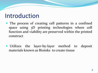
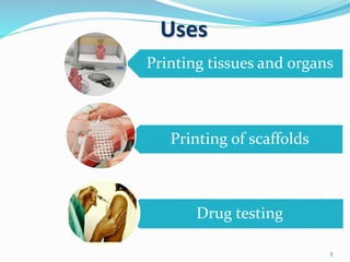
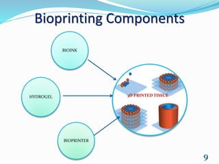
3D Bioprinting
Presented by
Kavya K N
Department of BT & BCE
Roll no : 728
Date : 01-08-2017
Guided by
Sri. Jickson G H
Ass. Professor, BT & BCE
August 1
Introduction
 The process of creating cell patterns in a confined
space using 3D printing technologies where cell
function and viability are preserved within the printed
construct
 Utilizes the layer-by-layer method to deposit
materials known as Bioinks to create tissue
2
Uses
3
Printing tissues and organs
Printing of scaffolds
Drug testing
The first patent related to
this technology was filed
in the United States in
2003 and granted in 2006
(Thomas Boland of
Clemson University
patenting the use of inkjet
printing for cells in 2003)
4
Major Three steps
Pre-bioprinting
Bioprinting
Post-Bioprinting
5
Pre-Bioprinting
Creating a model that the printer will later create
Choosing the materials that will be used
One of the first steps is to obtain a biopsy of the organ
common technologies used are
 Computed Tomography (CT)
 Magnetic Resonance Imaging (MRI).
6
 To print with a layer-by-layer approach
 . The now-2D images are converted to 3D images and
it is later sliced then sent to the printer
 Once the image is created, certain cells are isolated
and multiplied
 These cells are then mixed with a special liquefied
material that provides oxygen and other nutrients
(bioink)
7
Bioprinting
 The liquid mixture of cells, matrix, and nutrients
known as Bioinks are placed in a printer cartridge and
deposited using the patient‘s medical scans
 Typically involves dispensing cells onto a
biocompatible scaffold using a successive layer-by-
layer approach to generate tissue-like three-
dimensional structures
8
Bioprinting Components
BIOINK
HYDROGEL
BIOPRINTER
9
3D PRINTED TISSUE
Post-Bioprinting
 The mechanical integrity and function of the 3D
printed object is necessary to create a stable structure
from the biological material
 The bioprinted tissue is kept in an incubator, this 3rd
phase of bioprinting helps cell to cell interaction.
 Tight junction
Both mechanical and chemical stimulations are needed
These stimulations send signals to the cells to control
the remodelling and growth of tissues.
10
Bioprinting Approach
Three main approaches
 Bio mimicry
 Autonomous self-assembly
 Mini-tissue building blocks
11
Biomimicry
 The main goal of this approach is to create fabricated
structures that are identical to the natural structure
 Requires duplication of the shape, framework, and the
microenvironment of the organs and tissue
 Creates both identical cellular and extracellular parts
of organs
12
Autonomous self-assembly
 This relies on the physical process of embryonic organ
development then replicates the tissues by using this
process as a model
 Demands specific information about the
developmental techniques of the tissues and organs of
the embryo
 Depends on the cell as the fundamental driver of
histogenesis, guiding the building blocks, structural
and functional properties of these tissues
13
Mini-tissue Building blocks
 Combination of both the biomimicry and self-
assembly approaches,
 approach takes these small pieces and manufacture
and arrange them into larger framework
Uses two different strategies
14
 First strategy ; Self-assembling cell spheres are
arranged into large scaled tissues by using natural
designs as a guide
 Second strategy ; Designing precise, high quality,
reproductions of a tissue and allowing them to self-
assemble into large scaled functional tissue
The mixture of these strategies is required to print a
complex three dimensional biological structure
15
Printers
16
NovoGen MMX Bioprinter
F i r s t B i o p r i n t e r
Inkjet inspired printers
Bioprinting experiments at Wake Forest University
were inspired by traditional Inkjet printers
 Printer allows multiple cell types and components to
be used
Earlier cells were placed in the actual wall of ink
cartridges and printers were programed to place the
cell in particular order
Today direct printing of skin cells is been adapted by
the university
17
Six-axis printer
At University of Louisville’s Cardiovascular
Innovation Institute Dr Stuart William is using a
robot printer
Instead of building the tissue from the ground up, as
traditional bioprinters do, can build multiple parts of
heart tissue he is making at the same time and move
them accordingly
18Dr StuartWilliam
Bio-ink
 Living cells +Hydrogels Bio ink
 Suitable hydrogels are used based on organs to be
made(collagen for bone)
 The cells are layered between water based layers until
the tissue is built
 Commonly used hydrogels are
collagen, algin and fibrin
19
Disadvantages
 Problems regarding printing of complex organs
 Problems regarding vascularisation
 Problems regarding cost
 Ethical issues
20
Recent Applications
ORGONOVO was the first company to
commercialize 3D bioprinting
technology. The company utilizes
its Novogen MMX Bioprinter for 3D
bioprinting . Able to print skin tissue,
heart tissue, and blood vessels among
other basic tissues that could be suitable
for surgical therapy and transplantation
At Swansea University in the UK is
using bioprinting technology to
produce soft tissues and artificial
bones for eventual use in
reconstructive surgery.
23
Further Advancements
Human ear Jaw scaffold Human kidney
Human Kidney
skin graft Beef
22
Reference
25
Thank you
𝟐6

3D BIOPRINTING

  • 1.
    3D Bioprinting Presented by KavyaK N Department of BT & BCE Roll no : 728 Date : 01-08-2017 Guided by Sri. Jickson G H Ass. Professor, BT & BCE August 1
  • 2.
    Introduction  The processof creating cell patterns in a confined space using 3D printing technologies where cell function and viability are preserved within the printed construct  Utilizes the layer-by-layer method to deposit materials known as Bioinks to create tissue 2
  • 3.
    Uses 3 Printing tissues andorgans Printing of scaffolds Drug testing
  • 4.
    The first patentrelated to this technology was filed in the United States in 2003 and granted in 2006 (Thomas Boland of Clemson University patenting the use of inkjet printing for cells in 2003) 4
  • 5.
  • 6.
    Pre-Bioprinting Creating a modelthat the printer will later create Choosing the materials that will be used One of the first steps is to obtain a biopsy of the organ common technologies used are  Computed Tomography (CT)  Magnetic Resonance Imaging (MRI). 6
  • 7.
     To printwith a layer-by-layer approach  . The now-2D images are converted to 3D images and it is later sliced then sent to the printer  Once the image is created, certain cells are isolated and multiplied  These cells are then mixed with a special liquefied material that provides oxygen and other nutrients (bioink) 7
  • 8.
    Bioprinting  The liquidmixture of cells, matrix, and nutrients known as Bioinks are placed in a printer cartridge and deposited using the patient‘s medical scans  Typically involves dispensing cells onto a biocompatible scaffold using a successive layer-by- layer approach to generate tissue-like three- dimensional structures 8
  • 9.
  • 10.
    Post-Bioprinting  The mechanicalintegrity and function of the 3D printed object is necessary to create a stable structure from the biological material  The bioprinted tissue is kept in an incubator, this 3rd phase of bioprinting helps cell to cell interaction.  Tight junction Both mechanical and chemical stimulations are needed These stimulations send signals to the cells to control the remodelling and growth of tissues. 10
  • 11.
    Bioprinting Approach Three mainapproaches  Bio mimicry  Autonomous self-assembly  Mini-tissue building blocks 11
  • 12.
    Biomimicry  The maingoal of this approach is to create fabricated structures that are identical to the natural structure  Requires duplication of the shape, framework, and the microenvironment of the organs and tissue  Creates both identical cellular and extracellular parts of organs 12
  • 13.
    Autonomous self-assembly  Thisrelies on the physical process of embryonic organ development then replicates the tissues by using this process as a model  Demands specific information about the developmental techniques of the tissues and organs of the embryo  Depends on the cell as the fundamental driver of histogenesis, guiding the building blocks, structural and functional properties of these tissues 13
  • 14.
    Mini-tissue Building blocks Combination of both the biomimicry and self- assembly approaches,  approach takes these small pieces and manufacture and arrange them into larger framework Uses two different strategies 14
  • 15.
     First strategy; Self-assembling cell spheres are arranged into large scaled tissues by using natural designs as a guide  Second strategy ; Designing precise, high quality, reproductions of a tissue and allowing them to self- assemble into large scaled functional tissue The mixture of these strategies is required to print a complex three dimensional biological structure 15
  • 16.
    Printers 16 NovoGen MMX Bioprinter Fi r s t B i o p r i n t e r
  • 17.
    Inkjet inspired printers Bioprintingexperiments at Wake Forest University were inspired by traditional Inkjet printers  Printer allows multiple cell types and components to be used Earlier cells were placed in the actual wall of ink cartridges and printers were programed to place the cell in particular order Today direct printing of skin cells is been adapted by the university 17
  • 18.
    Six-axis printer At Universityof Louisville’s Cardiovascular Innovation Institute Dr Stuart William is using a robot printer Instead of building the tissue from the ground up, as traditional bioprinters do, can build multiple parts of heart tissue he is making at the same time and move them accordingly 18Dr StuartWilliam
  • 19.
    Bio-ink  Living cells+Hydrogels Bio ink  Suitable hydrogels are used based on organs to be made(collagen for bone)  The cells are layered between water based layers until the tissue is built  Commonly used hydrogels are collagen, algin and fibrin 19
  • 20.
    Disadvantages  Problems regardingprinting of complex organs  Problems regarding vascularisation  Problems regarding cost  Ethical issues 20
  • 21.
    Recent Applications ORGONOVO wasthe first company to commercialize 3D bioprinting technology. The company utilizes its Novogen MMX Bioprinter for 3D bioprinting . Able to print skin tissue, heart tissue, and blood vessels among other basic tissues that could be suitable for surgical therapy and transplantation At Swansea University in the UK is using bioprinting technology to produce soft tissues and artificial bones for eventual use in reconstructive surgery. 23
  • 22.
    Further Advancements Human earJaw scaffold Human kidney Human Kidney skin graft Beef 22
  • 23.
  • 24.