SlideShare a Scribd company logo
Minireview
New roles for bacterial siderophores in metal transport
and toleranceemi_2556 2844..2854
Isabelle J. Schalk,* Mélissa Hannauer and
Armelle Braud
UMR7242, University of Strasbourg-CNRS, ESBS, Blvd
Sébastien Brant, F-67413 Illkirch, Strasbourg, France.
Summary
Siderophores are chelators with extremely strong
affinity for ferric iron and are best known for their
capacity to feed microorganisms with this metal.
Despite their preference for iron, they can also
chelate numerous other metals with variable affini-
ties. There is also increasing evidence that metals
other than iron can activate the production of sidero-
phores by bacteria, thereby implicating siderophores
in the homeostasis of metals other than iron and
especially heavy metal tolerance. This article consid-
ers this new concept that siderophores play a role in
protecting bacteria against metal toxicity and dis-
cusses the possible contribution of these chelators to
the transport of biological relevant metals in addition
to iron.
Introduction
Ions of the metals sodium, magnesium and potassium
participate in the basic metabolism of cells and are there-
fore absolutely essential for life. They are present in high
quantities in bacterial cells with concentrations in the mM
range (Heldal et al., 1985). The metals iron, zinc, nickel,
copper, manganese, cobalt and molybdenum are also
involved in cellular processes and are present in mM or
mM concentrations (Heldal et al., 1985). Most of the other
metals, including lead, mercury, aluminum and silver,
have absolutely no biological function and are therefore
termed abiotic elements. Bacteria have mechanisms to
protect themselves from these metals (Nies, 1999; 2003).
Metals involved in cellular processes are often cofactors
of enzymes. A systematic analysis of 1371 enzymes
showed that 47% contain a metal ion (Andreini et al.,
2008). Among metals found in enzymes, magnesium is by
far the most frequent, followed by zinc, iron and manga-
nese (Waldron and Robinson, 2009). In conditions of
limited availability of an essential metal, various enzymes
necessary for bacterial metabolism are inactive compro-
mising cell survival. On the opposite, the presence of an
excess of metal, even of a necessary metal, can be toxic
due to interactions with non-specific targets. In bacterial
cells, metalloproteins acquire the appropriate metal by
controlling and limiting the number of metals in the cyto-
plasm. Consequently, as proposed by Waldron and Rob-
inson: ‘metals are not in competition for a limited pool of
proteins, but rather the proteins compete for a limited pool
of metals’ (Robinson, 2007; Waldron and Robinson, 2009).
Metal occupancy of metalloproteins thus is a function of the
relative rather than the absolute affinities of the different
proteins for metals. In addition, high concentrations of
some metals, such as iron, copper, cobalt and chromium,
cause oxidative stress (production of reactive oxygen
species, including free radicals and peroxides), which can
lead to damage of cellular DNA (Valko et al., 2005). To
avoid such a process, the intracellular concentration of
every metal is tightly regulated and bacteria require precise
homeostatic mechanisms to balance uptake and storage
of the different essential metals and to avoid any uptake of
abiotic elements. It is generally accepted that siderophores
play key roles in iron homeostasis in microorganisms, but
there is increasing evidence implicating these chelators in
the homeostasis of other metals.
Siderophores and iron
Siderophores are low-molecular-weight chelating agents
(200–2000 Da) produced by bacteria, fungi and plants to
facilitate uptake of iron (Chu et al., 2010; Hider and Kong,
2010). This metal is essential for the growth and develop-
ment of almost all living organisms. It acts as a catalyst in
some of the most fundamental enzymatic processes,
including oxygen metabolism, electron transfer, and DNA
and RNA synthesis. In most aerobic microbial habitats,
iron is found in insoluble oxide hydrate complexes. During
infections, iron is similarly not freely accessible for the
Received 13 May, 2011; accepted 21 June, 2011. *For correspon-
dence. E-mail isabelle.schalk@unistra.fr; Tel. (+33) 3 68 85 47 19;
Fax (+33) 3 68 85 48 29.
Environmental Microbiology (2011) 13(11), 2844–2854 doi:10.1111/j.1462-2920.2011.02556.x
© 2011 Society for Applied Microbiology and Blackwell Publishing Ltd
pathogen, as it is tightly sequestered by proteins or
haems in the host. Therefore, the free iron concentrations
are always extremely low in the environment of micro-
organisms (10-7
to 10-24
M) (Raymond et al., 2003). The
range of siderophore concentrations found in soil is wide,
from tens of micromoles to a few millimoles per litre
(Hersman et al., 1995). The siderophores play an impor-
tant role in extracellular solubilization of iron from minerals
so as to make it available to microorganism(s). The sce-
nario during infection is often similar; siderophores are
produced by bacteria to extract iron from organic sub-
stances and are considered to be virulence factors
(Lamont et al., 2002; Dale et al., 2004).
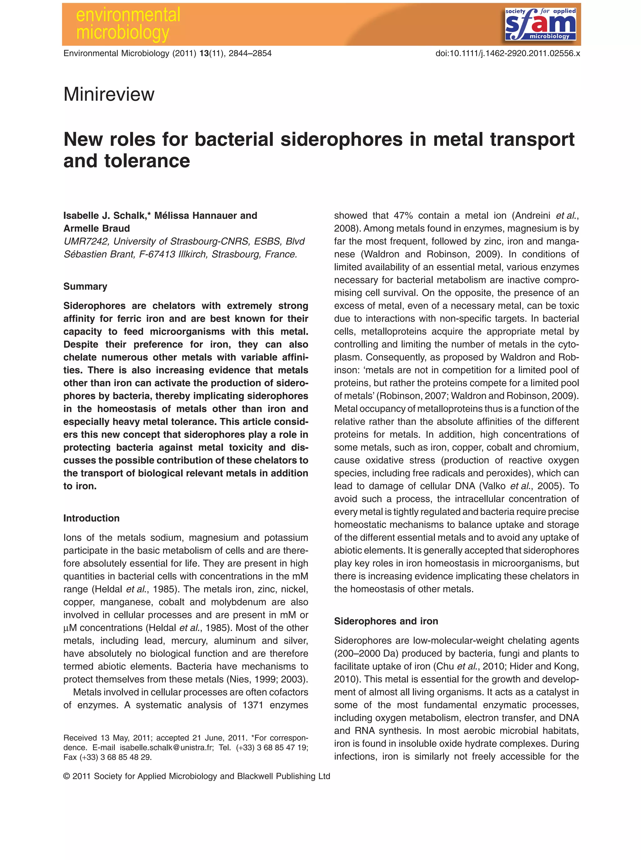
Siderophores have a variety of chemical structures and
form a family of at least 500 different compounds (Chu
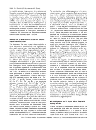
et al., 2010; Hider and Kong, 2010) (Fig. 1). They possess
up to six oxygen or nitrogen electron donor atoms, which
can bind metal cations. Some bacterial siderophores are
polycarboxylates, but most bacterial siderophores in soil
are catecholates, and most fungi produce hydroxamate
siderophores. Mixed siderophores, such as azotobactin
and yersiniabactin, are produced by Azotobacter vinelan-
dii and Yersinia pestis respectively (Knosp et al., 1984;
Haag et al., 1993), and pseudobactin and pyoverdine,
which carry both hydroxamate and catecholate groups,
are widespread among the pseudomonades (Abdallah
and Pattus, 2000; Boukhalfa and Crumbliss, 2002). All
siderophores are chelating agents with an extremely high
affinity for Fe3+
with stability constants in general around
1030
M-1
(Albrecht-Gary et al., 1994; Raymond et al.,
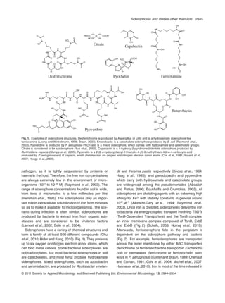
2003). Once iron is chelated, siderophores deliver the iron
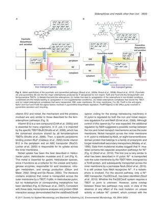
to bacteria via energy-coupled transport involving TBDTs
(TonB-Dependent Transporters) and the TonB complex,
an inner membrane complex composed of TonB, ExbB
and ExbD (Fig. 2) (Schalk, 2008; Noinaj et al., 2010).
Afterwards, ferrisiderophore fate in the periplasm is
dependent on the siderophore pathway and bacteria
(Fig. 2). For example, ferrisiderophores are transported
across the inner membrane by either ABC transporters
(ferrichrome or ferrienterobactine transport in Escherichia
coli) or permeases (ferrichrome or ferripyochelin path-
ways in P. aeruginosa) (Koster and Braun, 1989; Chenault
and Earhart, 1991; Cuiv et al., 2004; Michel et al., 2007;
Hannauer et al., 2010). Iron is most of the time released in
Fig. 1. Examples of siderophore structures. Desferrichrome is produced by Aspergillus or Ustil and is a hydroxamate siderophore like
ferrioxamine (Leong and Winkelmann, 1998; Braun, 2003). Enterobactin is a catechalote siderophore produced by E. coli (Raymond et al.,
2003). Pyoverdine is produced by P. aeruginosa PAO1 and is a mixed siderophore, which carries both hydroxamate and catecholate groups.
Citrate is considered to be a siderophore (Yue et al., 2003). Cepabactin is a 1-hydroxy-2-pyridinone bidentate siderophore produced by
Burkholderia cepacia (Klumpp et al., 2005). Pyochelin is a 2-(2-o-hydroxyphenyl-2-thiazolin-4-yl)-3-methylthiazo-lidine-4-carboxylic acid
produced by P. aeruginosa and B. cepacia, which chelates iron via oxygen and nitrogen electron donor atoms (Cox et al., 1981; Youard et al.,
2007; Hoegy et al., 2009).
Siderophores and metals other than iron 2845
© 2011 Society for Applied Microbiology and Blackwell Publishing Ltd, Environmental Microbiology, 13, 2844–2854
the cytoplasm by mechanisms involving either enzymatic
degradation or chemical modification of the siderophore
and/or iron reduction (Hartman and Braun, 1980; Brick-
man and McIntosh, 1992; Matzanke et al., 2004).
However, iron can also be released from the siderophore
in the periplasm. For pyoverdine pathway in P. aerugi-
nosa, iron is released from the siderophore by a me-
chanism involving no chemical modification of the
siderophore but apparently iron reduction (Schalk et al.,
2002; Greenwald et al., 2007; Imperi et al., 2009; Yeterian
et al., 2010).
Siderophores and their ability to chelate metals
other than iron
Stable complexes are also formed between siderophores
and metal cations other than iron (Hernlem et al., 1996;
Neubauer et al., 2000). For example, the formation con-
stants for the hydroxamate siderophore desferrioxamine
B with Ga3+
, Al3+
and In3+
are between 1020
and 1028
M-1
,
whereas that with Fe3+
is 1030
M-1
(Evers et al., 1989;
Hernlem et al., 1996). Screening with 16 different metals
(Ag+
, Al3+
, Cd2+
, Co2+
, Cr2+
, Cu2+
, Eu3+
, Ga3+
, Hg2+
, Mn2+
,
Ni2+
, Pb2+
, Sn2+
, Tb3+
, Tl+
and Zn2+
) revealed that pyover-
dine and pyochelin (Fig. 1), the two major siderophores
produced by Pseudomonas aeruginosa, are able to
chelate all these metals (Braud et al., 2009a,b). However,
competition experiments between iron and other metals
for pyoverdine showed a clear preference for iron (Braud
et al., 2009b). Formation constants for pyoverdine with
Zn2+
, Cu2+
and Mn2+
are between 1017
and 1022
M-1
,
whereas that with Fe3+
is 1032
M-1
(Chen et al., 1994).
Vanadium is a metal that under physiological conditions
can exist in two oxidation states, V(IV) (vanadyl ion) and
V(V) (vanadate ion). It was demonstrated that both ions
can form complexes with pyochelin and putrebactin, but
only vanadyl ion was found in complex with pyoverdine
and enterobactin, and vanadate ion with desferrioxamine
B (Lutoretti and Grdinic, 1986; Karpishin et al., 1993;
Baysse et al., 2000; Pakchung et al., 2011). Interactions
between pyoverdine released by Pseudomonas fluore-
scens (CCUG 32456) isolated from the granitic rock aqui-
Fig. 2. Different mechanisms and strategies (A, B and C) for the ferrisiderophore pathways in Gram-negative bacteria. Uptake across the
outer membrane always involves a specific TonB-dependent transporter (TBDT). A bacterium able to use several siderophores to sequester
iron expresses a TBDT specific for each siderophore. The energy required for this first uptake process is provided by the proton-motive force
(pmf) of the inner membrane by means of an inner membrane complex comprising TonB, ExbB and ExbD (Postle and Kadner, 2003; Wiener,
2005; Gumbart et al., 2007). This TonB–ExbB–ExbD complex interacts with the N-terminal region of the TBDT (Schalk et al., 2009). Uptake of
ferrisiderophore across the inner membrane involves either specific ABC transporters (B, ferrichrome and ferrienterobactin pathways in E. coli;
Koster and Braun, 1989; Staudenmaier et al., 1989; Chenault and Earhart, 1991; Koster, 1991; Köster, 1997; Braun and Herrmann, 2007) or
specific permeases (A, ferripyochelin or ferrichrome pathways in P. aeruginosa; Cuiv et al., 2004; 2007). Iron may be released from the
siderophore either in the cytoplasm (A and B, ferrichrome and ferrientreobactin pathways in E. coli) or periplasm (C, ferripyoverdine pathway in
P. aeruginosa) depending on the ferrisiderophore pathway. The mechanism of ferrisiderophore dissociation in the cytoplasm has been
investigated for the ferrichrome and ferrienterobactin pathways in E. coli. Iron is released from ferrichrome in the cytoplasm in a process
probably involving iron reduction (Matzanke et al., 2004) followed by acetylation of the siderophore and its recycling into the growth media
(Hartman and Braun, 1980). For the ferrienterobactin pathway, a cytoplasmic esterase hydrolyses the siderophore (Brickman and McIntosh,
1992). Iron release from the siderophore in the periplasm (panel C) has only been described for the ferripyoverdine pathway in P. aeruginosa
and involves no chemical modification of the siderophore, but the iron is reduced and the siderophore recycled into the extracellular medium
by the efflux pump PvdRT-OpmQ (Schalk et al., 2002; Greenwald et al., 2007; Imperi et al., 2009; Yeterian et al., 2010). OM, outer membrane;
IN, inner membrane.
2846 I. J. Schalk, M. Hannauer and A. Braud
© 2011 Society for Applied Microbiology and Blackwell Publishing Ltd, Environmental Microbiology, 13, 2844–2854
fers and curium(III) have also been described (Moll et al.,
2008). The complexation of curium(III) with this sidero-
phore is stronger than the complexation with EDTA,
hydroxide or carbonate. Desferrioxamine has the ability to
coordinate plutonium(V), a much larger and structurally
diverse metal centre than iron (Neu et al., 2000). All these
data show that the influence of siderophores secreted by
microorganisms on the migration processes of any toxic
metal in our environment must be taken into account in
strategies for the risk assessment of potential waste dis-
posal sites.
Siderophore production is regulated by the iron
concentration, but other metals seem also
have a role
It is well known that siderophore biosynthesis by bacteria
is regulated by both the amount of iron present in the
environment and the amount of iron already acquired by
the bacteria. For example, pyoverdine and pyochelin pro-
duction in P. aeruginosa is repressed by the cytoplasmic
Fur protein in the presence of iron (Lee and Helmann,
2007). In the absence of iron, the upregulation of the
expression of genes related to pyoverdine synthesis and
uptake involves a transmembrane signalling cascade.
The system is induced by the binding of the siderophore
to the TBDT FpvA at the cell surface and involves at the
level of the inner membrane and cytoplasm the extracy-
toplasmic function (ECF) sigma factor/anti-sigma factor
pairs, FpvI/FpvR and PvdS/FpvR (Wilson and Lamont,
2000; Wilson et al., 2001; Lamont et al., 2002; Shen et al.,
2002; Visca et al., 2002; Beare et al., 2003; Redly and
Poole, 2003; 2005). Equivalent sigma/anti-sigma factors
have been described for ferrichrome, ferrioxamine and
mycobactin uptake by P. aeruginosa, and citrate uptake
by P. aeruginosa and E. coli (Braun et al., 2003; Llamas
et al., 2006; 2008; Mettrick and Lamont, 2009). The bio-
synthesis of pyochelin is positively regulated by a different
mechanism involving an AraC-like regulatory protein (the
PchR proteins) (Michel et al., 2005) as also observed for
the alcaligin siderophore in Bordetella alcaligin (Brickman
et al., 2001). In this case, the pyochelin–Fe complex, after
its uptake across the outer and inner membranes, inter-
acts with the AraC-like regulator in the bacterial cytoplasm
(Michel et al., 2007).
Metals other than iron are also able to stimulate or
inhibit siderophore production in a number of bacteria,
even in the presence of high iron concentrations. For
instance, the presence of molybdenum in the environment
of Azotobacter vinelandii regulates the production of azo-
tochelin, a catecholate siderophore able to chelate this
metal. At concentrations up to 100 mM of molybdenum,
azotochelin production is activated, whereas at higher
metal concentrations the synthesis of the siderophore is
completely repressed (Duhme et al., 1998). High concen-
trations of aluminum increase the production of schi-
zokinen and N-deoxyschizokinen (two hydroxamate
siderophores) in iron-limited cultures but not iron-replete
cultures of Bacillus megaterium (Hu and Boyer, 1996). In
iron-limited succinate medium, pyoverdine production is
induced by 10 mM Al3+
, addition of Cu2+
, Ga3+
, Mn2+
and
Ni2+
(Braud et al., 2009b). Still under iron-limited condi-
tions, exposure to 10 mM Cu2+
upregulates genes
involved in the synthesis of pyoverdine and downregu-
lates those involved in the synthesis of pyochelin (Teitzel
et al., 2006). Interestingly, even in the presence of iron
(100 mM), pyoverdine production is increased by 10 and
100 mM Cu2+
and Ni2+
(290 and 380% respectively) and to
a lesser extent by 100 mM Cr2+
(134%) (Fig. 3B) (Braud
et al., 2010). No activation of pyochelin production was
observed, but a repression for 10 mM Co2+
and 100 mM
Cu2+
, Mo+6
and Ni2+
in iron-limited medium (Visca et al.,
1992). When culture media were supplemented with
100 mM Fe3+
, Cd2+
, Cu2+
, Eu3+
, Ga3+
, Mn2+
, Ni2+
and Tb3+
decreased pyochelin production (Braud et al., 2010). In all
these experiments, metals were added at the beginning of
the cultures. Unfortunately, most of these studies have
been carried out with only a single or in some cases two
metal concentrations and not for a large range of concen-
trations. Probably, as for iron, siderophore production can
be both activated and inhibited by many of these metals,
depending on their concentrations. Therefore, it would be
useful to test large ranges of concentrations of each metal
to establish their exact roles in regulation of siderophore
production in more detail.
It is still unclear how metals other than iron stimulate
siderophore production. One possible explanation is that
the free siderophore concentration in the medium is
reduced in the presence of other metals as a result of
complex formation. Such a decrease in the siderophore
concentration may be sufficient to activate secretion of
additional siderophore into the medium. However, pyover-
dine production after an overnight culture was about
120 mM (in succinate medium), a concentration much
higher than the 10 mM of metal added to the culture
(Braud et al., 2009b), indicating clearly that the amount of
siderophore was not limiting, at least in this experimental
system. The same study revealed that all metals increas-
ing pyoverdine production (Al3+
, Cu2+
, Cr2+
, Ga3+
, Mn2+
and
Ni2+
) were able either to inhibit pyoverdine–55
Fe uptake or
to be incorporated into P. aeruginosa via the pyoverdine
pathway (Braud et al., 2009b). Presumably, therefore,
these pyoverdine–metal complexes are able to interact
with FpvA, at the cell surface. Thus, the stimulation of
pyoverdine production by metals other than iron may
involve the FpvR/PvdS signalling cascade being activated
by the pyoverdine–metal complex binding to FpvA at the
cell surface. This mechanism does not need any uptake of
Siderophores and metals other than iron 2847
© 2011 Society for Applied Microbiology and Blackwell Publishing Ltd, Environmental Microbiology, 13, 2844–2854
the metal to activate the production of the siderophore.
Regulation of pyochelin biosynthesis involves a cytoplas-
mic interaction between PchR and pyocheline–Fe. Such
an interaction requires uptake of the siderophore–metal
complex into the cytoplasm, which is dangerous for the
cell if the metal is toxic. This presumably explains why no
metals have been found to activate the biosynthesis of
this siderophore, and indeed, they all inhibit its synthesis
in iron-limited and iron-supplemented media. Stimulation
of siderophore production by metals other than iron as
observed for pyoverdine in P. aeruginosa, azotochelin in
A. vinelandii and schizokinen in B. megaterium raises the
question of the purpose of such a process.
Another role for siderophores: protecting bacteria
against metal toxicities
The observation that toxic metals induce production of
some siderophores suggests that these chelators may
play a role in bacterial heavy metal tolerance. Toxic metals
enter the periplasm of Gram-negative bacteria mostly by
diffusion across the porins (Lutkenhaus, 1977; Pugsley
and Schnaitman, 1978; Li et al., 1997). Thus, the binding
of metals to siderophores in the extracellular medium
reduces the free metal concentration, probably affect-
ing diffusion (the molecular mass of the resulting
siderophore–metal complex is to great for diffusion via
porins) and therefore their toxicity. Growth assays showed
that P. aeruginosa strains capable of producing pyover-
dine and pyochelin appeared to be more resistant to metal
toxicity than a siderophore non-producing strain (Braud
et al., 2010). The presence of the siderophores decreases
metal accumulation in bacteria as monitored by Induc-
tively Coupled Plasma-Atomic Emission Spectrometry
(ICP-AES): pyochelin reduces the uptake of Al3+
, Co2+
,
Cu2+
, Ni2+
, Pb2+
and Zn2+
(uptake inhibition greater than
80% for Al3+
, Co2+
, Cu2+
and Zn2+
); and pyoverdine
decreases the uptake of Al3+
, Co2+
, Cu2+
, Eu3+
, Ni2+
, Pb2+
,
Tb3+
and Zn2+
(uptake inhibition around 80% for Al3+
and
Cu2+
and 40% for the other metals) (Braud et al., 2010).
Pyoverdine was more effective than pyochelin in inhibiting
the diffusion of Eu3+
, Ga3+
, Pb2+
, Sn2+
and Tb3+
(all non-
biological metals), whereas pyochelin was more effective
against Co2+
, Fe3+
, Ni2+
and Zn2+
(biological metals). These
studies with P. aeruginosa clearly show that siderophores
are able to sequester metals in the extracellular medium
and probably prevent their diffusion across the bacterial
membranes. The transcriptional profile of P. aeruginosa
exposed to Cu2+
is consistent with siderophores having
this function in metal resistance (Teitzel et al., 2006).
Indeed, the genes involved in the synthesis of pyoverdine
are upregulated and fpvA, involved in pyoverdine–Fe
uptake, is downregulated. So Cu2+
induces the production
of pyoverdine and not of the transporter of pyoverdine–
Fe, such that the metal will be sequestered in the extra-
cellular medium but not transported. Such a mechanism
must involve a different mechanism and sensitivity to the
presence of metals for the two sigma factor/anti sigma
factor pairs, FpvR/FpvI and FpvR/PvdS. Sequestration of
toxic metals by siderophores in the extracellular medium
has also been proposed for Streptomyces tendae F4,
a Cd-resistant bacterium, which can simultaneously
produce a variety of hydroxamate siderophores (desferri-
oxamine B, desferrioxamine E and coelichelin) that bind
Cd2+
. The production of these siderophores is upregulated
by Cd2+
, both in the presence and absence of iron. Fur-
thermore, the production of the siderophores reduces
uptake of Cd2+
by Streptomyces tendae F4 while supply-
ing it with iron (Dimkpa et al., 2009). Gilis et al. have
shown that alcaligin E, a siderophore produced by Alcali-
genes eutrophus strain CH34, markedly stimulates the
growth of the bacteria in the presence of Cd2+
(Gilis et al.,
1998). Bacillus megaterium, a Gram-positive bacteria,
produces two hydroxamate siderophores under con-
ditions of iron limitation, schizokinen and N-
deoxyschizokinen. At high concentration of aluminum and
low concentrations of iron, siderophore production slowed
the passive transport of aluminum in B. megaterium (Hu
and Boyer, 1996).
All these data suggest a role of siderophores in bacte-
rial metal resistance and such a function must involve an
induction of siderophore production by toxic metals.
However, do all siderophores have a role in metal resis-
tance? For P. aeruginosa, pyoverdine is more involved
than pyochelin in the protection against toxic metals.
Indeed, pyoverdine is more efficient than pyochelin in
heavy metals sequestration outside the bacteria (Braud
et al., 2010). In addition, toxic metals (at least at the
concentrations studied) activate only the production of
pyoverdine and not pyochelin (Braud et al., 2010). At last,
the molecular mechanism involved in the activation of
pyoverdine production starts from the cell surface with an
interaction between the PVD-metal complexes and FpvA,
while in the pyocheline pathway it occurs in the cytoplasm
and involves pyochelin–metal uptake and its interaction
with the cytoplasmic PchR regulator. Such a regulation
mechanism with cytoplasmic uptake of siderophore–
metal complexes is unfavourable to a strategy of metal
resistance involving siderophores.
Are siderophores able to import metals other than
iron by TBDTs?
Specific TBDTs for the translocation of metals across the
outer membrane have been well described for iron in
complex with siderophores (Fig. 3), for vitamin B12, which
contains a cobalt ion in a corrin ring, and more recently for
nickel (Shultis et al., 2006; Schauer et al., 2007). For
2848 I. J. Schalk, M. Hannauer and A. Braud
© 2011 Society for Applied Microbiology and Blackwell Publishing Ltd, Environmental Microbiology, 13, 2844–2854
vitamin B12 and nickel, the mechanism and the proteins
involved are very similar to those described for the ferri-
siderophore pathways (Fig. 3).
Vitamin B12 is a rare compound (Croft et al., 2005)) and
is essential for many organisms. In E. coli, it is imported
by the specific TBDT BtuB (Shultis et al., 2006), which has
the conserved structure shared by all ferrisiderophore
TBDTs (Shultis et al., 2006). Then, a specific periplasmic
binding protein BtuF (Cadieux et al., 2002) binds vitamin
B12 in the periplasm and an ABC transporter (BtuCD;
Locher et al., 2002) is responsible for its uptake across
the inner membrane.
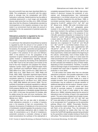
Nickel uptake has been the best described in Helico-
bacter pylori, Helicobacter mustelae and E. coli (Fig. 4).
This metal is essential for gastric Helicobacter species,
since it functions as a cofactor for the urease and hydro-
genase enzymes, responsible for acid resistance, nitro-
gen metabolism, and energy production (Olson and
Maier, 2002; Stingl and De Reuse, 2005). The literature
contains evidence that nickel is transported across the
outer membrane by a TBDT, FrpB4, in Helicobacter pylori,
but no nickelophore or corresponding metalophore has
been identified (Fig. 4) (Schauer et al., 2007). Consistent
with these data, transcriptome analyses and protein–DNA
interaction assays demonstrated that the exbB/exbD/tonB
operon coding for the energy transducing machinery in
H. pylori is regulated by both the iron and nickel respon-
sive regulators Fur and NikR (Ernst et al., 2006). Although
control of this operon by Fur was expected, the additional
regulation by NikR suggested a possible overlap between
the iron and nickel transport mechanisms across the outer
membrane. Nickel transport across the inner membrane
in H. pylori is mediated by NixA, an eight-transmembrane-
domain protein that belongs to a family of prokaryotic and
fungal nickel/cobalt secondary transporters (Mobley et al.,
1995). Data from mutational studies suggest that H. mus-
telae contains two separate acquisition pathways for Ni2+
(Fig. 4) (Stoof et al., 2010). The first one is very similar to
what has been described in H. pylori. Ni2+
is transported
over the outer membrane by the TBDT NikH, energized by
a TonB protein, and subsequently transported across the
inner membrane by a permease NixA (Stoof et al., 2010).
It is still unclear how NikH recognizes Ni2+
, if a nickelo-
phore is involved. For the second pathway, only a Ni2+
ABC transporter, FecDECeuE, has been identified (Stoof
et al., 2010). Whether the FecDECeuE system requires a
TBDT or porin is unknown. However, some overlap
between these two pathways may exist, in view of the
absence of any effect of the nixA mutation on urease
activity or cellular Ni2+
content, which contrast with the
A B
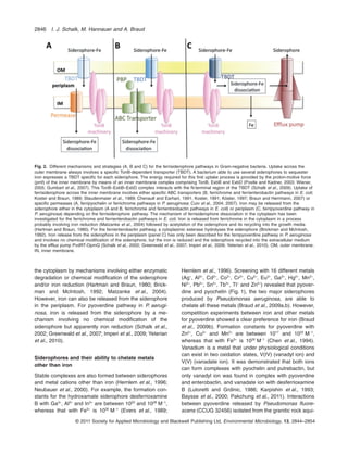
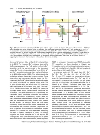
Fig. 3. Metal specificities of the pyochelin and pyoverdine pathways (Braud et al., 2009a; Braud et al., 2009b; Braud et al., 2010). Pyochelin
(A) and pyoverdine (B) are the two major siderophores produced by P. aeruginosa for iron import. FptA and FpvA are the ferripyochelin and
ferripyoverdine TBDTs. (i) Metals that bind the siderophore; (ii) metals that lead to upregulation of the siderophore in iron-limited conditions
(underlined are the metals leading to upregulation in iron-supplemented conditions); (iii) metal–siderophore complexes that bound the TBDT;
and (iv) metal–siderophore complexes that were transported. OM, outer membrane; IN, inner membrane. For (B), FpvR is the anti-sigma
factor and FpvI and PvdS the sigma factors involved in pyoverdine biosynthesis regulation. PvdRT-OpmQ is the efflux pump involved in
pyoverdine secretion and recycling.
Siderophores and metals other than iron 2849
© 2011 Society for Applied Microbiology and Blackwell Publishing Ltd, Environmental Microbiology, 13, 2844–2854
decrease Ni2+
content of the tonB and nikH mutants (Stoof
et al., 2010). The increased Co2+
resistance observed for
fecD mutants suggests that FecD is also involved in Co2+
acquisition, and this was confirmed by the decreased
cellular Co2+
levels in this mutant (Stoof et al., 2010). Ni2+
and Co2+
acquisition pathways often overlap (Rodionov
et al., 2006; Zhang et al., 2009). This is likely due to the
similarities between these two transition metals. These
observations are in agreement with the finding that Co2+
-
cofactored NikRs (Ni2+
-dependent transcriptional regula-
tor) from H. pylori and E. coli are able to bind target DNA
in vitro although with less affinity than Ni2+
-cofactored
NikR (Zambelli et al., 2008; Wang et al., 2009; Dian et al.,
2011). Escherichia coli uses the NikABCDE transporter
for nickel uptake across the cytoplasmic membrane and
no specific TBDT was identified (Fig. 4) (Mulrooney and
Hausinger, 2003). NikABCDE comprises five proteins:
NikA is a periplasmic nickel-binding protein, NikB and
NikC are integral inner membrane proteins; and NikD and
NikE are membrane-associated proteins with intrinsic
ATPase activity. The transport across the outer mem-
brane may involve diffusion via porins, but because sid-
erophores are able to chelate this metal, it is plausible that
a TBDT operates in situations of nickel limitation. Indeed,
a metal–EDTA complex has been observed in the binding
site of the E. coli NikA (Cherrier et al., 2005). This strongly
suggests that nickel transport into E. coli requires the
binding of this metal ion to a metallophore and an uptake
mechanism across the outer membrane, which requires a
TBDT. In conclusion, the existence of TBDTs involved in
Ni2+
acquisition has been described in H. pylori and
H. mustelae, but the only evidence that these transporters
work with nickelophores is the ability of E. coli periplasmic
protein NikA to bind metal–EDTA.
A screening study with 16 metals (Ag+
, Al3+
, Cd2+
,
Co2+
, Cr2+
, Cu2+
, Eu3+
, Ga3+
, Hg2+
, Mn2+
, Ni2+
, Pb2+
, Sn2+
,
Tb3+
, Tl+
and Zn2+
) showed that a siderophore-deficient
strain of P. aeruginosa imported Co2+
, Ga3+
and Ni2+
in
complex with the siderophore pyochelin, but with uptake
rates 23- to 35-fold lower than for pyocheline–Fe (Fig. 3)
(Braud et al., 2009a). Equivalent data have been
obtained for the pyoverdine pathway (Fig. 3): Cu2+
, Ga3+
,
Mn2+
and Ni2+
in complex with pyoverdine accumulated
into the cells but with lower uptake rates compared with
pyoverdine–Fe (7- to 42-fold lower). In both assays,
metal uptakes were dependent on the presence of the
siderophore and were TonB-dependent, suggesting that
they occurred via the siderophores and their correspond-
ing TBDT. None of the other metals tested was accumu-
lated through either the pyoverdine or pyochelin
pathways (Braud et al., 2009b). Note that all the metals
transported are biological metals except Ga3+
. Ga3+
has
no known biological function, but has the same coordi-
nation as iron and is often used as a tool to investigate
iron uptake pathways (Folschweiller et al., 2002; Green-
wald et al., 2007). No accumulation of toxic metals sug-
gests that the import of Co2+
, Cu2+
, Mn2+
and Ni2+
could
have a biological significance.
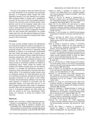
Fig. 4. Different mechanisms and strategies for Ni2+
uptake in Gram-negative bacteria. In H. pylori, Ni2+
uptake pathway involves a TBDT Frp4
and a permease NixA for the transports across the outer and inner membranes respectively (Mobley et al., 1995; Schauer et al., 2007). In
H. mustelae, two separate acquisition pathways are present (Stoof et al., 2010). The first one involves as for H. pylori a TBDT NikH and a
permease NixA. For the second, only the inner membrane ABC transporter FecDE-CeuE has been identified and nothing is known about the
mechanism involved in the uptake of the metal across the outer. In E. coli, only an ABC transporter NikABCDE has been identified and no
TBDT (Mulrooney and Hausinger, 2003). Interestingly, the periplasmic binding protein NikA is able to bind Ni2+
in complex with EDTA,
suggesting the involvement of a nickelophore in this pathway (Cherrier et al., 2005).
2850 I. J. Schalk, M. Hannauer and A. Braud
© 2011 Society for Applied Microbiology and Blackwell Publishing Ltd, Environmental Microbiology, 13, 2844–2854
The work on the uptake of nickel and vitamin B12 and
the metal specificities of the pyochelin and pyoverdine
pathways in P. aeruginosa clearly indicate that TonB-
dependent transport is not only reserved to iron uptake.
Other biological metals in complex with a metallophore
may also use this route to enter Gram-negative bacteria.
However, this raises the issue of whether specific metal-
lophores exist for all the biological metals able to be
transported by TonB-dependent pathways. There is cur-
rently no data indicating the existence of specific metal-
lophores for nickel, zinc, copper or manganese. However,
there are data showing that siderophores can chelate
metals other than iron with different affinities and there-
fore they may have the potential to transport other metals
present in their environment.
Conclusion
The data currently available indicate that siderophores
are produced by bacteria not solely to scavenge for iron.
They may have wider and more complex functions that
previously thought. Their presence is apparently highly
beneficial for bacteria in an environment contaminated
with toxic metals and bacteria able to produce sidero-
phores are more resistant to heavy metals than those not
producing siderophores. With their high affinity for iron
and other metals and their substantial production and
secretion into the extracellular medium, siderophores
clearly provide an extracellular protection for bacteria by
sequestering heavy metals outside the bacteria and
avoiding their diffusion through porins into the bacteria.
However, it is possible that siderophore production in
response to heavy metal exposure could also have detri-
mental effects. If the siderophore uptake receptor does
not distinguish between the metal–siderophore and the
ferri–siderophore complexes, siderophores may provide a
secondary mechanism for the uptake of toxic metals.
Therefore, the ferrisiderophore pathways must have high
metal specificity to transport or accumulate only the
appropriate metal(s). Is the right metal only iron, or are
other biological metals like nickel, copper or manganese,
included? Work with the pyoverdine and pyochelin path-
ways in P. aeruginosa suggests that other metals can
enter the bacteria by this route. Nevertheless, further
studies are necessary to answer this question more
rigorously.
References
Abdallah, M.A., and Pattus, F. (2000) Siderophores and iron-
transport in microorganisms. J Chin Chem Soc 47: 1–20.
Albrecht-Gary, A.M., Blanc, S., Rochel, N., Ocacktan, A.Z.,
and Abdallah, M.A. (1994) Bacterial iron transport: coordi-
nation properties of pyoverdin PaA, a peptidic siderophore
of Pseudomonas aeruginosa. Inorg Chem 33: 6391–6402.
Andreini, C., Bertini, I., Cavallaro, G., Holliday, G.L., and
Thornton, J.M. (2008) Metal ions in biological catalysis:
from enzyme databases to general principles. J Biol Inorg
Chem 13: 1205–1218.
Baysse, C., De Vos, D., Naudet, Y., Vandermonde, A.,
Ochsner, U., Meyer, J.M., et al. (2000) Vanadium interferes
with siderophore-mediated iron uptake in Pseudomonas
aeruginosa. Microbiology 146 (Pt 10): 2425–2434.
Beare, P.A., For, R.J., Martin, L.W., and Lamont, I.L. (2003)
Siderophore-mediated cell signalling in Pseudomonas
aeruginosa: divergent pathways regulate virulence factor
production and siderophore receptor synthesis. Mol Micro-
biol 47: 195–207.
Boukhalfa, H., and Crumbliss, A.L. (2002) Chemical aspects
of siderophore mediated iron transport. Biometals 15: 325–
339.
Braud, A., Hannauer, M., Milsin, G.L.A., and Schalk, I.J.
(2009a) The Pseudomonas aeruginosa pyochelin-iron
uptake pathway and its metal specificity. J Bacteriol 191:
5317–5325.
Braud, A., Hoegy, F., Jezequel, K., Lebeau, T., and Schalk,
I.J. (2009b) New insights into the metal specificity of
the Pseudomonas aeruginosa pyoverdine-iron uptake
pathway. Environ Microbiol 11: 1079–1091.
Braud, A., Geoffroy, V., Hoegy, F., Mislin, G.L.A., and Schalk,
I.J. (2010) The siderophores pyoverdine and pyochelin are
involved in Pseudomonas aeruginosa resistence against
metals: Another biological function of these two sidero-
phores. Environ Microbiol Report 2: 419–425.
Braun, V. (2003) Iron uptake by Escherichia coli. Front Biosci
8: 1409–1421.
Braun, V., and Herrmann, C. (2007) Docking of the periplas-
mic FecB binding protein to the FecCD transmembrane
proteins in the ferric citrate transport system of Escherichia
coli. J Bacteriol 189: 6913–6918.
Braun, V., Mahren, S., and Ogierman, M. (2003) Regulation
of the FecI-type ECF sigma factor by transmembrane sig-
nalling. Curr Opin Microbiol 6: 173–180.
Brickman, T.J., and McIntosh, M.A. (1992) Overexpression
and purification of ferric enterobactin esterase from
Escherichia coli. Demonstration of enzymatic hydrolysis of
enterobactin and its iron complex. J Biol Chem 267:
12350–12355.
Brickman, T.J., Kang, H.Y., and Armstrong, S.K. (2001) Tran-
scriptional activation of Bordetella alcaligin siderophore
genes requires the AlcR regulator with alcaligin as inducer.
J Bacteriol 183: 483–489.
Cadieux, N., Bradbeer, C., Reeger-Schneider, E., Koster, W.,
Mohanty, A.K., Wiener, M.C., and Kadner, R.J. (2002)
Identification of the periplasmic cobalamin-binding protein
BtuF of Escherichia coli. J Bacteriol 184: 706–717.
Chen, Y., Jurkewitch, E., Bar-Ness, E., and Hadar, Y. (1994)
Stability constants of pseudobactin complexes with transi-
tion metals. Soil Sci Soc Am J 58: 390–396.
Chenault, S.S., and Earhart, C.F. (1991) Organization
of genes encoding membrane proteins of the Escherichia
coli ferrienterobactin permease. Mol Microbiol 5: 1405–
1413.
Cherrier, M.V., Martin, L., Cavazza, C., Jacquamet, L.,
Lemaire, D., Gaillard, J., and Fontecilla-Camps, J.C.
(2005) Crystallographic and spectroscopic evidence for
Siderophores and metals other than iron 2851
© 2011 Society for Applied Microbiology and Blackwell Publishing Ltd, Environmental Microbiology, 13, 2844–2854
high affinity binding of FeEDTA(H2O)- to the periplasmic
nickel transporter NikA. J Am Chem Soc 127: 10075–
10082.
Chu, B.C., Garcia-Herrero, A., Johanson, T.H., Krewulak,
K.D., Lau, C.K., Peacock, R.S., et al. (2010) Siderophore
uptake in bacteria and the battle for iron with the host; a
bird’s eye view. Biometals 23: 601–611.
Cox, C.D., Rinehart, K.L., Jr, Moore, M.L., and Cook, J.C., Jr
(1981) Pyochelin: novel structure of an iron-chelating
growth promoter for Pseudomonas aeruginosa. Proc Natl
Acad Sci USA 78: 4256–4260.
Croft, M.T., Lawrence, A.D., Raux-Deery, E., Warren,
M.J., and Smith, A.G. (2005) Algae acquire vitamin B12
through a symbiotic relationship with bacteria. Nature 438:
90–93.
Cuiv, P.O., Clarke, P., Lynch, D., and O’Connell, M. (2004)
Identification of rhtX and fptX, novel genes encoding pro-
teins that show homology and function in the utilization of
the siderophores rhizobactin 1021 by Sinorhizobium
meliloti and pyochelin by Pseudomonas aeruginosa,
respectively. J Bacteriol 186: 2996–3005.
Cuiv, P.O., Keogh, D., Clarke, P., and O’Connell, M. (2007)
FoxB of Pseudomonas aeruginosa functions in the utiliza-
tion of the xenosiderophores ferrichrome, ferrioxamine B,
and schizokinen: evidence for transport redundancy at the
inner membrane. J Bacteriol 189: 284–287.
Dale, S.E., Doherty-Kirby, A., Lajoie, G., and Heinrichs, D.E.
(2004) Role of siderophore biosynthesis in virulence of
Staphylococcus aureus: identification and characterization
of genes involved in production of a siderophore. Infect
Immun 72: 29–37.
Dian, C., Vitale, S., Leonard, G.A., Bahlawane, C., Fauquant,
C., Leduc, D., et al. (2011) The structure of the
Helicobacter pylori ferric uptake regulator Fur reveals three
functional metal binding sites. Mol Microbiol 79: 1260–
1275.
Dimkpa, C.O., Merten, D., Svatos, A., Buchel, G., and Kothe,
E. (2009) Siderophores mediate reduced and increased
uptake of cadmium by Streptomyces tendae F4 and sun-
flower (Helianthus annuus), respectively. J Appl Microbiol
107: 1687–1696.
Duhme, R.C., Hider, M.J., Naldrett, M.J., and Pau, R.N.
(1998) The stability of the molybdenum-azotochelin
complex and its effect on siderophore production in Azoto-
bacter vinelandii. J Biol Inorg Chem 3: 520–526.
Ernst, F.D., Stoof, J., Horrevoets, W.M., Kuipers, E.J.,
Kusters, J.G., and van Vliet, A.H. (2006) NikR mediates
nickel-responsive transcriptional repression of the Helico-
bacter pylori outer membrane proteins FecA3 (HP1400)
and FrpB4 (HP1512). Infect Immun 74: 6821–6828.
Evers, A., Hancock, R., Martell, A., and Motekaitis, R. (1989)
Metal ion recognition in ligands with negatively charged
oxygen donor groups. Complexation of Fe (III), Ga(III),
In(III), Al(III) and other highly charged metal ions. Inorg
Chem 28: 2189–2195.
Folschweiller, N., Gallay, J., Vincent, M., Abdallah, M.A.,
Pattus, F., and Schalk, I.J. (2002) The interaction between
pyoverdin and its outer membrane receptor in Pseudo-
monas aeruginosa leads to different conformers: a
time-resolved fluorescence study. Biochemistry 41:
14591–14601.
Gilis, A., Corbisier, P., Baeyens, W., Taghavi, S., Mergeay,
M., and van der Lelie, D. (1998) Effect of the siderophore
alcaligin E on the bioavailability of Cd to Alcaligenes eutro-
phus CH34. J Ind Microbiol Biotechnol 20: 61–68.
Greenwald, J., Hoegy, F., Nader, M., Journet, L., Mislin,
G.L.A., Graumann, P.L., and Schalk, I.J. (2007) Real-time
FRET visualization of ferric-pyoverdine uptake in
Pseudomonas aeruginosa: a role for ferrous iron. J Biol
Chem 282: 2987–2995.
Gumbart, J., Wiener, M.C., and Tajkhorshid, E. (2007)
Mechanics of force propagation in TonB-dependent outer
membrane transport. Biophys J 93: 496–504.
Haag, H., Hantke, K., Drechsel, H., Stojiljkovic, I., Jung, G.,
and Zahner, H. (1993) Purification of yersiniabactin: a sid-
erophore and possible virulence factor of Yersinia entero-
colitica. J Gen Microbiol 139: 2159–2165.
Hannauer, M., Barda, Y., Mislin, G.L., Shanzer, A., and
Schalk, I.J. (2010) The ferrichrome uptake pathway in
Pseudomonas aeruginosa involves an iron release
mechansim with acylation of the siderophore and a recy-
cling of the modified desferrichrome. J Bacteriol 192:
1212–1220.
Hartman, A., and Braun, V. (1980) Iron transport in Escheri-
chia coli: uptake and modification of ferrichrome.
J Bacteriol 143: 246–255.
Heldal, M., Norland, S., and Tumyr, O. (1985) X-ray
microanalytic method for measurement of dry matter and
elemental content of individual bacteria. Appl Environ
Microbiol 50: 1251–1257.
Hernlem, B.J., Vane, L.M., and Sayles, G.D. (1996) Stability
constants for complexes of the siderophore desferrioxam-
ine B with selected heavy metal cations. Inorganic Chimica
Acta 244: 179–184.
Hersman, L., Lloyd, T., and Sposito, G. (1995) Siderophore-
promoted dissolution of hematite. Geochimica Cosmo-
chimica Acta 59: 3327–3330.
Hider, R.C., and Kong, X. (2010) Chemistry and biology of
siderophores. Nat Prod Rep 27: 637–657.
Hoegy, F., Lee, X., Noël, S., Mislin, G.L., Rognan, D., Reim-
mann, C., and Schalk, I.J. (2009) Stereospecificity of the
siderophore pyochelin outer membrane transporters in
fluorescent Pseudomonads. J Biol Chem 284: 14949–
14957.
Hu, X., and Boyer, G.L. (1996) Siderophore-mediated alumi-
num uptake by Bacillus megaterium ATCC 19213. Appl
Environ Microbiol 62: 4044–4048.
Imperi, F., Tiburzi, F., and Visca, P. (2009) Molecular basis of
pyoverdine siderophore recycling in Pseudomonas aerugi-
nosa. Proc Natl Acad Sci USA 106: 20440–20445.
Karpishin, T.B., Dewey, T.M., and Raymond, K.N. (1993) The
vanadium(IV) enterobactin complex: structural, spectro-
scopic, and electrochemical characterization. J Am Chem
Soc 115: 1842–1851.
Klumpp, C., Burger, A., Mislin, G.L., and Abdallah, M.A.
(2005) From a total synthesis of cepabactin and its 3:1
ferric complex to the isolation of a 1:1:1 mixed complex
between iron (III), cepabactin and pyochelin. Bioorg Med
Chem Lett 15: 1721–1724.
Knosp, O., von Tigerstrom, M., and Page, W.J. (1984)
Siderophore-mediated uptake of iron in Azotobacter vine-
landii. J Bacteriol 159: 341–347.
2852 I. J. Schalk, M. Hannauer and A. Braud
© 2011 Society for Applied Microbiology and Blackwell Publishing Ltd, Environmental Microbiology, 13, 2844–2854
Koster, W. (1991) Iron(III) hydroxamate transport across the
cytoplasmic membrane of Escherichia coli. Biol Met 4:
23–32.
Köster, W. (1997) Transport of iron(III) hydroxamates across
the cytoplasmic membrane of Escherichia coli. In Biorganic
Chemistry, Transition Metals in Biology and Their Coordi-
nation Chemistry. In Trautwein, A.X. (ed.). Weinheim,
Germany: VCH Verlagsgesellschaft, pp. 56–68.
Koster, W., and Braun, V. (1989) Iron-hydroxamate transport
into Escherichia coli K12: localization of FhuD in the peri-
plasm and of FhuB in the cytoplasmic membrane. Mol Gen
Genet 217: 233–239.
Lamont, I.L., Beare, P.A., Ochsner, U., Vasil, A.I., and Vasil,
M.L. (2002) Siderophore-mediated signaling regulates
virulence factor production in Pseudomonas aeruginosa.
Proc Natl Acad Sci USA 99: 7072–7077.
Lee, J.W., and Helmann, J.D. (2007) Functional specializa-
tion within the Fur family of metalloregulators. Biometals
20: 485–499.
Leong, S.A., and Winkelmann, G. (1998) Molecular biology of
iron transport in fungi. Met Ions Biol Syst 35: 147–186.
Li, X.Z., Nikaido, H., and Williams, K.E. (1997) Silver-
resistant mutants of Escherichia coli display active efflux of
Ag+
and are deficient in porins. J Bacteriol 179: 6127–
6132.
Llamas, M.A., Sparrius, M., Kloet, R., Jimenez, C.R.,
Vandenbroucke-Grauls, C., and Bitter, W. (2006) The het-
erologous siderophores ferrioxamine B and ferrichrome
activate signaling pathways in Pseudomonas aeruginosa.
J Bacteriol 188: 1882–1891.
Llamas, M.A., Mooij, M.J., Sparrius, M., Vandenbroucke-
Grauls, C.M., Ratledge, C., and Bitter, W. (2008) Charac-
terization of five novel Pseudomonas aeruginosa cell-
surface signalling systems. Mol Microbiol 67: 458–472.
Locher, K.P., Lee, A.T., and Rees, D.C. (2002) The E. coli
BtuCD structure: a framework for ABC transporter archi-
tecture and mechanism. Science 296: 1793–1800.
Lutkenhaus, J.F. (1977) Role of a major outer membrane
protein in Escherichia coli. J Bacteriol 131: 631–637.
Lutoretti, S., and Grdinic, V. (1986) Spectrophotometric deter-
mination of vanadium(V) with desferrioxamine B. Analyst
111: 1163–1165.
Matzanke, B.F., Anemuller, S., Schunemann, V., Trautwein,
A.X., and Hantke, K. (2004) FhuF, part of a siderophore-
reductase system. Biochemistry 43: 1386–1392.
Mettrick, K.A., and Lamont, I.L. (2009) Different roles for
anti-sigma factors in siderophore signalling pathways of
Pseudomonas aeruginosa. Mol Microbiol 74: 1257–1271.
Michel, L., Gonzalez, N., Jagdeep, S., Nguyen-Ngoc, T., and
Reimmann, C. (2005) PchR-box recognition by the AraC-
type regulator PchR of Pseudomonas aeruginosa requires
the siderophore pyochelin as an effector. Mol Microbiol 58:
495–509.
Michel, L., Bachelard, A., and Reimmann, C. (2007) Ferripy-
ochelin uptake genes are involved in pyochelin-mediated
signalling in Pseudomonas aeruginosa. Microbiology 153:
1508–1518.
Mobley, H.L., Garner, R.M., and Bauerfeind, P. (1995) Heli-
cobacter pylori nickel-transport gene nixA: synthesis of
catalytically active urease in Escherichia coli independent
of growth conditions. Mol Microbiol 16: 97–109.
Moll, H., Johnsson, A., Schafer, M., Pedersen, K., Budzik-
iewicz, H., and Bernhard, G. (2008) Curium(III) complex-
ation with pyoverdins secreted by a groundwater strain of
Pseudomonas fluorescens. Biometals 21: 219–228.
Mulrooney, S.B., and Hausinger, R.P. (2003) Nickel uptake
and utilization by microorganisms. FEMS Microbiol Rev 27:
239–261.
Neu, M.P., Matonic, J.H., Ruggiero, C.E., and Scott, B.L.
(2000) Structural characterization of a Plutonium(IV)
siderophore complex: single-crystal structure of
Pu-Desferrioxamine E. Angew Chem Int Ed Engl 39:
1442–1444.
Neubauer, U., Nowack, B., Furrer, G., and Schulin, R. (2000)
Heavy metal sorption on clay minerals affected by the
siderophore desferrioxamine B. Environ Sci Technol 34:
2749–2755.
Nies, D.H. (1999) Microbial heavy-metal resistance. Appl
Microbiol Biotechnol 51: 730–750.
Nies, D.H. (2003) Efflux-mediated heavy metal resistance in
prokaryotes. FEMS Microbiol Rev 27: 313–339.
Noinaj, N., Guillier, M., Barnard, T.J., and Buchanan, S.K.
(2010) TonB-dependent transporters: regulation, structure,
and function. Annu Rev Microbiol 64: 43–60.
Olson, J.W., and Maier, R.J. (2002) Molecular hydrogen as
an energy source for Helicobacter pylori. Science 298:
1788–1790.
Pakchung, A.A., Soe, C.Z., Lifa, T., and Codd, R. (2011)
Complexes formed in solution between Vanadium(IV)/(V)
and the cyclic dihydroxamic acid putrebactin or linear sub-
erodihydroxamic acid. Inorg Chem 50: 5978–5989.
Postle, K., and Kadner, R.J. (2003) Touch and go: tying TonB
to transport. Mol Microbiol 49: 869–882.
Pugsley, A.P., and Schnaitman, C.A. (1978) Outer membrane
proteins of Escherichia coli. VII. Evidence that
bacteriophage-directed protein 2 functions as a pore.
J Bacteriol 133: 1181–1189.
Raymond, K.N., Dertz, E.A., and Kim, S.S. (2003) Enterobac-
tin: an archetype for microbial iron transport. Proc Natl
Acad Sci USA 100: 3584–3588.
Redly, G.A., and Poole, K. (2003) Pyoverdine-mediated regu-
lation of FpvA synthesis in Pseudomonas aeruginosa:
involvement of a probable extracytoplasmic-function sigma
factor, FpvI. J Bacteriol 185: 1261–1265.
Redly, G.A., and Poole, K. (2005) FpvIR control of fpvA
ferric pyoverdine receptor gene expression in
Pseudomonas aeruginosa: demonstration of an interaction
between FpvI and FpvR and identification of mutations in
each compromising this interaction. J Bacteriol 187: 5648–
5657.
Robinson, N.J. (2007) A more discerning zinc exporter. Nat
Chem Biol 3: 692–693.
Rodionov, D.A., Hebbeln, P., Gelfand, M.S., and Eitinger, T.
(2006) Comparative and functional genomic analysis of
prokaryotic nickel and cobalt uptake transporters: evidence
for a novel group of ATP-binding cassette transporters.
J Bacteriol 188: 317–327.
Schalk, I.J. (2008) Metal trafficking via siderophores in Gram-
negative bacteria: specificities and characteristics of the
pyoverdine pathway. J Inorg Biochemi 102: 1159–1169.
Schalk, I.J., Abdallah, M.A., and Pattus, F. (2002) Recycling
of pyoverdin on the FpvA receptor after ferric pyoverdin
Siderophores and metals other than iron 2853
© 2011 Society for Applied Microbiology and Blackwell Publishing Ltd, Environmental Microbiology, 13, 2844–2854
uptake and dissociation in Pseudomonas aeruginosa. Bio-
chemistry 41: 1663–1671.
Schalk, I.J., Lamont, I.L., and Cobessi, D. (2009) Structure-
function relationships in the bifunctional ferrisiderophore
FpvA receptor from Pseudomonas aeruginosa. Biometals
22: 671–678.
Schauer, K., Gouget, B., Carriere, M., Labigne, A., and de
Reuse, H. (2007) Novel nickel transport mechanism across
the bacterial outer membrane energized by the TonB/ExbB/
ExbD machinery. Mol Microbiol 63: 1054–1068.
Shen, J., Meldrum, A., and Poole, K. (2002) FpvA receptor
involvement in pyoverdine biosynthesis in Pseudomonas
aeruginosa. J Bacteriol 184: 3268–3275.
Shultis, D.D., Purdy, M.D., Banchs, C.N., and Wiener, M.C.
(2006) Outer membrane active transport: structure of the
BtuB:TonB complex. Science 312: 1396–1399.
Staudenmaier, H., Van Hove, B., Yaraghi, Z., and Braun, V.
(1989) Nucleotide sequences of the fecBCDE
genes and locations of the proteins suggest a periplasmic-
binding-protein-dependent transport mechanism for
iron(III) dicitrate in Escherichia coli. J Bacteriol 171: 2626–
2633.
Stingl, K., and De Reuse, H. (2005) Staying alive overdosed:
how does Helicobacter pylori control urease activity? Int J
Med Microbiol 295: 307–315.
Stoof, J., Kuipers, E.J., Klaver, G., and van Vliet, A.H. (2010)
An ABC transporter and a TonB ortholog contribute to Heli-
cobacter mustelae nickel and cobalt acquisition. Infect
Immun 78: 4261–4267.
Teitzel, G.M., Geddie, A., De Long, S.K., Kirisits, M.J., White-
ley, M., and Parsek, M.R. (2006) Survival and growth in the
presence of elevated copper: transcriptional profiling of
copper-stressed Pseudomonas aeruginosa. J Bacteriol
188: 7242–7256.
Valko, M., Morris, H., and Cronin, M.T. (2005) Metals, toxicity
and oxidative stress. Curr Med Chem 12: 1161–1208.
Visca, P., Colotti, G., Serino, L., Verzili, D., Orsi, N., and
Chiancone, E. (1992) Metal regulation of siderophore syn-
thesis in Pseudomonas aeruginosa and functional effects
of siderophore–metal complexes. Appl Environ Microbiol
58: 2886–2893.
Visca, P., Leoni, L., Wilson, M.J., and Lamont, I.L. (2002) Iron
transport and regulation, cell signalling and genomics:
lessons from Escherichia coli and Pseudomonas. Mol
Microbiol 45: 1177–1190.
Waldron, K.J., and Robinson, N.J. (2009) How do bacterial
cells ensure that metalloproteins get the correct metal? Nat
Rev Microbiol 7: 25–35.
Wang, S.C., Dias, A.V., and Zamble, D.B. (2009) The metallo-
specific response of proteins: a perspective based on the
Escherichia coli transcriptional regulator NikR. Dalton
Trans 14: 2459–2466.
Wiener, M.C. (2005) TonB-dependent outer membrane trans-
port: going for Baroque? Curr Opin Struct Biol 15: 394–
400.
Wilson, M.J., and Lamont, I.L. (2000) Characterization of an
ECF sigma factor protein from Pseudomonas aeruginosa.
Biochem Biophys Res Commun 273: 578–583.
Wilson, M.J., McMorran, B.J., and Lamont, I.L. (2001) Analy-
sis of promoters recognized by PvdS, an extracytoplasmic-
function sigma factor protein from Pseudomonas
aeruginosa. J Bacteriol 183: 2151–2155.
Yeterian, E., Martin, L.W., Lamont, I.L., and Schalk, I.J.
(2010) An efflux pump is required for siderophore recycling
by Pseudomonas aeruginosa. Environ Microbiol Report 2:
412–418.
Youard, Z.A., Mislin, G.L., Majcherczyk, P.A., Schalk, I.J., and
Reimmann, C. (2007) Pseudomonas fluorescens CHA0
produces enantio-pyochelin, the optical antipode of the
Pseudomonas aeruginosa siderophore pyochelin. J Biol
Chem 282: 35546–35553.
Yue, W.W., Grizot, S., and Buchanan, S.K. (2003) Structural
evidence for iron-free citrate and ferric citrate binding to the
TonB-dependent outer membrane transporter FecA. J Mol
Biol 332: 353–368.
Zambelli, B., Danielli, A., Romagnoli, S., Neyroz, P., Ciurli, S.,
and Scarlato, V. (2008) High-affinity Ni2+
binding selectively
promotes binding of Helicobacter pylori NikR to its target
urease promoter. J Mol Biol 383: 1129–1143.
Zhang, Y., Rodionov, D.A., Gelfand, M.S., and Gladyshev,
V.N. (2009) Comparative genomic analyses of nickel,
cobalt and vitamin B12 utilization. BMC Genomics 10: 78.
2854 I. J. Schalk, M. Hannauer and A. Braud
© 2011 Society for Applied Microbiology and Blackwell Publishing Ltd, Environmental Microbiology, 13, 2844–2854

More Related Content

What's hot

1 s2.0-s0016703713000161-main
1 s2.0-s0016703713000161-main1 s2.0-s0016703713000161-main
1 s2.0-s0016703713000161-mainCarlos Bella
 
Asbestos bodies formation studies in human lung
Asbestos bodies formation studies in human lungAsbestos bodies formation studies in human lung
Asbestos bodies formation studies in human lung
Sanjay Dubey
 
Metal tolerance evolution_ms
Metal tolerance evolution_msMetal tolerance evolution_ms
Metal tolerance evolution_ms
Michal Slota
 
Isolation of serratia liquefaciens as metal resistant bacteria from industria...
Isolation of serratia liquefaciens as metal resistant bacteria from industria...Isolation of serratia liquefaciens as metal resistant bacteria from industria...
Isolation of serratia liquefaciens as metal resistant bacteria from industria...
IJARIIT
 
Risk in the use of silver nanoparticles on humain
Risk in the use of silver nanoparticles on humainRisk in the use of silver nanoparticles on humain
Risk in the use of silver nanoparticles on humain
Pierre Basmaji
 
M.Tech. thesis presentation
M.Tech. thesis presentationM.Tech. thesis presentation
M.Tech. thesis presentationSayan Sarkar
 
26248 28893-1-pb
26248 28893-1-pb26248 28893-1-pb
26248 28893-1-pb
Taghreed Al-Noor
 
Heavy Metal Concentrations in Feathers of Critically Endangered Long-Billed V...
Heavy Metal Concentrations in Feathers of Critically Endangered Long-Billed V...Heavy Metal Concentrations in Feathers of Critically Endangered Long-Billed V...
Heavy Metal Concentrations in Feathers of Critically Endangered Long-Billed V...
SSR Institute of International Journal of Life Sciences
 
Isolation and Characterization of Nickel Tolerant Bacterial Strains from Elec...
Isolation and Characterization of Nickel Tolerant Bacterial Strains from Elec...Isolation and Characterization of Nickel Tolerant Bacterial Strains from Elec...
Isolation and Characterization of Nickel Tolerant Bacterial Strains from Elec...
Agriculture Journal IJOEAR
 
Nanoparticles and production of reactive oxygen species
Nanoparticles and production of reactive oxygen speciesNanoparticles and production of reactive oxygen species
Nanoparticles and production of reactive oxygen speciesAdwaith P. Suratkar
 
Christian Mueller ABIO 399 paper 12 16 15
Christian Mueller ABIO 399 paper 12 16 15Christian Mueller ABIO 399 paper 12 16 15
Christian Mueller ABIO 399 paper 12 16 15Christian Mueller
 
Biominerals and waxes of Calamagrostis epigejos and Phragmites australis leav...
Biominerals and waxes of Calamagrostis epigejos and Phragmites australis leav...Biominerals and waxes of Calamagrostis epigejos and Phragmites australis leav...
Biominerals and waxes of Calamagrostis epigejos and Phragmites australis leav...
EdytaSierka
 
BIOTRANSFORMATION OF MANGANESE_Biogenic Metals in Water Treatment and Managem...
BIOTRANSFORMATION OF MANGANESE_Biogenic Metals in Water Treatment and Managem...BIOTRANSFORMATION OF MANGANESE_Biogenic Metals in Water Treatment and Managem...
BIOTRANSFORMATION OF MANGANESE_Biogenic Metals in Water Treatment and Managem...zurielle16
 
Biofilm Formation asignment
Biofilm Formation asignmentBiofilm Formation asignment
Biofilm Formation asignment
punjab university
 
Research Inventy : International Journal of Engineering and Science
Research Inventy : International Journal of Engineering and ScienceResearch Inventy : International Journal of Engineering and Science
Research Inventy : International Journal of Engineering and Science
researchinventy
 
CMBHEngineeringandPurificationPEDS
CMBHEngineeringandPurificationPEDSCMBHEngineeringandPurificationPEDS
CMBHEngineeringandPurificationPEDSPatrick McTernan
 
Heavy Metals Level Evaluation in Lycopersicon Esculentum Tomato and Daucus Ca...
Heavy Metals Level Evaluation in Lycopersicon Esculentum Tomato and Daucus Ca...Heavy Metals Level Evaluation in Lycopersicon Esculentum Tomato and Daucus Ca...
Heavy Metals Level Evaluation in Lycopersicon Esculentum Tomato and Daucus Ca...
ijtsrd
 
The International Journal of Engineering and Science (The IJES)
The International Journal of Engineering and Science (The IJES)The International Journal of Engineering and Science (The IJES)
The International Journal of Engineering and Science (The IJES)
theijes
 

What's hot (20)

1 s2.0-s0016703713000161-main
1 s2.0-s0016703713000161-main1 s2.0-s0016703713000161-main
1 s2.0-s0016703713000161-main
 
Asbestos bodies formation studies in human lung
Asbestos bodies formation studies in human lungAsbestos bodies formation studies in human lung
Asbestos bodies formation studies in human lung
 
Metal tolerance evolution_ms
Metal tolerance evolution_msMetal tolerance evolution_ms
Metal tolerance evolution_ms
 
Isolation of serratia liquefaciens as metal resistant bacteria from industria...
Isolation of serratia liquefaciens as metal resistant bacteria from industria...Isolation of serratia liquefaciens as metal resistant bacteria from industria...
Isolation of serratia liquefaciens as metal resistant bacteria from industria...
 
Risk in the use of silver nanoparticles on humain
Risk in the use of silver nanoparticles on humainRisk in the use of silver nanoparticles on humain
Risk in the use of silver nanoparticles on humain
 
M.Tech. thesis presentation
M.Tech. thesis presentationM.Tech. thesis presentation
M.Tech. thesis presentation
 
26248 28893-1-pb
26248 28893-1-pb26248 28893-1-pb
26248 28893-1-pb
 
Heavy Metal Concentrations in Feathers of Critically Endangered Long-Billed V...
Heavy Metal Concentrations in Feathers of Critically Endangered Long-Billed V...Heavy Metal Concentrations in Feathers of Critically Endangered Long-Billed V...
Heavy Metal Concentrations in Feathers of Critically Endangered Long-Billed V...
 
Isolation and Characterization of Nickel Tolerant Bacterial Strains from Elec...
Isolation and Characterization of Nickel Tolerant Bacterial Strains from Elec...Isolation and Characterization of Nickel Tolerant Bacterial Strains from Elec...
Isolation and Characterization of Nickel Tolerant Bacterial Strains from Elec...
 
Nanoparticles and production of reactive oxygen species
Nanoparticles and production of reactive oxygen speciesNanoparticles and production of reactive oxygen species
Nanoparticles and production of reactive oxygen species
 
CV_Edmiston
CV_EdmistonCV_Edmiston
CV_Edmiston
 
Christian Mueller ABIO 399 paper 12 16 15
Christian Mueller ABIO 399 paper 12 16 15Christian Mueller ABIO 399 paper 12 16 15
Christian Mueller ABIO 399 paper 12 16 15
 
Inorg Chem 2003 Grotjahn
Inorg Chem 2003 GrotjahnInorg Chem 2003 Grotjahn
Inorg Chem 2003 Grotjahn
 
Biominerals and waxes of Calamagrostis epigejos and Phragmites australis leav...
Biominerals and waxes of Calamagrostis epigejos and Phragmites australis leav...Biominerals and waxes of Calamagrostis epigejos and Phragmites australis leav...
Biominerals and waxes of Calamagrostis epigejos and Phragmites australis leav...
 
BIOTRANSFORMATION OF MANGANESE_Biogenic Metals in Water Treatment and Managem...
BIOTRANSFORMATION OF MANGANESE_Biogenic Metals in Water Treatment and Managem...BIOTRANSFORMATION OF MANGANESE_Biogenic Metals in Water Treatment and Managem...
BIOTRANSFORMATION OF MANGANESE_Biogenic Metals in Water Treatment and Managem...
 
Biofilm Formation asignment
Biofilm Formation asignmentBiofilm Formation asignment
Biofilm Formation asignment
 
Research Inventy : International Journal of Engineering and Science
Research Inventy : International Journal of Engineering and ScienceResearch Inventy : International Journal of Engineering and Science
Research Inventy : International Journal of Engineering and Science
 
CMBHEngineeringandPurificationPEDS
CMBHEngineeringandPurificationPEDSCMBHEngineeringandPurificationPEDS
CMBHEngineeringandPurificationPEDS
 
Heavy Metals Level Evaluation in Lycopersicon Esculentum Tomato and Daucus Ca...
Heavy Metals Level Evaluation in Lycopersicon Esculentum Tomato and Daucus Ca...Heavy Metals Level Evaluation in Lycopersicon Esculentum Tomato and Daucus Ca...
Heavy Metals Level Evaluation in Lycopersicon Esculentum Tomato and Daucus Ca...
 
The International Journal of Engineering and Science (The IJES)
The International Journal of Engineering and Science (The IJES)The International Journal of Engineering and Science (The IJES)
The International Journal of Engineering and Science (The IJES)
 

Similar to 3

Tech seminar
Tech seminarTech seminar
Tech seminar
ri1236
 
Heavy metal water pollution.pdf
Heavy metal water pollution.pdfHeavy metal water pollution.pdf
Heavy metal water pollution.pdf
ProximaCentauri15
 
The Use Of Pistia stratiotes To Remove Some Heavy Metals From Romi Stream: A ...
The Use Of Pistia stratiotes To Remove Some Heavy Metals From Romi Stream: A ...The Use Of Pistia stratiotes To Remove Some Heavy Metals From Romi Stream: A ...
The Use Of Pistia stratiotes To Remove Some Heavy Metals From Romi Stream: A ...
iosrjce
 
Effects of heavy metals and radiation pollution on biological system.pptx
Effects of heavy metals and radiation pollution on biological system.pptxEffects of heavy metals and radiation pollution on biological system.pptx
Effects of heavy metals and radiation pollution on biological system.pptx
LuckyJutt4
 
The Synthesis Of Nmofs And Other Ligand And Metal...
The Synthesis Of Nmofs And Other Ligand And Metal...The Synthesis Of Nmofs And Other Ligand And Metal...
The Synthesis Of Nmofs And Other Ligand And Metal...
Jennifer Lopez
 
Eight forms of corrosion
Eight  forms of corrosionEight  forms of corrosion
Eight forms of corrosion
Gulfam Hussain
 
Recent advances in the study of biocorrosion an overview iwona b. beech
Recent advances in the study of biocorrosion   an overview iwona b. beechRecent advances in the study of biocorrosion   an overview iwona b. beech
Recent advances in the study of biocorrosion an overview iwona b. beech
panaderia
 
Role of Copper in Environment as a Potential and Challenges
Role of Copper in Environment as a Potential and ChallengesRole of Copper in Environment as a Potential and Challenges
Role of Copper in Environment as a Potential and Challenges
ijtsrd
 
In-Vitro Evaluation of Heavy Metal Tolerance and Biosorptive Potential of Two...
In-Vitro Evaluation of Heavy Metal Tolerance and Biosorptive Potential of Two...In-Vitro Evaluation of Heavy Metal Tolerance and Biosorptive Potential of Two...
In-Vitro Evaluation of Heavy Metal Tolerance and Biosorptive Potential of Two...
semualkaira
 
In-Vitro Evaluation of Heavy Metal Tolerance and Biosorptive Potential of Two...
In-Vitro Evaluation of Heavy Metal Tolerance and Biosorptive Potential of Two...In-Vitro Evaluation of Heavy Metal Tolerance and Biosorptive Potential of Two...
In-Vitro Evaluation of Heavy Metal Tolerance and Biosorptive Potential of Two...
semualkaira
 
K0356778
K0356778K0356778
K0356778
IOSR Journals
 
International Journal of Engineering and Science Invention (IJESI)
International Journal of Engineering and Science Invention (IJESI)International Journal of Engineering and Science Invention (IJESI)
International Journal of Engineering and Science Invention (IJESI)
inventionjournals
 
Microbial life in heavy metal environment
Microbial life in heavy metal environmentMicrobial life in heavy metal environment
Microbial life in heavy metal environment
sankarshankarpillai
 
Oladipupo okebunmi's project
Oladipupo okebunmi's projectOladipupo okebunmi's project
Oladipupo okebunmi's project
OladipupoOkebunmi
 
Biosorption of heavy metals
Biosorption of heavy metals Biosorption of heavy metals
Biosorption of heavy metals
Parvathy A
 
Microbial nutrient requirements (part 2)
Microbial nutrient requirements  (part 2)Microbial nutrient requirements  (part 2)
Microbial nutrient requirements (part 2)
VijayMScNET
 

Similar to 3 (20)

Tech seminar
Tech seminarTech seminar
Tech seminar
 
Heavy metal water pollution.pdf
Heavy metal water pollution.pdfHeavy metal water pollution.pdf
Heavy metal water pollution.pdf
 
The Use Of Pistia stratiotes To Remove Some Heavy Metals From Romi Stream: A ...
The Use Of Pistia stratiotes To Remove Some Heavy Metals From Romi Stream: A ...The Use Of Pistia stratiotes To Remove Some Heavy Metals From Romi Stream: A ...
The Use Of Pistia stratiotes To Remove Some Heavy Metals From Romi Stream: A ...
 
Effects of heavy metals and radiation pollution on biological system.pptx
Effects of heavy metals and radiation pollution on biological system.pptxEffects of heavy metals and radiation pollution on biological system.pptx
Effects of heavy metals and radiation pollution on biological system.pptx
 
Role of metal ions
Role of metal ionsRole of metal ions
Role of metal ions
 
The Synthesis Of Nmofs And Other Ligand And Metal...
The Synthesis Of Nmofs And Other Ligand And Metal...The Synthesis Of Nmofs And Other Ligand And Metal...
The Synthesis Of Nmofs And Other Ligand And Metal...
 
Eight forms of corrosion
Eight  forms of corrosionEight  forms of corrosion
Eight forms of corrosion
 
Recent advances in the study of biocorrosion an overview iwona b. beech
Recent advances in the study of biocorrosion   an overview iwona b. beechRecent advances in the study of biocorrosion   an overview iwona b. beech
Recent advances in the study of biocorrosion an overview iwona b. beech
 
Role of Copper in Environment as a Potential and Challenges
Role of Copper in Environment as a Potential and ChallengesRole of Copper in Environment as a Potential and Challenges
Role of Copper in Environment as a Potential and Challenges
 
In-Vitro Evaluation of Heavy Metal Tolerance and Biosorptive Potential of Two...
In-Vitro Evaluation of Heavy Metal Tolerance and Biosorptive Potential of Two...In-Vitro Evaluation of Heavy Metal Tolerance and Biosorptive Potential of Two...
In-Vitro Evaluation of Heavy Metal Tolerance and Biosorptive Potential of Two...
 
In-Vitro Evaluation of Heavy Metal Tolerance and Biosorptive Potential of Two...
In-Vitro Evaluation of Heavy Metal Tolerance and Biosorptive Potential of Two...In-Vitro Evaluation of Heavy Metal Tolerance and Biosorptive Potential of Two...
In-Vitro Evaluation of Heavy Metal Tolerance and Biosorptive Potential of Two...
 
K0356778
K0356778K0356778
K0356778
 
International Journal of Engineering and Science Invention (IJESI)
International Journal of Engineering and Science Invention (IJESI)International Journal of Engineering and Science Invention (IJESI)
International Journal of Engineering and Science Invention (IJESI)
 
Microbial life in heavy metal environment
Microbial life in heavy metal environmentMicrobial life in heavy metal environment
Microbial life in heavy metal environment
 
Microbes at the extreme
Microbes at the extremeMicrobes at the extreme
Microbes at the extreme
 
Oladipupo okebunmi's project
Oladipupo okebunmi's projectOladipupo okebunmi's project
Oladipupo okebunmi's project
 
Biosorption of heavy metals
Biosorption of heavy metals Biosorption of heavy metals
Biosorption of heavy metals
 
Paper
PaperPaper
Paper
 
Microbial nutrient requirements (part 2)
Microbial nutrient requirements  (part 2)Microbial nutrient requirements  (part 2)
Microbial nutrient requirements (part 2)
 
Biocorrosion 1
Biocorrosion 1Biocorrosion 1
Biocorrosion 1
 

More from Naidilene Aguilar

Aula_1_Atomo e modelos atômicos.ppt lalala
Aula_1_Atomo e modelos atômicos.ppt lalalaAula_1_Atomo e modelos atômicos.ppt lalala
Aula_1_Atomo e modelos atômicos.ppt lalala
Naidilene Aguilar
 
fracionamento.pdffqfqfqqvfavvdvaefefqefava
fracionamento.pdffqfqfqqvfavvdvaefefqefavafracionamento.pdffqfqfqqvfavvdvaefefqefava
fracionamento.pdffqfqfqqvfavvdvaefefqefava
Naidilene Aguilar
 
_Conceito_Assist_Farmac_Atividade_Ciclo.pptx
_Conceito_Assist_Farmac_Atividade_Ciclo.pptx_Conceito_Assist_Farmac_Atividade_Ciclo.pptx
_Conceito_Assist_Farmac_Atividade_Ciclo.pptx
Naidilene Aguilar
 
atencao-farmaceutica farmacia clinica.pdf
atencao-farmaceutica farmacia clinica.pdfatencao-farmaceutica farmacia clinica.pdf
atencao-farmaceutica farmacia clinica.pdf
Naidilene Aguilar
 
PRM para aula 2.pdf
PRM para aula 2.pdfPRM para aula 2.pdf
PRM para aula 2.pdf
Naidilene Aguilar
 
PRM caso clinico.pdf
PRM caso clinico.pdfPRM caso clinico.pdf
PRM caso clinico.pdf
Naidilene Aguilar
 
Gama módulo 26
Gama   módulo 26Gama   módulo 26
Gama módulo 26
Naidilene Aguilar
 
Descrição do equipamento
Descrição do equipamentoDescrição do equipamento
Descrição do equipamento
Naidilene Aguilar
 
Aldeidos cetonas 2014_hb
Aldeidos cetonas 2014_hbAldeidos cetonas 2014_hb
Aldeidos cetonas 2014_hb
Naidilene Aguilar
 
Manual de aplica o e descarte seguro de
Manual de aplica  o e descarte seguro deManual de aplica  o e descarte seguro de
Manual de aplica o e descarte seguro deNaidilene Aguilar
 

More from Naidilene Aguilar (13)

Aula_1_Atomo e modelos atômicos.ppt lalala
Aula_1_Atomo e modelos atômicos.ppt lalalaAula_1_Atomo e modelos atômicos.ppt lalala
Aula_1_Atomo e modelos atômicos.ppt lalala
 
fracionamento.pdffqfqfqqvfavvdvaefefqefava
fracionamento.pdffqfqfqqvfavvdvaefefqefavafracionamento.pdffqfqfqqvfavvdvaefefqefava
fracionamento.pdffqfqfqqvfavvdvaefefqefava
 
_Conceito_Assist_Farmac_Atividade_Ciclo.pptx
_Conceito_Assist_Farmac_Atividade_Ciclo.pptx_Conceito_Assist_Farmac_Atividade_Ciclo.pptx
_Conceito_Assist_Farmac_Atividade_Ciclo.pptx
 
atencao-farmaceutica farmacia clinica.pdf
atencao-farmaceutica farmacia clinica.pdfatencao-farmaceutica farmacia clinica.pdf
atencao-farmaceutica farmacia clinica.pdf
 
PRM para aula 2.pdf
PRM para aula 2.pdfPRM para aula 2.pdf
PRM para aula 2.pdf
 
PRM caso clinico.pdf
PRM caso clinico.pdfPRM caso clinico.pdf
PRM caso clinico.pdf
 
Gama módulo 26
Gama   módulo 26Gama   módulo 26
Gama módulo 26
 
Descrição do equipamento
Descrição do equipamentoDescrição do equipamento
Descrição do equipamento
 
Aldeidos cetonas 2014_hb
Aldeidos cetonas 2014_hbAldeidos cetonas 2014_hb
Aldeidos cetonas 2014_hb
 
óTica profissional
óTica profissionalóTica profissional
óTica profissional
 
Aula 1
Aula 1Aula 1
Aula 1
 
Manual de aplica o e descarte seguro de
Manual de aplica  o e descarte seguro deManual de aplica  o e descarte seguro de
Manual de aplica o e descarte seguro de
 
Sífilis
SífilisSífilis
Sífilis
 

Recently uploaded

The Electrocardiogram - Physiologic Principles
The Electrocardiogram - Physiologic PrinciplesThe Electrocardiogram - Physiologic Principles
The Electrocardiogram - Physiologic Principles
MedicoseAcademics
 
Light House Retreats: Plant Medicine Retreat Europe
Light House Retreats: Plant Medicine Retreat EuropeLight House Retreats: Plant Medicine Retreat Europe
Light House Retreats: Plant Medicine Retreat Europe
Lighthouse Retreat
 
How STIs Influence the Development of Pelvic Inflammatory Disease.pptx
How STIs Influence the Development of Pelvic Inflammatory Disease.pptxHow STIs Influence the Development of Pelvic Inflammatory Disease.pptx
How STIs Influence the Development of Pelvic Inflammatory Disease.pptx
FFragrant
 
Top-Vitamin-Supplement-Brands-in-India.pptx
Top-Vitamin-Supplement-Brands-in-India.pptxTop-Vitamin-Supplement-Brands-in-India.pptx
Top-Vitamin-Supplement-Brands-in-India.pptx
SwisschemDerma
 
ANATOMY AND PHYSIOLOGY OF URINARY SYSTEM.pptx
ANATOMY AND PHYSIOLOGY OF URINARY SYSTEM.pptxANATOMY AND PHYSIOLOGY OF URINARY SYSTEM.pptx
ANATOMY AND PHYSIOLOGY OF URINARY SYSTEM.pptx
Swetaba Besh
 
Ozempic: Preoperative Management of Patients on GLP-1 Receptor Agonists
Ozempic: Preoperative Management of Patients on GLP-1 Receptor Agonists  Ozempic: Preoperative Management of Patients on GLP-1 Receptor Agonists
Ozempic: Preoperative Management of Patients on GLP-1 Receptor Agonists
Saeid Safari
 
Knee anatomy and clinical tests 2024.pdf
Knee anatomy and clinical tests 2024.pdfKnee anatomy and clinical tests 2024.pdf
Knee anatomy and clinical tests 2024.pdf
vimalpl1234
 
Pictures of Superficial & Deep Fascia.ppt.pdf
Pictures of Superficial & Deep Fascia.ppt.pdfPictures of Superficial & Deep Fascia.ppt.pdf
Pictures of Superficial & Deep Fascia.ppt.pdf
Dr. Rabia Inam Gandapore
 
Maxilla, Mandible & Hyoid Bone & Clinical Correlations by Dr. RIG.pptx
Maxilla, Mandible & Hyoid Bone & Clinical Correlations by Dr. RIG.pptxMaxilla, Mandible & Hyoid Bone & Clinical Correlations by Dr. RIG.pptx
Maxilla, Mandible & Hyoid Bone & Clinical Correlations by Dr. RIG.pptx
Dr. Rabia Inam Gandapore
 
SURGICAL ANATOMY OF THE RETROPERITONEUM, ADRENALS, KIDNEYS AND URETERS.pptx
SURGICAL ANATOMY OF THE RETROPERITONEUM, ADRENALS, KIDNEYS AND URETERS.pptxSURGICAL ANATOMY OF THE RETROPERITONEUM, ADRENALS, KIDNEYS AND URETERS.pptx
SURGICAL ANATOMY OF THE RETROPERITONEUM, ADRENALS, KIDNEYS AND URETERS.pptx
Bright Chipili
 
KDIGO 2024 guidelines for diabetologists
KDIGO 2024 guidelines for diabetologistsKDIGO 2024 guidelines for diabetologists
KDIGO 2024 guidelines for diabetologists
د.محمود نجيب
 
Cervical & Brachial Plexus By Dr. RIG.pptx
Cervical & Brachial Plexus By Dr. RIG.pptxCervical & Brachial Plexus By Dr. RIG.pptx
Cervical & Brachial Plexus By Dr. RIG.pptx
Dr. Rabia Inam Gandapore
 
Colonic and anorectal physiology with surgical implications
Colonic and anorectal physiology with surgical implicationsColonic and anorectal physiology with surgical implications
Colonic and anorectal physiology with surgical implications
Dr Maria Tamanna
 
Cardiac Assessment for B.sc Nursing Student.pdf
Cardiac Assessment for B.sc Nursing Student.pdfCardiac Assessment for B.sc Nursing Student.pdf
Cardiac Assessment for B.sc Nursing Student.pdf
shivalingatalekar1
 
Role of Mukta Pishti in the Management of Hyperthyroidism
Role of Mukta Pishti in the Management of HyperthyroidismRole of Mukta Pishti in the Management of Hyperthyroidism
Role of Mukta Pishti in the Management of Hyperthyroidism
Dr. Jyothirmai Paindla
 
Thyroid Gland- Gross Anatomy by Dr. Rabia Inam Gandapore.pptx
Thyroid Gland- Gross Anatomy by Dr. Rabia Inam Gandapore.pptxThyroid Gland- Gross Anatomy by Dr. Rabia Inam Gandapore.pptx
Thyroid Gland- Gross Anatomy by Dr. Rabia Inam Gandapore.pptx
Dr. Rabia Inam Gandapore
 
Dehradun #ℂall #gIRLS Oyo Hotel 9719300533 #ℂall #gIRL in Dehradun
Dehradun #ℂall #gIRLS Oyo Hotel 9719300533 #ℂall #gIRL in DehradunDehradun #ℂall #gIRLS Oyo Hotel 9719300533 #ℂall #gIRL in Dehradun
Dehradun #ℂall #gIRLS Oyo Hotel 9719300533 #ℂall #gIRL in Dehradun
chandankumarsmartiso
 
Basavarajeeyam - Ayurvedic heritage book of Andhra pradesh
Basavarajeeyam - Ayurvedic heritage book of Andhra pradeshBasavarajeeyam - Ayurvedic heritage book of Andhra pradesh
Basavarajeeyam - Ayurvedic heritage book of Andhra pradesh
Dr. Madduru Muni Haritha
 
BRACHYTHERAPY OVERVIEW AND APPLICATORS
BRACHYTHERAPY OVERVIEW  AND  APPLICATORSBRACHYTHERAPY OVERVIEW  AND  APPLICATORS
BRACHYTHERAPY OVERVIEW AND APPLICATORS
Krishan Murari
 
Hemodialysis: Chapter 4, Dialysate Circuit - Dr.Gawad
Hemodialysis: Chapter 4, Dialysate Circuit - Dr.GawadHemodialysis: Chapter 4, Dialysate Circuit - Dr.Gawad
Hemodialysis: Chapter 4, Dialysate Circuit - Dr.Gawad
NephroTube - Dr.Gawad
 

Recently uploaded (20)

The Electrocardiogram - Physiologic Principles
The Electrocardiogram - Physiologic PrinciplesThe Electrocardiogram - Physiologic Principles
The Electrocardiogram - Physiologic Principles
 
Light House Retreats: Plant Medicine Retreat Europe
Light House Retreats: Plant Medicine Retreat EuropeLight House Retreats: Plant Medicine Retreat Europe
Light House Retreats: Plant Medicine Retreat Europe
 
How STIs Influence the Development of Pelvic Inflammatory Disease.pptx
How STIs Influence the Development of Pelvic Inflammatory Disease.pptxHow STIs Influence the Development of Pelvic Inflammatory Disease.pptx
How STIs Influence the Development of Pelvic Inflammatory Disease.pptx
 
Top-Vitamin-Supplement-Brands-in-India.pptx
Top-Vitamin-Supplement-Brands-in-India.pptxTop-Vitamin-Supplement-Brands-in-India.pptx
Top-Vitamin-Supplement-Brands-in-India.pptx
 
ANATOMY AND PHYSIOLOGY OF URINARY SYSTEM.pptx
ANATOMY AND PHYSIOLOGY OF URINARY SYSTEM.pptxANATOMY AND PHYSIOLOGY OF URINARY SYSTEM.pptx
ANATOMY AND PHYSIOLOGY OF URINARY SYSTEM.pptx
 
Ozempic: Preoperative Management of Patients on GLP-1 Receptor Agonists
Ozempic: Preoperative Management of Patients on GLP-1 Receptor Agonists  Ozempic: Preoperative Management of Patients on GLP-1 Receptor Agonists
Ozempic: Preoperative Management of Patients on GLP-1 Receptor Agonists
 
Knee anatomy and clinical tests 2024.pdf
Knee anatomy and clinical tests 2024.pdfKnee anatomy and clinical tests 2024.pdf
Knee anatomy and clinical tests 2024.pdf
 
Pictures of Superficial & Deep Fascia.ppt.pdf
Pictures of Superficial & Deep Fascia.ppt.pdfPictures of Superficial & Deep Fascia.ppt.pdf
Pictures of Superficial & Deep Fascia.ppt.pdf
 
Maxilla, Mandible & Hyoid Bone & Clinical Correlations by Dr. RIG.pptx
Maxilla, Mandible & Hyoid Bone & Clinical Correlations by Dr. RIG.pptxMaxilla, Mandible & Hyoid Bone & Clinical Correlations by Dr. RIG.pptx
Maxilla, Mandible & Hyoid Bone & Clinical Correlations by Dr. RIG.pptx
 
SURGICAL ANATOMY OF THE RETROPERITONEUM, ADRENALS, KIDNEYS AND URETERS.pptx
SURGICAL ANATOMY OF THE RETROPERITONEUM, ADRENALS, KIDNEYS AND URETERS.pptxSURGICAL ANATOMY OF THE RETROPERITONEUM, ADRENALS, KIDNEYS AND URETERS.pptx
SURGICAL ANATOMY OF THE RETROPERITONEUM, ADRENALS, KIDNEYS AND URETERS.pptx
 
KDIGO 2024 guidelines for diabetologists
KDIGO 2024 guidelines for diabetologistsKDIGO 2024 guidelines for diabetologists
KDIGO 2024 guidelines for diabetologists
 
Cervical & Brachial Plexus By Dr. RIG.pptx
Cervical & Brachial Plexus By Dr. RIG.pptxCervical & Brachial Plexus By Dr. RIG.pptx
Cervical & Brachial Plexus By Dr. RIG.pptx
 
Colonic and anorectal physiology with surgical implications
Colonic and anorectal physiology with surgical implicationsColonic and anorectal physiology with surgical implications
Colonic and anorectal physiology with surgical implications
 
Cardiac Assessment for B.sc Nursing Student.pdf
Cardiac Assessment for B.sc Nursing Student.pdfCardiac Assessment for B.sc Nursing Student.pdf
Cardiac Assessment for B.sc Nursing Student.pdf
 
Role of Mukta Pishti in the Management of Hyperthyroidism
Role of Mukta Pishti in the Management of HyperthyroidismRole of Mukta Pishti in the Management of Hyperthyroidism
Role of Mukta Pishti in the Management of Hyperthyroidism
 
Thyroid Gland- Gross Anatomy by Dr. Rabia Inam Gandapore.pptx
Thyroid Gland- Gross Anatomy by Dr. Rabia Inam Gandapore.pptxThyroid Gland- Gross Anatomy by Dr. Rabia Inam Gandapore.pptx
Thyroid Gland- Gross Anatomy by Dr. Rabia Inam Gandapore.pptx
 
Dehradun #ℂall #gIRLS Oyo Hotel 9719300533 #ℂall #gIRL in Dehradun
Dehradun #ℂall #gIRLS Oyo Hotel 9719300533 #ℂall #gIRL in DehradunDehradun #ℂall #gIRLS Oyo Hotel 9719300533 #ℂall #gIRL in Dehradun
Dehradun #ℂall #gIRLS Oyo Hotel 9719300533 #ℂall #gIRL in Dehradun
 
Basavarajeeyam - Ayurvedic heritage book of Andhra pradesh
Basavarajeeyam - Ayurvedic heritage book of Andhra pradeshBasavarajeeyam - Ayurvedic heritage book of Andhra pradesh
Basavarajeeyam - Ayurvedic heritage book of Andhra pradesh
 
BRACHYTHERAPY OVERVIEW AND APPLICATORS
BRACHYTHERAPY OVERVIEW  AND  APPLICATORSBRACHYTHERAPY OVERVIEW  AND  APPLICATORS
BRACHYTHERAPY OVERVIEW AND APPLICATORS
 
Hemodialysis: Chapter 4, Dialysate Circuit - Dr.Gawad
Hemodialysis: Chapter 4, Dialysate Circuit - Dr.GawadHemodialysis: Chapter 4, Dialysate Circuit - Dr.Gawad
Hemodialysis: Chapter 4, Dialysate Circuit - Dr.Gawad
 

3

  • 1. Minireview New roles for bacterial siderophores in metal transport and toleranceemi_2556 2844..2854 Isabelle J. Schalk,* Mélissa Hannauer and Armelle Braud UMR7242, University of Strasbourg-CNRS, ESBS, Blvd Sébastien Brant, F-67413 Illkirch, Strasbourg, France. Summary Siderophores are chelators with extremely strong affinity for ferric iron and are best known for their capacity to feed microorganisms with this metal. Despite their preference for iron, they can also chelate numerous other metals with variable affini- ties. There is also increasing evidence that metals other than iron can activate the production of sidero- phores by bacteria, thereby implicating siderophores in the homeostasis of metals other than iron and especially heavy metal tolerance. This article consid- ers this new concept that siderophores play a role in protecting bacteria against metal toxicity and dis- cusses the possible contribution of these chelators to the transport of biological relevant metals in addition to iron. Introduction Ions of the metals sodium, magnesium and potassium participate in the basic metabolism of cells and are there- fore absolutely essential for life. They are present in high quantities in bacterial cells with concentrations in the mM range (Heldal et al., 1985). The metals iron, zinc, nickel, copper, manganese, cobalt and molybdenum are also involved in cellular processes and are present in mM or mM concentrations (Heldal et al., 1985). Most of the other metals, including lead, mercury, aluminum and silver, have absolutely no biological function and are therefore termed abiotic elements. Bacteria have mechanisms to protect themselves from these metals (Nies, 1999; 2003). Metals involved in cellular processes are often cofactors of enzymes. A systematic analysis of 1371 enzymes showed that 47% contain a metal ion (Andreini et al., 2008). Among metals found in enzymes, magnesium is by far the most frequent, followed by zinc, iron and manga- nese (Waldron and Robinson, 2009). In conditions of limited availability of an essential metal, various enzymes necessary for bacterial metabolism are inactive compro- mising cell survival. On the opposite, the presence of an excess of metal, even of a necessary metal, can be toxic due to interactions with non-specific targets. In bacterial cells, metalloproteins acquire the appropriate metal by controlling and limiting the number of metals in the cyto- plasm. Consequently, as proposed by Waldron and Rob- inson: ‘metals are not in competition for a limited pool of proteins, but rather the proteins compete for a limited pool of metals’ (Robinson, 2007; Waldron and Robinson, 2009). Metal occupancy of metalloproteins thus is a function of the relative rather than the absolute affinities of the different proteins for metals. In addition, high concentrations of some metals, such as iron, copper, cobalt and chromium, cause oxidative stress (production of reactive oxygen species, including free radicals and peroxides), which can lead to damage of cellular DNA (Valko et al., 2005). To avoid such a process, the intracellular concentration of every metal is tightly regulated and bacteria require precise homeostatic mechanisms to balance uptake and storage of the different essential metals and to avoid any uptake of abiotic elements. It is generally accepted that siderophores play key roles in iron homeostasis in microorganisms, but there is increasing evidence implicating these chelators in the homeostasis of other metals. Siderophores and iron Siderophores are low-molecular-weight chelating agents (200–2000 Da) produced by bacteria, fungi and plants to facilitate uptake of iron (Chu et al., 2010; Hider and Kong, 2010). This metal is essential for the growth and develop- ment of almost all living organisms. It acts as a catalyst in some of the most fundamental enzymatic processes, including oxygen metabolism, electron transfer, and DNA and RNA synthesis. In most aerobic microbial habitats, iron is found in insoluble oxide hydrate complexes. During infections, iron is similarly not freely accessible for the Received 13 May, 2011; accepted 21 June, 2011. *For correspon- dence. E-mail isabelle.schalk@unistra.fr; Tel. (+33) 3 68 85 47 19; Fax (+33) 3 68 85 48 29. Environmental Microbiology (2011) 13(11), 2844–2854 doi:10.1111/j.1462-2920.2011.02556.x © 2011 Society for Applied Microbiology and Blackwell Publishing Ltd
  • 2. pathogen, as it is tightly sequestered by proteins or haems in the host. Therefore, the free iron concentrations are always extremely low in the environment of micro- organisms (10-7 to 10-24 M) (Raymond et al., 2003). The range of siderophore concentrations found in soil is wide, from tens of micromoles to a few millimoles per litre (Hersman et al., 1995). The siderophores play an impor- tant role in extracellular solubilization of iron from minerals so as to make it available to microorganism(s). The sce- nario during infection is often similar; siderophores are produced by bacteria to extract iron from organic sub- stances and are considered to be virulence factors (Lamont et al., 2002; Dale et al., 2004). Siderophores have a variety of chemical structures and form a family of at least 500 different compounds (Chu et al., 2010; Hider and Kong, 2010) (Fig. 1). They possess up to six oxygen or nitrogen electron donor atoms, which can bind metal cations. Some bacterial siderophores are polycarboxylates, but most bacterial siderophores in soil are catecholates, and most fungi produce hydroxamate siderophores. Mixed siderophores, such as azotobactin and yersiniabactin, are produced by Azotobacter vinelan- dii and Yersinia pestis respectively (Knosp et al., 1984; Haag et al., 1993), and pseudobactin and pyoverdine, which carry both hydroxamate and catecholate groups, are widespread among the pseudomonades (Abdallah and Pattus, 2000; Boukhalfa and Crumbliss, 2002). All siderophores are chelating agents with an extremely high affinity for Fe3+ with stability constants in general around 1030 M-1 (Albrecht-Gary et al., 1994; Raymond et al., 2003). Once iron is chelated, siderophores deliver the iron to bacteria via energy-coupled transport involving TBDTs (TonB-Dependent Transporters) and the TonB complex, an inner membrane complex composed of TonB, ExbB and ExbD (Fig. 2) (Schalk, 2008; Noinaj et al., 2010). Afterwards, ferrisiderophore fate in the periplasm is dependent on the siderophore pathway and bacteria (Fig. 2). For example, ferrisiderophores are transported across the inner membrane by either ABC transporters (ferrichrome or ferrienterobactine transport in Escherichia coli) or permeases (ferrichrome or ferripyochelin path- ways in P. aeruginosa) (Koster and Braun, 1989; Chenault and Earhart, 1991; Cuiv et al., 2004; Michel et al., 2007; Hannauer et al., 2010). Iron is most of the time released in Fig. 1. Examples of siderophore structures. Desferrichrome is produced by Aspergillus or Ustil and is a hydroxamate siderophore like ferrioxamine (Leong and Winkelmann, 1998; Braun, 2003). Enterobactin is a catechalote siderophore produced by E. coli (Raymond et al., 2003). Pyoverdine is produced by P. aeruginosa PAO1 and is a mixed siderophore, which carries both hydroxamate and catecholate groups. Citrate is considered to be a siderophore (Yue et al., 2003). Cepabactin is a 1-hydroxy-2-pyridinone bidentate siderophore produced by Burkholderia cepacia (Klumpp et al., 2005). Pyochelin is a 2-(2-o-hydroxyphenyl-2-thiazolin-4-yl)-3-methylthiazo-lidine-4-carboxylic acid produced by P. aeruginosa and B. cepacia, which chelates iron via oxygen and nitrogen electron donor atoms (Cox et al., 1981; Youard et al., 2007; Hoegy et al., 2009). Siderophores and metals other than iron 2845 © 2011 Society for Applied Microbiology and Blackwell Publishing Ltd, Environmental Microbiology, 13, 2844–2854
  • 3. the cytoplasm by mechanisms involving either enzymatic degradation or chemical modification of the siderophore and/or iron reduction (Hartman and Braun, 1980; Brick- man and McIntosh, 1992; Matzanke et al., 2004). However, iron can also be released from the siderophore in the periplasm. For pyoverdine pathway in P. aerugi- nosa, iron is released from the siderophore by a me- chanism involving no chemical modification of the siderophore but apparently iron reduction (Schalk et al., 2002; Greenwald et al., 2007; Imperi et al., 2009; Yeterian et al., 2010). Siderophores and their ability to chelate metals other than iron Stable complexes are also formed between siderophores and metal cations other than iron (Hernlem et al., 1996; Neubauer et al., 2000). For example, the formation con- stants for the hydroxamate siderophore desferrioxamine B with Ga3+ , Al3+ and In3+ are between 1020 and 1028 M-1 , whereas that with Fe3+ is 1030 M-1 (Evers et al., 1989; Hernlem et al., 1996). Screening with 16 different metals (Ag+ , Al3+ , Cd2+ , Co2+ , Cr2+ , Cu2+ , Eu3+ , Ga3+ , Hg2+ , Mn2+ , Ni2+ , Pb2+ , Sn2+ , Tb3+ , Tl+ and Zn2+ ) revealed that pyover- dine and pyochelin (Fig. 1), the two major siderophores produced by Pseudomonas aeruginosa, are able to chelate all these metals (Braud et al., 2009a,b). However, competition experiments between iron and other metals for pyoverdine showed a clear preference for iron (Braud et al., 2009b). Formation constants for pyoverdine with Zn2+ , Cu2+ and Mn2+ are between 1017 and 1022 M-1 , whereas that with Fe3+ is 1032 M-1 (Chen et al., 1994). Vanadium is a metal that under physiological conditions can exist in two oxidation states, V(IV) (vanadyl ion) and V(V) (vanadate ion). It was demonstrated that both ions can form complexes with pyochelin and putrebactin, but only vanadyl ion was found in complex with pyoverdine and enterobactin, and vanadate ion with desferrioxamine B (Lutoretti and Grdinic, 1986; Karpishin et al., 1993; Baysse et al., 2000; Pakchung et al., 2011). Interactions between pyoverdine released by Pseudomonas fluore- scens (CCUG 32456) isolated from the granitic rock aqui- Fig. 2. Different mechanisms and strategies (A, B and C) for the ferrisiderophore pathways in Gram-negative bacteria. Uptake across the outer membrane always involves a specific TonB-dependent transporter (TBDT). A bacterium able to use several siderophores to sequester iron expresses a TBDT specific for each siderophore. The energy required for this first uptake process is provided by the proton-motive force (pmf) of the inner membrane by means of an inner membrane complex comprising TonB, ExbB and ExbD (Postle and Kadner, 2003; Wiener, 2005; Gumbart et al., 2007). This TonB–ExbB–ExbD complex interacts with the N-terminal region of the TBDT (Schalk et al., 2009). Uptake of ferrisiderophore across the inner membrane involves either specific ABC transporters (B, ferrichrome and ferrienterobactin pathways in E. coli; Koster and Braun, 1989; Staudenmaier et al., 1989; Chenault and Earhart, 1991; Koster, 1991; Köster, 1997; Braun and Herrmann, 2007) or specific permeases (A, ferripyochelin or ferrichrome pathways in P. aeruginosa; Cuiv et al., 2004; 2007). Iron may be released from the siderophore either in the cytoplasm (A and B, ferrichrome and ferrientreobactin pathways in E. coli) or periplasm (C, ferripyoverdine pathway in P. aeruginosa) depending on the ferrisiderophore pathway. The mechanism of ferrisiderophore dissociation in the cytoplasm has been investigated for the ferrichrome and ferrienterobactin pathways in E. coli. Iron is released from ferrichrome in the cytoplasm in a process probably involving iron reduction (Matzanke et al., 2004) followed by acetylation of the siderophore and its recycling into the growth media (Hartman and Braun, 1980). For the ferrienterobactin pathway, a cytoplasmic esterase hydrolyses the siderophore (Brickman and McIntosh, 1992). Iron release from the siderophore in the periplasm (panel C) has only been described for the ferripyoverdine pathway in P. aeruginosa and involves no chemical modification of the siderophore, but the iron is reduced and the siderophore recycled into the extracellular medium by the efflux pump PvdRT-OpmQ (Schalk et al., 2002; Greenwald et al., 2007; Imperi et al., 2009; Yeterian et al., 2010). OM, outer membrane; IN, inner membrane. 2846 I. J. Schalk, M. Hannauer and A. Braud © 2011 Society for Applied Microbiology and Blackwell Publishing Ltd, Environmental Microbiology, 13, 2844–2854
  • 4. fers and curium(III) have also been described (Moll et al., 2008). The complexation of curium(III) with this sidero- phore is stronger than the complexation with EDTA, hydroxide or carbonate. Desferrioxamine has the ability to coordinate plutonium(V), a much larger and structurally diverse metal centre than iron (Neu et al., 2000). All these data show that the influence of siderophores secreted by microorganisms on the migration processes of any toxic metal in our environment must be taken into account in strategies for the risk assessment of potential waste dis- posal sites. Siderophore production is regulated by the iron concentration, but other metals seem also have a role It is well known that siderophore biosynthesis by bacteria is regulated by both the amount of iron present in the environment and the amount of iron already acquired by the bacteria. For example, pyoverdine and pyochelin pro- duction in P. aeruginosa is repressed by the cytoplasmic Fur protein in the presence of iron (Lee and Helmann, 2007). In the absence of iron, the upregulation of the expression of genes related to pyoverdine synthesis and uptake involves a transmembrane signalling cascade. The system is induced by the binding of the siderophore to the TBDT FpvA at the cell surface and involves at the level of the inner membrane and cytoplasm the extracy- toplasmic function (ECF) sigma factor/anti-sigma factor pairs, FpvI/FpvR and PvdS/FpvR (Wilson and Lamont, 2000; Wilson et al., 2001; Lamont et al., 2002; Shen et al., 2002; Visca et al., 2002; Beare et al., 2003; Redly and Poole, 2003; 2005). Equivalent sigma/anti-sigma factors have been described for ferrichrome, ferrioxamine and mycobactin uptake by P. aeruginosa, and citrate uptake by P. aeruginosa and E. coli (Braun et al., 2003; Llamas et al., 2006; 2008; Mettrick and Lamont, 2009). The bio- synthesis of pyochelin is positively regulated by a different mechanism involving an AraC-like regulatory protein (the PchR proteins) (Michel et al., 2005) as also observed for the alcaligin siderophore in Bordetella alcaligin (Brickman et al., 2001). In this case, the pyochelin–Fe complex, after its uptake across the outer and inner membranes, inter- acts with the AraC-like regulator in the bacterial cytoplasm (Michel et al., 2007). Metals other than iron are also able to stimulate or inhibit siderophore production in a number of bacteria, even in the presence of high iron concentrations. For instance, the presence of molybdenum in the environment of Azotobacter vinelandii regulates the production of azo- tochelin, a catecholate siderophore able to chelate this metal. At concentrations up to 100 mM of molybdenum, azotochelin production is activated, whereas at higher metal concentrations the synthesis of the siderophore is completely repressed (Duhme et al., 1998). High concen- trations of aluminum increase the production of schi- zokinen and N-deoxyschizokinen (two hydroxamate siderophores) in iron-limited cultures but not iron-replete cultures of Bacillus megaterium (Hu and Boyer, 1996). In iron-limited succinate medium, pyoverdine production is induced by 10 mM Al3+ , addition of Cu2+ , Ga3+ , Mn2+ and Ni2+ (Braud et al., 2009b). Still under iron-limited condi- tions, exposure to 10 mM Cu2+ upregulates genes involved in the synthesis of pyoverdine and downregu- lates those involved in the synthesis of pyochelin (Teitzel et al., 2006). Interestingly, even in the presence of iron (100 mM), pyoverdine production is increased by 10 and 100 mM Cu2+ and Ni2+ (290 and 380% respectively) and to a lesser extent by 100 mM Cr2+ (134%) (Fig. 3B) (Braud et al., 2010). No activation of pyochelin production was observed, but a repression for 10 mM Co2+ and 100 mM Cu2+ , Mo+6 and Ni2+ in iron-limited medium (Visca et al., 1992). When culture media were supplemented with 100 mM Fe3+ , Cd2+ , Cu2+ , Eu3+ , Ga3+ , Mn2+ , Ni2+ and Tb3+ decreased pyochelin production (Braud et al., 2010). In all these experiments, metals were added at the beginning of the cultures. Unfortunately, most of these studies have been carried out with only a single or in some cases two metal concentrations and not for a large range of concen- trations. Probably, as for iron, siderophore production can be both activated and inhibited by many of these metals, depending on their concentrations. Therefore, it would be useful to test large ranges of concentrations of each metal to establish their exact roles in regulation of siderophore production in more detail. It is still unclear how metals other than iron stimulate siderophore production. One possible explanation is that the free siderophore concentration in the medium is reduced in the presence of other metals as a result of complex formation. Such a decrease in the siderophore concentration may be sufficient to activate secretion of additional siderophore into the medium. However, pyover- dine production after an overnight culture was about 120 mM (in succinate medium), a concentration much higher than the 10 mM of metal added to the culture (Braud et al., 2009b), indicating clearly that the amount of siderophore was not limiting, at least in this experimental system. The same study revealed that all metals increas- ing pyoverdine production (Al3+ , Cu2+ , Cr2+ , Ga3+ , Mn2+ and Ni2+ ) were able either to inhibit pyoverdine–55 Fe uptake or to be incorporated into P. aeruginosa via the pyoverdine pathway (Braud et al., 2009b). Presumably, therefore, these pyoverdine–metal complexes are able to interact with FpvA, at the cell surface. Thus, the stimulation of pyoverdine production by metals other than iron may involve the FpvR/PvdS signalling cascade being activated by the pyoverdine–metal complex binding to FpvA at the cell surface. This mechanism does not need any uptake of Siderophores and metals other than iron 2847 © 2011 Society for Applied Microbiology and Blackwell Publishing Ltd, Environmental Microbiology, 13, 2844–2854
  • 5. the metal to activate the production of the siderophore. Regulation of pyochelin biosynthesis involves a cytoplas- mic interaction between PchR and pyocheline–Fe. Such an interaction requires uptake of the siderophore–metal complex into the cytoplasm, which is dangerous for the cell if the metal is toxic. This presumably explains why no metals have been found to activate the biosynthesis of this siderophore, and indeed, they all inhibit its synthesis in iron-limited and iron-supplemented media. Stimulation of siderophore production by metals other than iron as observed for pyoverdine in P. aeruginosa, azotochelin in A. vinelandii and schizokinen in B. megaterium raises the question of the purpose of such a process. Another role for siderophores: protecting bacteria against metal toxicities The observation that toxic metals induce production of some siderophores suggests that these chelators may play a role in bacterial heavy metal tolerance. Toxic metals enter the periplasm of Gram-negative bacteria mostly by diffusion across the porins (Lutkenhaus, 1977; Pugsley and Schnaitman, 1978; Li et al., 1997). Thus, the binding of metals to siderophores in the extracellular medium reduces the free metal concentration, probably affect- ing diffusion (the molecular mass of the resulting siderophore–metal complex is to great for diffusion via porins) and therefore their toxicity. Growth assays showed that P. aeruginosa strains capable of producing pyover- dine and pyochelin appeared to be more resistant to metal toxicity than a siderophore non-producing strain (Braud et al., 2010). The presence of the siderophores decreases metal accumulation in bacteria as monitored by Induc- tively Coupled Plasma-Atomic Emission Spectrometry (ICP-AES): pyochelin reduces the uptake of Al3+ , Co2+ , Cu2+ , Ni2+ , Pb2+ and Zn2+ (uptake inhibition greater than 80% for Al3+ , Co2+ , Cu2+ and Zn2+ ); and pyoverdine decreases the uptake of Al3+ , Co2+ , Cu2+ , Eu3+ , Ni2+ , Pb2+ , Tb3+ and Zn2+ (uptake inhibition around 80% for Al3+ and Cu2+ and 40% for the other metals) (Braud et al., 2010). Pyoverdine was more effective than pyochelin in inhibiting the diffusion of Eu3+ , Ga3+ , Pb2+ , Sn2+ and Tb3+ (all non- biological metals), whereas pyochelin was more effective against Co2+ , Fe3+ , Ni2+ and Zn2+ (biological metals). These studies with P. aeruginosa clearly show that siderophores are able to sequester metals in the extracellular medium and probably prevent their diffusion across the bacterial membranes. The transcriptional profile of P. aeruginosa exposed to Cu2+ is consistent with siderophores having this function in metal resistance (Teitzel et al., 2006). Indeed, the genes involved in the synthesis of pyoverdine are upregulated and fpvA, involved in pyoverdine–Fe uptake, is downregulated. So Cu2+ induces the production of pyoverdine and not of the transporter of pyoverdine– Fe, such that the metal will be sequestered in the extra- cellular medium but not transported. Such a mechanism must involve a different mechanism and sensitivity to the presence of metals for the two sigma factor/anti sigma factor pairs, FpvR/FpvI and FpvR/PvdS. Sequestration of toxic metals by siderophores in the extracellular medium has also been proposed for Streptomyces tendae F4, a Cd-resistant bacterium, which can simultaneously produce a variety of hydroxamate siderophores (desferri- oxamine B, desferrioxamine E and coelichelin) that bind Cd2+ . The production of these siderophores is upregulated by Cd2+ , both in the presence and absence of iron. Fur- thermore, the production of the siderophores reduces uptake of Cd2+ by Streptomyces tendae F4 while supply- ing it with iron (Dimkpa et al., 2009). Gilis et al. have shown that alcaligin E, a siderophore produced by Alcali- genes eutrophus strain CH34, markedly stimulates the growth of the bacteria in the presence of Cd2+ (Gilis et al., 1998). Bacillus megaterium, a Gram-positive bacteria, produces two hydroxamate siderophores under con- ditions of iron limitation, schizokinen and N- deoxyschizokinen. At high concentration of aluminum and low concentrations of iron, siderophore production slowed the passive transport of aluminum in B. megaterium (Hu and Boyer, 1996). All these data suggest a role of siderophores in bacte- rial metal resistance and such a function must involve an induction of siderophore production by toxic metals. However, do all siderophores have a role in metal resis- tance? For P. aeruginosa, pyoverdine is more involved than pyochelin in the protection against toxic metals. Indeed, pyoverdine is more efficient than pyochelin in heavy metals sequestration outside the bacteria (Braud et al., 2010). In addition, toxic metals (at least at the concentrations studied) activate only the production of pyoverdine and not pyochelin (Braud et al., 2010). At last, the molecular mechanism involved in the activation of pyoverdine production starts from the cell surface with an interaction between the PVD-metal complexes and FpvA, while in the pyocheline pathway it occurs in the cytoplasm and involves pyochelin–metal uptake and its interaction with the cytoplasmic PchR regulator. Such a regulation mechanism with cytoplasmic uptake of siderophore– metal complexes is unfavourable to a strategy of metal resistance involving siderophores. Are siderophores able to import metals other than iron by TBDTs? Specific TBDTs for the translocation of metals across the outer membrane have been well described for iron in complex with siderophores (Fig. 3), for vitamin B12, which contains a cobalt ion in a corrin ring, and more recently for nickel (Shultis et al., 2006; Schauer et al., 2007). For 2848 I. J. Schalk, M. Hannauer and A. Braud © 2011 Society for Applied Microbiology and Blackwell Publishing Ltd, Environmental Microbiology, 13, 2844–2854
  • 6. vitamin B12 and nickel, the mechanism and the proteins involved are very similar to those described for the ferri- siderophore pathways (Fig. 3). Vitamin B12 is a rare compound (Croft et al., 2005)) and is essential for many organisms. In E. coli, it is imported by the specific TBDT BtuB (Shultis et al., 2006), which has the conserved structure shared by all ferrisiderophore TBDTs (Shultis et al., 2006). Then, a specific periplasmic binding protein BtuF (Cadieux et al., 2002) binds vitamin B12 in the periplasm and an ABC transporter (BtuCD; Locher et al., 2002) is responsible for its uptake across the inner membrane. Nickel uptake has been the best described in Helico- bacter pylori, Helicobacter mustelae and E. coli (Fig. 4). This metal is essential for gastric Helicobacter species, since it functions as a cofactor for the urease and hydro- genase enzymes, responsible for acid resistance, nitro- gen metabolism, and energy production (Olson and Maier, 2002; Stingl and De Reuse, 2005). The literature contains evidence that nickel is transported across the outer membrane by a TBDT, FrpB4, in Helicobacter pylori, but no nickelophore or corresponding metalophore has been identified (Fig. 4) (Schauer et al., 2007). Consistent with these data, transcriptome analyses and protein–DNA interaction assays demonstrated that the exbB/exbD/tonB operon coding for the energy transducing machinery in H. pylori is regulated by both the iron and nickel respon- sive regulators Fur and NikR (Ernst et al., 2006). Although control of this operon by Fur was expected, the additional regulation by NikR suggested a possible overlap between the iron and nickel transport mechanisms across the outer membrane. Nickel transport across the inner membrane in H. pylori is mediated by NixA, an eight-transmembrane- domain protein that belongs to a family of prokaryotic and fungal nickel/cobalt secondary transporters (Mobley et al., 1995). Data from mutational studies suggest that H. mus- telae contains two separate acquisition pathways for Ni2+ (Fig. 4) (Stoof et al., 2010). The first one is very similar to what has been described in H. pylori. Ni2+ is transported over the outer membrane by the TBDT NikH, energized by a TonB protein, and subsequently transported across the inner membrane by a permease NixA (Stoof et al., 2010). It is still unclear how NikH recognizes Ni2+ , if a nickelo- phore is involved. For the second pathway, only a Ni2+ ABC transporter, FecDECeuE, has been identified (Stoof et al., 2010). Whether the FecDECeuE system requires a TBDT or porin is unknown. However, some overlap between these two pathways may exist, in view of the absence of any effect of the nixA mutation on urease activity or cellular Ni2+ content, which contrast with the A B Fig. 3. Metal specificities of the pyochelin and pyoverdine pathways (Braud et al., 2009a; Braud et al., 2009b; Braud et al., 2010). Pyochelin (A) and pyoverdine (B) are the two major siderophores produced by P. aeruginosa for iron import. FptA and FpvA are the ferripyochelin and ferripyoverdine TBDTs. (i) Metals that bind the siderophore; (ii) metals that lead to upregulation of the siderophore in iron-limited conditions (underlined are the metals leading to upregulation in iron-supplemented conditions); (iii) metal–siderophore complexes that bound the TBDT; and (iv) metal–siderophore complexes that were transported. OM, outer membrane; IN, inner membrane. For (B), FpvR is the anti-sigma factor and FpvI and PvdS the sigma factors involved in pyoverdine biosynthesis regulation. PvdRT-OpmQ is the efflux pump involved in pyoverdine secretion and recycling. Siderophores and metals other than iron 2849 © 2011 Society for Applied Microbiology and Blackwell Publishing Ltd, Environmental Microbiology, 13, 2844–2854
  • 7. decrease Ni2+ content of the tonB and nikH mutants (Stoof et al., 2010). The increased Co2+ resistance observed for fecD mutants suggests that FecD is also involved in Co2+ acquisition, and this was confirmed by the decreased cellular Co2+ levels in this mutant (Stoof et al., 2010). Ni2+ and Co2+ acquisition pathways often overlap (Rodionov et al., 2006; Zhang et al., 2009). This is likely due to the similarities between these two transition metals. These observations are in agreement with the finding that Co2+ - cofactored NikRs (Ni2+ -dependent transcriptional regula- tor) from H. pylori and E. coli are able to bind target DNA in vitro although with less affinity than Ni2+ -cofactored NikR (Zambelli et al., 2008; Wang et al., 2009; Dian et al., 2011). Escherichia coli uses the NikABCDE transporter for nickel uptake across the cytoplasmic membrane and no specific TBDT was identified (Fig. 4) (Mulrooney and Hausinger, 2003). NikABCDE comprises five proteins: NikA is a periplasmic nickel-binding protein, NikB and NikC are integral inner membrane proteins; and NikD and NikE are membrane-associated proteins with intrinsic ATPase activity. The transport across the outer mem- brane may involve diffusion via porins, but because sid- erophores are able to chelate this metal, it is plausible that a TBDT operates in situations of nickel limitation. Indeed, a metal–EDTA complex has been observed in the binding site of the E. coli NikA (Cherrier et al., 2005). This strongly suggests that nickel transport into E. coli requires the binding of this metal ion to a metallophore and an uptake mechanism across the outer membrane, which requires a TBDT. In conclusion, the existence of TBDTs involved in Ni2+ acquisition has been described in H. pylori and H. mustelae, but the only evidence that these transporters work with nickelophores is the ability of E. coli periplasmic protein NikA to bind metal–EDTA. A screening study with 16 metals (Ag+ , Al3+ , Cd2+ , Co2+ , Cr2+ , Cu2+ , Eu3+ , Ga3+ , Hg2+ , Mn2+ , Ni2+ , Pb2+ , Sn2+ , Tb3+ , Tl+ and Zn2+ ) showed that a siderophore-deficient strain of P. aeruginosa imported Co2+ , Ga3+ and Ni2+ in complex with the siderophore pyochelin, but with uptake rates 23- to 35-fold lower than for pyocheline–Fe (Fig. 3) (Braud et al., 2009a). Equivalent data have been obtained for the pyoverdine pathway (Fig. 3): Cu2+ , Ga3+ , Mn2+ and Ni2+ in complex with pyoverdine accumulated into the cells but with lower uptake rates compared with pyoverdine–Fe (7- to 42-fold lower). In both assays, metal uptakes were dependent on the presence of the siderophore and were TonB-dependent, suggesting that they occurred via the siderophores and their correspond- ing TBDT. None of the other metals tested was accumu- lated through either the pyoverdine or pyochelin pathways (Braud et al., 2009b). Note that all the metals transported are biological metals except Ga3+ . Ga3+ has no known biological function, but has the same coordi- nation as iron and is often used as a tool to investigate iron uptake pathways (Folschweiller et al., 2002; Green- wald et al., 2007). No accumulation of toxic metals sug- gests that the import of Co2+ , Cu2+ , Mn2+ and Ni2+ could have a biological significance. Fig. 4. Different mechanisms and strategies for Ni2+ uptake in Gram-negative bacteria. In H. pylori, Ni2+ uptake pathway involves a TBDT Frp4 and a permease NixA for the transports across the outer and inner membranes respectively (Mobley et al., 1995; Schauer et al., 2007). In H. mustelae, two separate acquisition pathways are present (Stoof et al., 2010). The first one involves as for H. pylori a TBDT NikH and a permease NixA. For the second, only the inner membrane ABC transporter FecDE-CeuE has been identified and nothing is known about the mechanism involved in the uptake of the metal across the outer. In E. coli, only an ABC transporter NikABCDE has been identified and no TBDT (Mulrooney and Hausinger, 2003). Interestingly, the periplasmic binding protein NikA is able to bind Ni2+ in complex with EDTA, suggesting the involvement of a nickelophore in this pathway (Cherrier et al., 2005). 2850 I. J. Schalk, M. Hannauer and A. Braud © 2011 Society for Applied Microbiology and Blackwell Publishing Ltd, Environmental Microbiology, 13, 2844–2854
  • 8. The work on the uptake of nickel and vitamin B12 and the metal specificities of the pyochelin and pyoverdine pathways in P. aeruginosa clearly indicate that TonB- dependent transport is not only reserved to iron uptake. Other biological metals in complex with a metallophore may also use this route to enter Gram-negative bacteria. However, this raises the issue of whether specific metal- lophores exist for all the biological metals able to be transported by TonB-dependent pathways. There is cur- rently no data indicating the existence of specific metal- lophores for nickel, zinc, copper or manganese. However, there are data showing that siderophores can chelate metals other than iron with different affinities and there- fore they may have the potential to transport other metals present in their environment. Conclusion The data currently available indicate that siderophores are produced by bacteria not solely to scavenge for iron. They may have wider and more complex functions that previously thought. Their presence is apparently highly beneficial for bacteria in an environment contaminated with toxic metals and bacteria able to produce sidero- phores are more resistant to heavy metals than those not producing siderophores. With their high affinity for iron and other metals and their substantial production and secretion into the extracellular medium, siderophores clearly provide an extracellular protection for bacteria by sequestering heavy metals outside the bacteria and avoiding their diffusion through porins into the bacteria. However, it is possible that siderophore production in response to heavy metal exposure could also have detri- mental effects. If the siderophore uptake receptor does not distinguish between the metal–siderophore and the ferri–siderophore complexes, siderophores may provide a secondary mechanism for the uptake of toxic metals. Therefore, the ferrisiderophore pathways must have high metal specificity to transport or accumulate only the appropriate metal(s). Is the right metal only iron, or are other biological metals like nickel, copper or manganese, included? Work with the pyoverdine and pyochelin path- ways in P. aeruginosa suggests that other metals can enter the bacteria by this route. Nevertheless, further studies are necessary to answer this question more rigorously. References Abdallah, M.A., and Pattus, F. (2000) Siderophores and iron- transport in microorganisms. J Chin Chem Soc 47: 1–20. Albrecht-Gary, A.M., Blanc, S., Rochel, N., Ocacktan, A.Z., and Abdallah, M.A. (1994) Bacterial iron transport: coordi- nation properties of pyoverdin PaA, a peptidic siderophore of Pseudomonas aeruginosa. Inorg Chem 33: 6391–6402. Andreini, C., Bertini, I., Cavallaro, G., Holliday, G.L., and Thornton, J.M. (2008) Metal ions in biological catalysis: from enzyme databases to general principles. J Biol Inorg Chem 13: 1205–1218. Baysse, C., De Vos, D., Naudet, Y., Vandermonde, A., Ochsner, U., Meyer, J.M., et al. (2000) Vanadium interferes with siderophore-mediated iron uptake in Pseudomonas aeruginosa. Microbiology 146 (Pt 10): 2425–2434. Beare, P.A., For, R.J., Martin, L.W., and Lamont, I.L. (2003) Siderophore-mediated cell signalling in Pseudomonas aeruginosa: divergent pathways regulate virulence factor production and siderophore receptor synthesis. Mol Micro- biol 47: 195–207. Boukhalfa, H., and Crumbliss, A.L. (2002) Chemical aspects of siderophore mediated iron transport. Biometals 15: 325– 339. Braud, A., Hannauer, M., Milsin, G.L.A., and Schalk, I.J. (2009a) The Pseudomonas aeruginosa pyochelin-iron uptake pathway and its metal specificity. J Bacteriol 191: 5317–5325. Braud, A., Hoegy, F., Jezequel, K., Lebeau, T., and Schalk, I.J. (2009b) New insights into the metal specificity of the Pseudomonas aeruginosa pyoverdine-iron uptake pathway. Environ Microbiol 11: 1079–1091. Braud, A., Geoffroy, V., Hoegy, F., Mislin, G.L.A., and Schalk, I.J. (2010) The siderophores pyoverdine and pyochelin are involved in Pseudomonas aeruginosa resistence against metals: Another biological function of these two sidero- phores. Environ Microbiol Report 2: 419–425. Braun, V. (2003) Iron uptake by Escherichia coli. Front Biosci 8: 1409–1421. Braun, V., and Herrmann, C. (2007) Docking of the periplas- mic FecB binding protein to the FecCD transmembrane proteins in the ferric citrate transport system of Escherichia coli. J Bacteriol 189: 6913–6918. Braun, V., Mahren, S., and Ogierman, M. (2003) Regulation of the FecI-type ECF sigma factor by transmembrane sig- nalling. Curr Opin Microbiol 6: 173–180. Brickman, T.J., and McIntosh, M.A. (1992) Overexpression and purification of ferric enterobactin esterase from Escherichia coli. Demonstration of enzymatic hydrolysis of enterobactin and its iron complex. J Biol Chem 267: 12350–12355. Brickman, T.J., Kang, H.Y., and Armstrong, S.K. (2001) Tran- scriptional activation of Bordetella alcaligin siderophore genes requires the AlcR regulator with alcaligin as inducer. J Bacteriol 183: 483–489. Cadieux, N., Bradbeer, C., Reeger-Schneider, E., Koster, W., Mohanty, A.K., Wiener, M.C., and Kadner, R.J. (2002) Identification of the periplasmic cobalamin-binding protein BtuF of Escherichia coli. J Bacteriol 184: 706–717. Chen, Y., Jurkewitch, E., Bar-Ness, E., and Hadar, Y. (1994) Stability constants of pseudobactin complexes with transi- tion metals. Soil Sci Soc Am J 58: 390–396. Chenault, S.S., and Earhart, C.F. (1991) Organization of genes encoding membrane proteins of the Escherichia coli ferrienterobactin permease. Mol Microbiol 5: 1405– 1413. Cherrier, M.V., Martin, L., Cavazza, C., Jacquamet, L., Lemaire, D., Gaillard, J., and Fontecilla-Camps, J.C. (2005) Crystallographic and spectroscopic evidence for Siderophores and metals other than iron 2851 © 2011 Society for Applied Microbiology and Blackwell Publishing Ltd, Environmental Microbiology, 13, 2844–2854
  • 9. high affinity binding of FeEDTA(H2O)- to the periplasmic nickel transporter NikA. J Am Chem Soc 127: 10075– 10082. Chu, B.C., Garcia-Herrero, A., Johanson, T.H., Krewulak, K.D., Lau, C.K., Peacock, R.S., et al. (2010) Siderophore uptake in bacteria and the battle for iron with the host; a bird’s eye view. Biometals 23: 601–611. Cox, C.D., Rinehart, K.L., Jr, Moore, M.L., and Cook, J.C., Jr (1981) Pyochelin: novel structure of an iron-chelating growth promoter for Pseudomonas aeruginosa. Proc Natl Acad Sci USA 78: 4256–4260. Croft, M.T., Lawrence, A.D., Raux-Deery, E., Warren, M.J., and Smith, A.G. (2005) Algae acquire vitamin B12 through a symbiotic relationship with bacteria. Nature 438: 90–93. Cuiv, P.O., Clarke, P., Lynch, D., and O’Connell, M. (2004) Identification of rhtX and fptX, novel genes encoding pro- teins that show homology and function in the utilization of the siderophores rhizobactin 1021 by Sinorhizobium meliloti and pyochelin by Pseudomonas aeruginosa, respectively. J Bacteriol 186: 2996–3005. Cuiv, P.O., Keogh, D., Clarke, P., and O’Connell, M. (2007) FoxB of Pseudomonas aeruginosa functions in the utiliza- tion of the xenosiderophores ferrichrome, ferrioxamine B, and schizokinen: evidence for transport redundancy at the inner membrane. J Bacteriol 189: 284–287. Dale, S.E., Doherty-Kirby, A., Lajoie, G., and Heinrichs, D.E. (2004) Role of siderophore biosynthesis in virulence of Staphylococcus aureus: identification and characterization of genes involved in production of a siderophore. Infect Immun 72: 29–37. Dian, C., Vitale, S., Leonard, G.A., Bahlawane, C., Fauquant, C., Leduc, D., et al. (2011) The structure of the Helicobacter pylori ferric uptake regulator Fur reveals three functional metal binding sites. Mol Microbiol 79: 1260– 1275. Dimkpa, C.O., Merten, D., Svatos, A., Buchel, G., and Kothe, E. (2009) Siderophores mediate reduced and increased uptake of cadmium by Streptomyces tendae F4 and sun- flower (Helianthus annuus), respectively. J Appl Microbiol 107: 1687–1696. Duhme, R.C., Hider, M.J., Naldrett, M.J., and Pau, R.N. (1998) The stability of the molybdenum-azotochelin complex and its effect on siderophore production in Azoto- bacter vinelandii. J Biol Inorg Chem 3: 520–526. Ernst, F.D., Stoof, J., Horrevoets, W.M., Kuipers, E.J., Kusters, J.G., and van Vliet, A.H. (2006) NikR mediates nickel-responsive transcriptional repression of the Helico- bacter pylori outer membrane proteins FecA3 (HP1400) and FrpB4 (HP1512). Infect Immun 74: 6821–6828. Evers, A., Hancock, R., Martell, A., and Motekaitis, R. (1989) Metal ion recognition in ligands with negatively charged oxygen donor groups. Complexation of Fe (III), Ga(III), In(III), Al(III) and other highly charged metal ions. Inorg Chem 28: 2189–2195. Folschweiller, N., Gallay, J., Vincent, M., Abdallah, M.A., Pattus, F., and Schalk, I.J. (2002) The interaction between pyoverdin and its outer membrane receptor in Pseudo- monas aeruginosa leads to different conformers: a time-resolved fluorescence study. Biochemistry 41: 14591–14601. Gilis, A., Corbisier, P., Baeyens, W., Taghavi, S., Mergeay, M., and van der Lelie, D. (1998) Effect of the siderophore alcaligin E on the bioavailability of Cd to Alcaligenes eutro- phus CH34. J Ind Microbiol Biotechnol 20: 61–68. Greenwald, J., Hoegy, F., Nader, M., Journet, L., Mislin, G.L.A., Graumann, P.L., and Schalk, I.J. (2007) Real-time FRET visualization of ferric-pyoverdine uptake in Pseudomonas aeruginosa: a role for ferrous iron. J Biol Chem 282: 2987–2995. Gumbart, J., Wiener, M.C., and Tajkhorshid, E. (2007) Mechanics of force propagation in TonB-dependent outer membrane transport. Biophys J 93: 496–504. Haag, H., Hantke, K., Drechsel, H., Stojiljkovic, I., Jung, G., and Zahner, H. (1993) Purification of yersiniabactin: a sid- erophore and possible virulence factor of Yersinia entero- colitica. J Gen Microbiol 139: 2159–2165. Hannauer, M., Barda, Y., Mislin, G.L., Shanzer, A., and Schalk, I.J. (2010) The ferrichrome uptake pathway in Pseudomonas aeruginosa involves an iron release mechansim with acylation of the siderophore and a recy- cling of the modified desferrichrome. J Bacteriol 192: 1212–1220. Hartman, A., and Braun, V. (1980) Iron transport in Escheri- chia coli: uptake and modification of ferrichrome. J Bacteriol 143: 246–255. Heldal, M., Norland, S., and Tumyr, O. (1985) X-ray microanalytic method for measurement of dry matter and elemental content of individual bacteria. Appl Environ Microbiol 50: 1251–1257. Hernlem, B.J., Vane, L.M., and Sayles, G.D. (1996) Stability constants for complexes of the siderophore desferrioxam- ine B with selected heavy metal cations. Inorganic Chimica Acta 244: 179–184. Hersman, L., Lloyd, T., and Sposito, G. (1995) Siderophore- promoted dissolution of hematite. Geochimica Cosmo- chimica Acta 59: 3327–3330. Hider, R.C., and Kong, X. (2010) Chemistry and biology of siderophores. Nat Prod Rep 27: 637–657. Hoegy, F., Lee, X., Noël, S., Mislin, G.L., Rognan, D., Reim- mann, C., and Schalk, I.J. (2009) Stereospecificity of the siderophore pyochelin outer membrane transporters in fluorescent Pseudomonads. J Biol Chem 284: 14949– 14957. Hu, X., and Boyer, G.L. (1996) Siderophore-mediated alumi- num uptake by Bacillus megaterium ATCC 19213. Appl Environ Microbiol 62: 4044–4048. Imperi, F., Tiburzi, F., and Visca, P. (2009) Molecular basis of pyoverdine siderophore recycling in Pseudomonas aerugi- nosa. Proc Natl Acad Sci USA 106: 20440–20445. Karpishin, T.B., Dewey, T.M., and Raymond, K.N. (1993) The vanadium(IV) enterobactin complex: structural, spectro- scopic, and electrochemical characterization. J Am Chem Soc 115: 1842–1851. Klumpp, C., Burger, A., Mislin, G.L., and Abdallah, M.A. (2005) From a total synthesis of cepabactin and its 3:1 ferric complex to the isolation of a 1:1:1 mixed complex between iron (III), cepabactin and pyochelin. Bioorg Med Chem Lett 15: 1721–1724. Knosp, O., von Tigerstrom, M., and Page, W.J. (1984) Siderophore-mediated uptake of iron in Azotobacter vine- landii. J Bacteriol 159: 341–347. 2852 I. J. Schalk, M. Hannauer and A. Braud © 2011 Society for Applied Microbiology and Blackwell Publishing Ltd, Environmental Microbiology, 13, 2844–2854
  • 10. Koster, W. (1991) Iron(III) hydroxamate transport across the cytoplasmic membrane of Escherichia coli. Biol Met 4: 23–32. Köster, W. (1997) Transport of iron(III) hydroxamates across the cytoplasmic membrane of Escherichia coli. In Biorganic Chemistry, Transition Metals in Biology and Their Coordi- nation Chemistry. In Trautwein, A.X. (ed.). Weinheim, Germany: VCH Verlagsgesellschaft, pp. 56–68. Koster, W., and Braun, V. (1989) Iron-hydroxamate transport into Escherichia coli K12: localization of FhuD in the peri- plasm and of FhuB in the cytoplasmic membrane. Mol Gen Genet 217: 233–239. Lamont, I.L., Beare, P.A., Ochsner, U., Vasil, A.I., and Vasil, M.L. (2002) Siderophore-mediated signaling regulates virulence factor production in Pseudomonas aeruginosa. Proc Natl Acad Sci USA 99: 7072–7077. Lee, J.W., and Helmann, J.D. (2007) Functional specializa- tion within the Fur family of metalloregulators. Biometals 20: 485–499. Leong, S.A., and Winkelmann, G. (1998) Molecular biology of iron transport in fungi. Met Ions Biol Syst 35: 147–186. Li, X.Z., Nikaido, H., and Williams, K.E. (1997) Silver- resistant mutants of Escherichia coli display active efflux of Ag+ and are deficient in porins. J Bacteriol 179: 6127– 6132. Llamas, M.A., Sparrius, M., Kloet, R., Jimenez, C.R., Vandenbroucke-Grauls, C., and Bitter, W. (2006) The het- erologous siderophores ferrioxamine B and ferrichrome activate signaling pathways in Pseudomonas aeruginosa. J Bacteriol 188: 1882–1891. Llamas, M.A., Mooij, M.J., Sparrius, M., Vandenbroucke- Grauls, C.M., Ratledge, C., and Bitter, W. (2008) Charac- terization of five novel Pseudomonas aeruginosa cell- surface signalling systems. Mol Microbiol 67: 458–472. Locher, K.P., Lee, A.T., and Rees, D.C. (2002) The E. coli BtuCD structure: a framework for ABC transporter archi- tecture and mechanism. Science 296: 1793–1800. Lutkenhaus, J.F. (1977) Role of a major outer membrane protein in Escherichia coli. J Bacteriol 131: 631–637. Lutoretti, S., and Grdinic, V. (1986) Spectrophotometric deter- mination of vanadium(V) with desferrioxamine B. Analyst 111: 1163–1165. Matzanke, B.F., Anemuller, S., Schunemann, V., Trautwein, A.X., and Hantke, K. (2004) FhuF, part of a siderophore- reductase system. Biochemistry 43: 1386–1392. Mettrick, K.A., and Lamont, I.L. (2009) Different roles for anti-sigma factors in siderophore signalling pathways of Pseudomonas aeruginosa. Mol Microbiol 74: 1257–1271. Michel, L., Gonzalez, N., Jagdeep, S., Nguyen-Ngoc, T., and Reimmann, C. (2005) PchR-box recognition by the AraC- type regulator PchR of Pseudomonas aeruginosa requires the siderophore pyochelin as an effector. Mol Microbiol 58: 495–509. Michel, L., Bachelard, A., and Reimmann, C. (2007) Ferripy- ochelin uptake genes are involved in pyochelin-mediated signalling in Pseudomonas aeruginosa. Microbiology 153: 1508–1518. Mobley, H.L., Garner, R.M., and Bauerfeind, P. (1995) Heli- cobacter pylori nickel-transport gene nixA: synthesis of catalytically active urease in Escherichia coli independent of growth conditions. Mol Microbiol 16: 97–109. Moll, H., Johnsson, A., Schafer, M., Pedersen, K., Budzik- iewicz, H., and Bernhard, G. (2008) Curium(III) complex- ation with pyoverdins secreted by a groundwater strain of Pseudomonas fluorescens. Biometals 21: 219–228. Mulrooney, S.B., and Hausinger, R.P. (2003) Nickel uptake and utilization by microorganisms. FEMS Microbiol Rev 27: 239–261. Neu, M.P., Matonic, J.H., Ruggiero, C.E., and Scott, B.L. (2000) Structural characterization of a Plutonium(IV) siderophore complex: single-crystal structure of Pu-Desferrioxamine E. Angew Chem Int Ed Engl 39: 1442–1444. Neubauer, U., Nowack, B., Furrer, G., and Schulin, R. (2000) Heavy metal sorption on clay minerals affected by the siderophore desferrioxamine B. Environ Sci Technol 34: 2749–2755. Nies, D.H. (1999) Microbial heavy-metal resistance. Appl Microbiol Biotechnol 51: 730–750. Nies, D.H. (2003) Efflux-mediated heavy metal resistance in prokaryotes. FEMS Microbiol Rev 27: 313–339. Noinaj, N., Guillier, M., Barnard, T.J., and Buchanan, S.K. (2010) TonB-dependent transporters: regulation, structure, and function. Annu Rev Microbiol 64: 43–60. Olson, J.W., and Maier, R.J. (2002) Molecular hydrogen as an energy source for Helicobacter pylori. Science 298: 1788–1790. Pakchung, A.A., Soe, C.Z., Lifa, T., and Codd, R. (2011) Complexes formed in solution between Vanadium(IV)/(V) and the cyclic dihydroxamic acid putrebactin or linear sub- erodihydroxamic acid. Inorg Chem 50: 5978–5989. Postle, K., and Kadner, R.J. (2003) Touch and go: tying TonB to transport. Mol Microbiol 49: 869–882. Pugsley, A.P., and Schnaitman, C.A. (1978) Outer membrane proteins of Escherichia coli. VII. Evidence that bacteriophage-directed protein 2 functions as a pore. J Bacteriol 133: 1181–1189. Raymond, K.N., Dertz, E.A., and Kim, S.S. (2003) Enterobac- tin: an archetype for microbial iron transport. Proc Natl Acad Sci USA 100: 3584–3588. Redly, G.A., and Poole, K. (2003) Pyoverdine-mediated regu- lation of FpvA synthesis in Pseudomonas aeruginosa: involvement of a probable extracytoplasmic-function sigma factor, FpvI. J Bacteriol 185: 1261–1265. Redly, G.A., and Poole, K. (2005) FpvIR control of fpvA ferric pyoverdine receptor gene expression in Pseudomonas aeruginosa: demonstration of an interaction between FpvI and FpvR and identification of mutations in each compromising this interaction. J Bacteriol 187: 5648– 5657. Robinson, N.J. (2007) A more discerning zinc exporter. Nat Chem Biol 3: 692–693. Rodionov, D.A., Hebbeln, P., Gelfand, M.S., and Eitinger, T. (2006) Comparative and functional genomic analysis of prokaryotic nickel and cobalt uptake transporters: evidence for a novel group of ATP-binding cassette transporters. J Bacteriol 188: 317–327. Schalk, I.J. (2008) Metal trafficking via siderophores in Gram- negative bacteria: specificities and characteristics of the pyoverdine pathway. J Inorg Biochemi 102: 1159–1169. Schalk, I.J., Abdallah, M.A., and Pattus, F. (2002) Recycling of pyoverdin on the FpvA receptor after ferric pyoverdin Siderophores and metals other than iron 2853 © 2011 Society for Applied Microbiology and Blackwell Publishing Ltd, Environmental Microbiology, 13, 2844–2854
  • 11. uptake and dissociation in Pseudomonas aeruginosa. Bio- chemistry 41: 1663–1671. Schalk, I.J., Lamont, I.L., and Cobessi, D. (2009) Structure- function relationships in the bifunctional ferrisiderophore FpvA receptor from Pseudomonas aeruginosa. Biometals 22: 671–678. Schauer, K., Gouget, B., Carriere, M., Labigne, A., and de Reuse, H. (2007) Novel nickel transport mechanism across the bacterial outer membrane energized by the TonB/ExbB/ ExbD machinery. Mol Microbiol 63: 1054–1068. Shen, J., Meldrum, A., and Poole, K. (2002) FpvA receptor involvement in pyoverdine biosynthesis in Pseudomonas aeruginosa. J Bacteriol 184: 3268–3275. Shultis, D.D., Purdy, M.D., Banchs, C.N., and Wiener, M.C. (2006) Outer membrane active transport: structure of the BtuB:TonB complex. Science 312: 1396–1399. Staudenmaier, H., Van Hove, B., Yaraghi, Z., and Braun, V. (1989) Nucleotide sequences of the fecBCDE genes and locations of the proteins suggest a periplasmic- binding-protein-dependent transport mechanism for iron(III) dicitrate in Escherichia coli. J Bacteriol 171: 2626– 2633. Stingl, K., and De Reuse, H. (2005) Staying alive overdosed: how does Helicobacter pylori control urease activity? Int J Med Microbiol 295: 307–315. Stoof, J., Kuipers, E.J., Klaver, G., and van Vliet, A.H. (2010) An ABC transporter and a TonB ortholog contribute to Heli- cobacter mustelae nickel and cobalt acquisition. Infect Immun 78: 4261–4267. Teitzel, G.M., Geddie, A., De Long, S.K., Kirisits, M.J., White- ley, M., and Parsek, M.R. (2006) Survival and growth in the presence of elevated copper: transcriptional profiling of copper-stressed Pseudomonas aeruginosa. J Bacteriol 188: 7242–7256. Valko, M., Morris, H., and Cronin, M.T. (2005) Metals, toxicity and oxidative stress. Curr Med Chem 12: 1161–1208. Visca, P., Colotti, G., Serino, L., Verzili, D., Orsi, N., and Chiancone, E. (1992) Metal regulation of siderophore syn- thesis in Pseudomonas aeruginosa and functional effects of siderophore–metal complexes. Appl Environ Microbiol 58: 2886–2893. Visca, P., Leoni, L., Wilson, M.J., and Lamont, I.L. (2002) Iron transport and regulation, cell signalling and genomics: lessons from Escherichia coli and Pseudomonas. Mol Microbiol 45: 1177–1190. Waldron, K.J., and Robinson, N.J. (2009) How do bacterial cells ensure that metalloproteins get the correct metal? Nat Rev Microbiol 7: 25–35. Wang, S.C., Dias, A.V., and Zamble, D.B. (2009) The metallo- specific response of proteins: a perspective based on the Escherichia coli transcriptional regulator NikR. Dalton Trans 14: 2459–2466. Wiener, M.C. (2005) TonB-dependent outer membrane trans- port: going for Baroque? Curr Opin Struct Biol 15: 394– 400. Wilson, M.J., and Lamont, I.L. (2000) Characterization of an ECF sigma factor protein from Pseudomonas aeruginosa. Biochem Biophys Res Commun 273: 578–583. Wilson, M.J., McMorran, B.J., and Lamont, I.L. (2001) Analy- sis of promoters recognized by PvdS, an extracytoplasmic- function sigma factor protein from Pseudomonas aeruginosa. J Bacteriol 183: 2151–2155. Yeterian, E., Martin, L.W., Lamont, I.L., and Schalk, I.J. (2010) An efflux pump is required for siderophore recycling by Pseudomonas aeruginosa. Environ Microbiol Report 2: 412–418. Youard, Z.A., Mislin, G.L., Majcherczyk, P.A., Schalk, I.J., and Reimmann, C. (2007) Pseudomonas fluorescens CHA0 produces enantio-pyochelin, the optical antipode of the Pseudomonas aeruginosa siderophore pyochelin. J Biol Chem 282: 35546–35553. Yue, W.W., Grizot, S., and Buchanan, S.K. (2003) Structural evidence for iron-free citrate and ferric citrate binding to the TonB-dependent outer membrane transporter FecA. J Mol Biol 332: 353–368. Zambelli, B., Danielli, A., Romagnoli, S., Neyroz, P., Ciurli, S., and Scarlato, V. (2008) High-affinity Ni2+ binding selectively promotes binding of Helicobacter pylori NikR to its target urease promoter. J Mol Biol 383: 1129–1143. Zhang, Y., Rodionov, D.A., Gelfand, M.S., and Gladyshev, V.N. (2009) Comparative genomic analyses of nickel, cobalt and vitamin B12 utilization. BMC Genomics 10: 78. 2854 I. J. Schalk, M. Hannauer and A. Braud © 2011 Society for Applied Microbiology and Blackwell Publishing Ltd, Environmental Microbiology, 13, 2844–2854