Download to read offline

![likely to have been released into the geological environment through an undermining corrosion of the surrounding matrix,
thus affording localised sources of available water-soluble, chemically reactive P in the form of H-phosphite [H2POÀ
3 , Pi(III)
as determined by 31
P NMR spectroscopy]. This compound has been shown to have considerable prebiotic chemical potential
as a source of condensed P-oxyacids. Here we demonstrate that Pi(III) resulting from the hydrothermal modification of Sikh-
ote-Alin by sub-glacial geothermal fluids can be readily dehydrated into the condensed P-oxyacid pyrophosphite [H2P2O2À
5 ,
PPi(III)] by dry-heating under mild (85 °C) conditions. The potential significance of this latter condensed P-compound for
prebiotic chemistry is discussed in the light of its modified chemical properties compared to pyrophosphate [H2P2O2À
7 , PPi(V)].
Ó 2013 Elsevier Ltd. All rights reserved.
1. INTRODUCTION
The impact of meteoritic material to early planets, espe-
cially the Earth, may have played a key role in the emer-
gence of life. Meteoritic delivery has been demonstrated
to add volatiles such as water (Greenwood et al., 2011;
Alexander et al., 2012) and ammonia (Pizzarello et al.,
2011) to the early earth chemical inventory as well as organ-
ic compounds that are especially enriched within the carbo-
naceous chondrite class of impactors (Sephton, 2002). The
chemical behaviour of iron meteorites as a contributor to
the chemical inventory of the early Earth centres not only
on the presence of the potentially catalytically active metals
iron and nickel (Kress and Tielens, 2001) but through non-
metallic elements such as carbon (C), sulphur (S), nitrogen
(N) and phosphorus (P) which are widely present as acces-
sory phases within those materials (Benedix et al., 2000).
The latter have been subject to considerable recent investi-
gation as it has been shown that hydrothermal treatment of
siderophilic P-phases such as schreibersite (Fe,Ni)3P affords
water-soluble P-compounds which not only differ to those
normally found within the terrestrial geological record,
but are more chemically reactive (Pasek and Lauretta,
2005, 2008; Bryant and Kee, 2006; Pasek et al., 2007). It
has been suggested that these, more reactive forms of P
could have played a role in the emergence of phosphorus
energy currency molecules (specifically nucleotide triphos-
phates such as ATP) in contemporary biochemistry (Bryant
et al., 2010).
The corrosion of iron is usually a subject of interest to
modern day engineers concerned with the rusting of man-
made artefacts. Naturally occurring iron meteorites ex-
posed to wet, aerobic environments will similarly corrode
as meteorite curators and collectors are well aware. How-
ever, the present atmospheric conditions are very different
to those believed to have existed on the early (Hadean)
Earth when oxygen was present in only trace amounts
(Kasting, 1993), implying that meteoritic corrosion upon
the Hadean Earth would presumably have had to make
use of alternative electron acceptors to dioxygen, a scenario
common to biological systems (Harold, 1986). Galvanic
corrosion occurs when two dissimilar metals are in intimate
contact within an electrolyte and does not require the pres-
ence specifically of dioxygen but only of some electron
acceptor capable of initiating cathodic reactions to balance
the anodic oxidation of metallic iron to ferrous (Fe2+
) or
ferric (Fe3+
) ions. One suitable such acceptor is the proton
(H+
), which would have been an effective electron acceptor
within anoxic, low pH, Hadean environments such as
volcanic fumaroles and related fluids (Bortnikova et al.,
2010; Reigstad et al., 2010).
The slow cooling of iron–nickel mixtures within the par-
ent body from which meteorites are derived allows the iron
and nickel to crystallise into two principal forms, namely
nickel-rich (20–65% Ni) taenite and nickel-poor (5–10%
Ni) kamacite (Hutchison, 2004) and in theory there could
be Galvanic corrosion between these two at the grain
boundaries in the presence of a suitable electrolyte medium.
Indeed Galvanic corrosion is also implicated in oxidative
corrosion of present day samples (Buchwald and Clarke,
1989). Dissolution studies (Tackett et al., 1970; Tackett
and Goudy, 1972) show how kamacite dissolves preferen-
tially from a series of iron–nickel meteorites due to its lower
nickel content and that the overall dissolution rate of the
meteorite depends on its nickel content. Moreover, the
presence of siderophilic mineral inclusions such as carbides
[cohenite, (Fe,Ni,Co)3C], sulphides (troilite, FeS) and phos-
phides [such as schreibersite, (Fe,Ni)3P and allabogdanite,
(Fe,Ni)2P] (Britvin et al., 2002; Nazarov et al., 2009), lead-
ing to further local differences in metal composition has the
potential to establish local Galvanic couples. This could
lead to the non-metals such as carbon, sulphur and phos-
phorus being involved in corrosion electrochemistry, ulti-
mately to be released into the corroding fluid
environment and hence become available for early Earth
chemistry.
Schreibersite is an important, if minor, component of
iron meteorites within which it can be found in the 0–
20 vol% range (Geist et al., 2005). Its presence has been
linked to the crystallisation behaviour of kamacite and tae-
nite resulting in the well-known Widmansta¨tten patterns
characteristic of iron meteorites (Goldstein and Hopfe,
2001). The amount of nickel in schreibersite can vary just
as the amount in the matrix can vary and higher overall
nickel contents are frequently associated with higher phos-
phorus (P) contents (Kracher et al., 1980). Schreibersite
inclusions usually have higher nickel content than the sur-
rounding matrix and meteorites of low overall nickel con-
tent are often hexahedrites which have crystallised
primarily as kamacite that has subsequently lost nickel to
adjacent schreibersite inclusions (Mason, 1971). Two co-
existing schreibersite minerals, one a Ni-free variety and
the other containing 23 wt% Ni, were found in Fe–Ni metal
and troilite in lunar rocks (Hunter and Taylor 1982; Scott
et al., 2007; El Goresy et al., 1971), though meteoritic nickel
contents are more typically around 6–7%. The nickel con-
tent of terrestrial, natural iron found on Disko Island,
Greenland was lower than average meteoritic values at less
D.E. Bryant et al. / Geochimica et Cosmochimica Acta 109 (2013) 90–112 91](https://image.slidesharecdn.com/1-s2-0-s0016703713000161-main-130424133654-phpapp02/85/1-s2-0-s0016703713000161-main-2-320.jpg)
![than 3% (Holtstam, 2006). Therefore, given the electro-
chemical potential differences between meteoritic matrix
and schreibersite inclusions due to varying nickel composi-
tions, it would be expected that local anodes and cathodes
would be more closely identified with matrix and inclusion
regions respectively during hydrothermal modification. In-
deed, such electrochemical potential differences have been
suggested by the scanning Kelvin probe analysis of the
Seymchan pallasite (Bryant et al., 2009), the first time such
techniques have been employed to analyse the surface of a
meteorite. Whilst our studies have focused more closely on
schreibersitic inclusions within iron meteorities, it is worth
noting that phosphorus-bearing Fe and Ni sulphides have
been identified and characterised as primary mineral phases
within type CM carbonaceous chondrites (Nazarov et al.,
2009) thus opening up the potential for chemistries derived
from metal phases to converge with those centred on organ-
ic molecules (vide infra).
Corrosion of iron meteorites in an oxygenic atmosphere
usually proceeds via goethite (a-FeOOH) through to hae-
matite and magnetite (Grokhovsky et al., 2006). Alterna-
tively, it has been shown that akaganeite (b-FeOOH) can
also be a corrosion intermediate and one which facilitates
corrosion acceleration in the presence of the chloride anion
(Buchwald and Clarke, 1989). The schreibersite inclusions
within iron meteorites also undergo hydrothermal modifi-
cation (Pasek and Lauretta, 2005, 2008; Pasek et al.,
2007) to afford a range of P-oxyanion species including
dihydrogenphosphate (H2POÀ
4 ) and hypophosphate
(H2P2O2À
6 ) but principally the lower oxidation state P-oxy-
anion, H-phosphonate (aka phosphite) [P(III); HPO2À
3 ] as
well as iron nickel hydroxides similar to those derived from
matrix material. Subsequently, it has been demonstrated
that, in the presence of UV light, even lower oxidation state
P species such as H-phosphinate [P(I); H2POÀ
2 ] can be ob-
tained from such inclusions (Bryant and Kee, 2006), and
that this compound may be present even under hydrother-
mal modification at concentrations 10–20 times lower than
phosphite (Pech et al., 2011). Natural weathering of schre-
ibersite under aerobic conditions has been shown to pro-
duce arupite-vivianite (Fe,Ni)3(PO4)2Á8H2O in Australian
meteorite samples (Tilley and Bevan, 2010), undoubtedly
the result of oxygenic corrosion processes favouring the
highest, and most thermodynamically stable, oxidation
state of phosphorus. Given that schreibersite has a higher
nickel content than the surrounding matrix it would be ex-
pected to act as the cathode in a Galvanic cell between the
two and this was indeed found to be the case using a scan-
ning Kelvin probe to map the surface of a pallasite (Bryant
et al., 2009).
Described here are our studies on the hydrothermal
modification of the type IIAB iron meteorite Sikhote Alin,
which fell in eastern Siberia in 1947, under low pH condi-
tions which simulate those prevalent within geothermally
heated volcanic environments (Dessert et al., 2009). In addi-
tion to these simulations, we also report here in situ hydro-
thermal studies of Sikhote-Alin in sub-glacial, geothermally
heated fluids from the Kverkfjo¨ll volcanic field in the north-
ern region of the Icelandic Vatnajoku¨ll glacier during a re-
cent field expedition, between 8th and 21st June 2011. This
region was selected as an ideal low pH geothermal site as
the region is dominated by basaltic volcanism and near-sur-
face hydrothermal activity affording localised, out-of-equi-
librium environments which provide low pH, sulphur-rich
(to support a sulphuric acid hydrosphere) and high temper-
ature fluids. We have explored the changes which occur to
the surface morphology, corrosion and solution chemistry
via a unique combination of elemental (inductively coupled
plasma) analysis, mapping Raman spectroscopy, scanning
electrochemistry, X-ray photoelectron spectroscopy
(XPS), 31
P NMR spectroscopy and infinite focus micros-
copy (IFM) techniques. Collectively, these tools have al-
lowed us to draw important conclusions on the following
problems: (i) where, upon an iron meteoritic surface, anaer-
obic corrosion is most likely to occur; (ii) what P-containing
products result from the hydrothermal modification of P-
containing inclusions within the meteorite Sikhote Alin
and (iii) suggest possible consequences of such corrosion
for primitive Earth solution chemistry. We have focused
on the relative anodic potentials of meteoritic matrix and
schreibersite inclusions which allow us to comment upon,
the nature, location and release of reactive, water-soluble
P during surface corrosion under putative early Earth envi-
ronments. Our key conclusions therefore centre around the
availability of reactive P-species resulting from hydrother-
mal modification of meteoritic surfaces and also how these
P-species can be readily converted to condensed P-oxyacids,
specifically pyrophosphite [H2P2O2À
5 , PPi(III)] which has re-
cently been demonstrated to have properties commensurate
with an ability to act as an energy currency molecule within
putative early Earth environments (Bryant et al., 2010).
2. MATERIALS, LOCATION AND EXPERIMENTAL
METHODS
2.1. Materials and location
2.1.1. Chemicals
Water was purified by ion exchange on a Purite Select
Analyst (PSA) reverse osmosis-deionisation system (Purite
Ltd., Oxford, UK). D2O for NMR analyses was used as re-
ceived from Sigma–Aldrich. Solutions of aqueous HCl were
prepared by dilution of commercial samples in PSA deion-
ised water. Similarly, aqueous NaOH, Na2S, Zn(NO3)2,
Cu(NO3)2 and Pb(NO3)2 were prepared by dissolution of
commercial solids in PSA water to the appropriate concen-
tration. Solution pH measurements were made on a Scho-
chem pH meter buffered to pH 4 and 7 with commercial
(Fisher Chemicals) standards.
2.1.2. Meteorites
Meteorite samples were provided by the Natural History
Museum, London (BM.1992,M39; BM.1992,M42 and two
separate samples from BM. 1992,M40). Particular emphasis
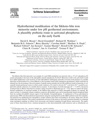
was placed on a sample of Sikhote-Alin (SA) which con-
tains a relatively large, well defined schreibersite inclusion
(Fig. 1). This cleaved, circular, polished SA fragment was
ca. 10 mm in diameter and the inclusion was ca. 3 mm in
length and 1 mm in width at its widest point.
92 D.E. Bryant et al. / Geochimica et Cosmochimica Acta 109 (2013) 90–112](https://image.slidesharecdn.com/1-s2-0-s0016703713000161-main-130424133654-phpapp02/85/1-s2-0-s0016703713000161-main-3-320.jpg)


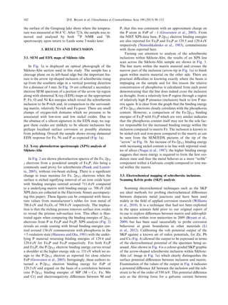
![nitrate salts (Fig. 4). The SKP output (u) can then be con-
verted to a potential value E via the calibration equation
E = f. u; where f = 0.456 from the calibration curve in
Fig. 4. The potential map illustrated in Fig. 4 is a montage
of two separate scans acquired sequentially.
2.2.4. Infinite focus microscopy (IFM)
Topographical analysis was carried out with an Alicona
Infinite Focus Microscope using a 5Â objective lens. Infi-
nite focus microscopy (IFM) is a non-contact optical tech-
nique that operates using focus-variation: two focal points,
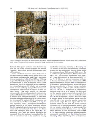
Fig. 10. Line-scan IFM image of the “arrow tip” region of the Sihote-Alin schreibersite inclusion, post-corrosion after cathodic
depolarisation cleaning of the surface. The image at right identifies the red line as traversing a crevice ca. 150 lm deep and 2 mm across.
Fig. 11. 31
P NMR spectrum (202.456 MHz; D2O) of water soluble extract from Sikhote-Alin following anaerobic digestion in the apparatus
of Fig. 6 (500 cm3
, 10% aqueous HCl; N2; 5 days 50 °C). [HPO4]2À
d 6.63 ppm. [HPO3]2À
d 4.22 ppm; 1
JPH = 566.9 Hz; [DPO3]2À
d 3.89;
1
JPD = 85 Hz (1:1:1 triplet).
D.E. Bryant et al. / Geochimica et Cosmochimica Acta 109 (2013) 90–112 97](https://image.slidesharecdn.com/1-s2-0-s0016703713000161-main-130424133654-phpapp02/85/1-s2-0-s0016703713000161-main-8-320.jpg)
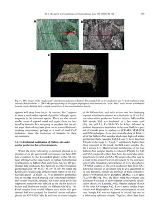
![Fig. 14. (a) Kverfjo¨ll volcanic region at the northern tip of the Vatnajoku¨ll glacier, SE Iceland. (b) Gengissig lake, Hveradalur geothermal
area (64° 40.1730
N; 16° 41.1000
W), Iceland. The geothermal site used for hydrothermal treatment of Sikhote-Alin is shown as a steaming, ice-
exposed region to the north of the lake. (c) Sikhote-Alin field sample SA1 incubating in fluid from liquid pool #1 (LP1; pH 3.1; T = 93–94 °C).
31
P NMR spectrum of post-incubation SA2 (d), SA4 (e), SA5 (f) and SA1 (g) fluids showing presence of both orthophosphate and and H-
phosphite [H2POÀ
4 ; d 0.08 ppm and H-phosphite, H2POÀ
3 ; d 2.71 ppm; 1
JPH 630 Hz for SA1]. (h) 31
P NMR spectrum of post-incubation SA1
fluid, evaporated and dry-heated to 85 °C under flowing dinitrogen atmosphere for 72 h. Present are pyrophosphite [PPi(III), H2P2O2À
5 ; d
À3.35 ppm and À6.64 ppm], Pi(III)-D [d 2.78 ppm; 1
JPD = 88 Hz], PPi(III)-D2 [multiplets at ca. d À4.7; À5.3 and À5.7 ppm], Pi(V) (d
1.85 ppm) and PPi(III–V) [multiplets at ca. d À2.6; À5.4 and À5.7 ppm].
100 D.E. Bryant et al. / Geochimica et Cosmochimica Acta 109 (2013) 90–112](https://image.slidesharecdn.com/1-s2-0-s0016703713000161-main-130424133654-phpapp02/85/1-s2-0-s0016703713000161-main-11-320.jpg)
![inclusion and matrix. The result of this galvanic couple
should be to polarise the matrix anodically, effectively mak-
ing the matrix more susceptible towards oxidation than the
inclusion. Whilst the SKP does not give information
regarding the time-evolution of the electrochemical reac-
tions that would occur in a corrosive environment, the tech-
nique is able to predict the likely location of the corrosive
attack that may occur. Such time-evolution information re-
quires linear polarisation resistance measurements (de Cris-
tofaro et al., 2012) which have been performed on a related
iron meteorite sample and will be described elsewhere. Con-
sequently, the large potential gradient between the two re-
gions suggests a strong tendency for the inclusion to
produce accelerated, localised corrosion of the matrix.
The ratio of the areas of the two components of this Gal-
vanic activity also have an effect upon the nature of the cor-
rosion. The consequence of a large cathodic matrix
containing a relatively small anodic inclusion would be ex-
pected to lead to accelerated dissolution of the inclusion,
whereas in the case of a comparatively small cathodic inclu-
sion, as is the situation with the sample studied, the corro-
sion due to the galvanic couple would be expected to occur
primarily on the matrix material around the interface with
the inclusion. This would result in a potential weakening of
physical attachment of the inclusion within the matrix and
hence ultimately a release of inclusion material which
would subsequently undergo, presumably slower, hydro-
thermal modification to release chemicals to the environ-
ment. To further probe this effect, we designed an
anaerobic hydrothermal reactor within which we could
probe accelerated corrosion of meteoritic fragments in the
presence of simulated low pH water environments. Follow-
ing corrosion and cathodic depolarisation to remove sur-
face oxide detritus, the sample could then be examined by
infinite focus microscopy to assess the validity of the above
electrochemical arguments.
3.4. Hydrothermal modification of Sikhote-Alin under
anaerobic simulated geothermal low pH environments
Infinite focus microscopy (IFM) is a relatively recently
developed technique for analysing surface morphology
and has found significant application in fields as diverse
as engineering corrosion (Jiang and Nesic, 2009) and bio-
materials (Winkler et al., 2010). The Sikhote-Alin sample
in Fig. 1 was analysed by IFM pre and post-acid corrosion
and the solution leachate analysed for Fe, Ni and P levels as
described in Section 2.2.9. Fig. 5 shows the Sikhote-Alin
inclusion imaged prior to the corrosion study. The surface
is essentially flat although a discontinuity can be seen at
the interface of inclusion with matrix. In addition, the inclu-
sion appears to show an increased level of roughness com-
pared to the matrix, a feature that we have seen and noted
previously through scanning electron microscopy and prob-
ably connected to the fact that schreibersitic inclusions have
increased levels of brittleness compared to the surrounding
matrix as indicated by their raised Vickers hardness num-
bers (Bryant et al., 2009).
The experimental arrangement for simulated anaerobic
corrosion used here is illustrated in Fig. 6, which shows
clearly the plastic coated cage in which the Sikhote-Alin
sample was suspended. Dinitrogen gas was bubbled
through a solution of 10% degassed hydrochloric acid for
a period of 5 days at 50 °C, so that the meteorite sample
would be subjected to condensed, low pH water within a
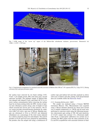
dynamic hydrothermal environment. Fig. 7 shows the
post-incubation surface of the Sikhote-Alin sample where
the inclusion is now difficult to see being largely obscured
by corrosion products except in the upper left corner where
the crust has flaked off to reveal a step. A line across this
step can be traced by IFM and the topographical change
along this line is plotted in Fig. 8. The topography of the
interface between the inclusion and the matrix shown in
Fig. 8 serves to reinforce the prediction of the SKP analysis
that corrosive attack would be most severe at the matrix-
inclusion boundary where local corrosion due to Galvanic
corrosion leads to accelerated anodic dissolution of the ma-
trix material over the inclusion. The presence of a steep,
sharp crevice at the inclusion side of the matrix-inclusion
boundary, with dimensions of ca. 40 lm depth and
120 lm wide, displays clear and preferential dissolution of
matrix material. Should such behaviour continue, one
would envisage weakening of the matrix-inclusion adhesion
to such a point that the inclusion may become sufficiently
weakened to allow it to be released from the surrounding
matrix. Indeed, this appears to be the case in practice as
illustrated by an IFM image of the arrow-tip inclusion
post-cathodic depolarisation to remove surface debris. In
this process the meteorite sample is rendered at a cathodic
potential in an electrochemical cell with the result that dihy-
drogen gas is produced from during reduction at the mete-
oritic electrode which serves to remove surface detritus. In
the process, a significant degree of corrosion appears to
have taken place surrounding the inclusion, which has
weakened its attachment to the encompassing matrix to
such an extent that a fragment of inclusion has also been
removed from its pre-incubation position resulting in a gap-
ing crevice (Figs. 9 and 10) visible in the line-scan trace with
dimensions ca. 150 mm deep and 2 mm across. The leachate
solution was analysed for dissolved Fe, Ni and P which
were measured to be present at concentrations of
440 ppm (Fe), 20 ppm (Ni) and 0.7 ppm (P). The latter
compared to a background in the distilled water of
0.007 ppm (see Electronic Annex for calibration and back-
grounds). Further analysis of the Fe-removed (addition of
Na2S) leachate using 31
P NMR spectroscopy identified
the major P-product to be the oxyacid H-phosphite,
[H2PO3]À
(Fig. 11) as compared against a known standard.
3.5. Raman mapping analysis of Sikhote-Alin post-anaerobic
corrosion
The relatively large ‘step’ across the ‘arrow head’ pre-
cluded efficient Raman mapping due to issues relating to
the depth of field at the appropriate (Â20) magnification.
Instead, two discrete sites of the corroded Sikote-Alin mete-
orite were chosen for micro-Raman mapping analysis. Site
1 incorporated a corroded ‘pit’, apparently with a relatively
large area of exposed metal surface; in contrast, Site 2 fea-
tured a substantial surface coverage of oxide material. In
D.E. Bryant et al. / Geochimica et Cosmochimica Acta 109 (2013) 90–112 103](https://image.slidesharecdn.com/1-s2-0-s0016703713000161-main-130424133654-phpapp02/85/1-s2-0-s0016703713000161-main-14-320.jpg)
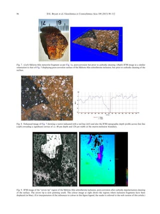
![common with other areas of the corroded meteorite surface,
Raman scattering was relatively weak and the resulting
spectra featured broadened peaks consistent with a largely
amorphous nature for the oxide deposits and/or several
microenvironments of the oxide materials. As iron oxides
have shown an ability to interconvert under laser irradia-
tion, care was taken to minimise the exposure of each site
through the use of relatively short exposure times (De Faria
et al., 1997). Comparison was made with reference spectra
for several iron oxide species implicated in oxidation,
including the Fe(III) oxides–hematite (a-Fe2O3), maghe-
mite (c-Fe2O3), the mixed Fe(III,II) oxides–magnetite,
and the oxyhydroxide series, goethite [a-Fe(OOH)], akag-
aneite [b-Fe(OOH)] and lepidocrocite (Remazeilles and Re-
fait, 2007; Re´guer et al., 2007; De Faria et al., 1997). While
the broadening of the spectra precluded specific identifica-
tion of the different morphological forms, e.g. the FeOOH
species, clear domains for Fe(III), Fe(II,III) oxides and
Fe(III) oxyhydroxides were identifiable in the spectra as
well as substantial areas of mixtures of these species
(Fig. 12). In this context, Raman images were generated
from multivariate analysis using Direct Classical Least
Squares and the best models for the image data were ob-
tained with three components: hematite, magnetite and goe-
thite. Other species were excluded from development
models on the basis of weak or absent correlation between
pixels in the images and the appropriate reference spectra
(Zhang et al., 2005). The resulting images are shown in
Fig. 13.
The distribution of oxide species in the maps are worthy
of note. At Site 1, the metal ‘pit’ appears to be bordered by
a preponderance of hematite, around which the lower oxi-
dation state magnetite appears. At this site, the major spe-
cies identified is Fe(II, III) although a small area of goethite
Fig. 15. SEM images and EDX maps of the “ducks head” schreibersite inclusion of Sikhote-Alin sample SA1 clearly displaying Ni-rich, Fe-
poor, P-rich nature of the inclusion against matrix. Some oxide materials are also clearly detectable on the surface; a mixture of iron oxides
and clay minerals from the geothermal fluids.
104 D.E. Bryant et al. / Geochimica et Cosmochimica Acta 109 (2013) 90–112](https://image.slidesharecdn.com/1-s2-0-s0016703713000161-main-130424133654-phpapp02/85/1-s2-0-s0016703713000161-main-15-320.jpg)
![bated image is a spur of schreibersite clearly visible at the
northern region of the principal inclusion which is not vis-
ible at all in the pre-incubated sample. Furthermore, a false-
colour topographic image (Fig. 19e) clearly shows the ma-
trix to have been corroded away from the, now sharply-de-
fined inclusion, in some regions to a depth of 100 lm and
some 300 lm wide (Fig. 19d) which is not present in the
pre-incubated sample.
The post-incubation fluid from SA1 above (containing
16.78 mg LÀ1
total P; Table 1) containing both orthophos-
phate and H-phosphite [Pi(III), H2POÀ
3 ; Fig. 14g] was pH
adjusted to 4 by addition of HClaq, followed by evapora-
tion and grinding of the resulting solid evaporate to a fine
powder. This was then heated to 85 °C in a sand bath under
a flowing atmosphere (ca. 1 cm3
sÀ1
flow) of dinitrogen for
a period of 72 h after which time the material was dissolved
in D2O, pH re-adjusted to ca. 7 and the P-components stud-
ies by 31
P NMR spectroscopy. The resulting spectrum
(Fig. 14h) reveals that a significant proportion (40+%) of
the total solution P is now present as the condensed oxy-
acid, pyrophosphite [PPi(III), H2P2O2À
5 ; d À3.35 and
À6.64 ppm] identified by comparison of its AA0
XX0
spin
system to an authentic sample (Bryant et al., 2010). Also
identifiable within this spectrum are products resulting
from H–D exchange within Pi(III) [d 2.78 ppm; 1
JPD = 88 -
Hz] and within PPi(III) [multiplets at ca. d À4.7; À5.3 and
À5.7 ppm] along with smaller signals due to Pi(V) (d
1.85 ppm) and mixed-valent species, isohypophosphate
PPi(III-V) [multiplets at ca. d À2.6; À5.4 and À5.7 ppm],
again identified by comparison to authentic samples (Car-
roll and Mesmer, 1967). That such a chemical condensation
of H-phosphite to pyrophosphite is potentially accessible
within a bona fide geological environment is illustrated by
the incubation of a dry sample of Ca(H2PO3)2.H2O, heated
to 94.4 °C in a Falcon tube inserted ca. 3 cm beneath the
sub-surface soil within the geothermal field at the edge of
the Gengissig lake (Fig. 20a). After ca. 3 days exposure,
analysis some 3 weeks later by both 31
P and 1
H NMR spec-
troscopy identified PPi(III) formation which was not pres-
ent in a reference sample of the same compound analysed
Fig. 18. IFM images of a schreibersite-matrix region of SA2; (a) pre-incubation and (b) post-incubation after cathodic depolarisation. (c and
d) Identify (red line) the vector across which IFM topological depth profiles (e) and (f) were recorded for both pre- and post-incubation
samples. (For interpretation of the references to colour in this figure legend, the reader is referred to the web version of this article.)
D.E. Bryant et al. / Geochimica et Cosmochimica Acta 109 (2013) 90–112 107](https://image.slidesharecdn.com/1-s2-0-s0016703713000161-main-130424133654-phpapp02/85/1-s2-0-s0016703713000161-main-18-320.jpg)
![without heating (Fig. 20). Pyrophosphite is an intriguing
material as it is structurally and chemically related to the
pyrophosphate [PPi(V)] moiety in nucleotide triphosphates
such as adenosine triphosphate (Fig. 21) the ubiquitous
suite of energy currency molecules of contemporary bio-
chemistry. Within a prebiotic context however, the advan-
tages of PPi(III) over PPi(V) are that it is, (i) formed
under far milder conditions than the latter and (ii) it is more
chemically reactive in the absence of sophisticated catalysis
(Bryant et al., 2010).
4. CONCLUSIONS
The emergence of phosphate-based biochemistry has
been a long-recognised problem in the field of abiogenesis
(Gulick, 1955). Phosphorus (P) in the fully oxidised +5 oxi-
dation state, as in contemporary biochemistry, has both
limited solubility in water in the presence of many common
metal ions (solubility products, Ksp at 25 °C for Ca3(PO4)2;
Mg3(PO4)2 and Fe(PO4)Á2H2O are 2.07 Â 10À33
;
1.04 Â 10À24
and 9.91 Â 10À16
respectively) and has rela-
tively low chemical reactivity in the absence of activating
agents (Steinman et al., 1965; Beck and Orgel, 1965; Oster-
berg and Orgel, 1972; Hermes-Lima and Vieyra, 1989). The
sophisticated enzymes of contemporary cellular life used to
activate P in energy currency molecules such as nucleoside
triphosphates (e.g. ATP), phosphocreatine and phosphoe-
nol pyruvate (Harold, 1986), are unlikely to have been
available within the Hadean period, however there is con-
siderable support for activated P-chemistry being central
Fig. 19. IFM images of a schreibersite-matrix region of SA4; (a) pre-incubation and (b) post-incubation after cathodic depolarisation with
highlighted vector (red line) line scans across which IFM topological depth profiles (c) and (d) were recorded for both pre- and post-
incubation samples. False colour image (e) shows a depth profile map where the more blue regions at the inclusion-matrix boundary represent
the deeper regions of the post-incubated sample. (For interpretation of the references to colour in this figure legend, the reader is referred to
the web version of this article.)
108 D.E. Bryant et al. / Geochimica et Cosmochimica Acta 109 (2013) 90–112](https://image.slidesharecdn.com/1-s2-0-s0016703713000161-main-130424133654-phpapp02/85/1-s2-0-s0016703713000161-main-19-320.jpg)
![to the bioenergetics of the early living organisms (Holm and
Baltscheffsky, 2011). The question then arises, how could
nature have activated geologically available P, predomi-
nantly in the form of orthophosphate (Bebie´ and Schoonen,
1999), in order to produce primitive energy currency mole-
cules? One suitable source of activated P on the early Earth
would have been siderophilic phosphide minerals, such as
schreibersite, (Fe,Ni)3P. Whilst such minerals are not com-
mon upon the Earth today, mainly due to their thermody-
namic instability with respect to oxidation to
orthophosphate, (Lauretta and Schmidt, 2009) they are
known to occur with natural metallic deposits on Disko Is-
land, Greenland (Klo¨ck et al., 1986) and to be produced by
chemical reduction of phosphates in soils during lightning
strikes (Pasek and Block, 2009). However, significant quan-
tities of schreibersite and related phosphide minerals would
likely have been delivered to the early Earth through mete-
oritic impacts and through interstellar dust particles
(IDP’s). Pasek and Lauretta have estimated such P-flux
rates during the putative late-heavy bombardment (between
ca. 4.0 and 3.8 Ga) and concluded that whilst both IDPs
and iron meteorites would likely have brought similar
quantities of siderophilic P to the early Earth (ca. 108
-
kg yrÀ1
), the far more localised impact events associated
with irons could have afforded very high local concentra-
tions of activated-P, in the region of 105
kg kmÀ2
(Pasek
and Lauretta, 2008). This process would require the interac-
tion of a hydrothermal system with a meteorite small
enough to impact and not destroy the system, yet large en-
ough to add enough reduced phosphorus to influence local
chemistry. The higher fall rate likely present on the early
earth (e.g. Johnson and Melosh, 2012; Bottke et al., 2012)
provided a greater frequency of meteorite falls to the early
earth. As most meteorites do not exceed a mass of about
50 tonnes, and have slowed significantly by ablation during
atmospheric entry, and have fragmented before impact,
meteorites in general should not destroy hydrothermal sys-
tems. An alternative to the random fall of a meteorite into a
hydrothermal pool is the de novo generation of hydrother-
mal systems after a large impact (Schwenzer and Kring,
2009; Osinski et al., in press), and the interaction of these
new systems with meteorite fragments from the impactor.
In this respect, an impact provides both the raw materials
(siderophilic phosphorus) and the environment (hydrother-
mal system) that has been investigated in the present work.
Our studies here on the low pH hydrothermal modifica-
tion of iron meteorites reveal that natural electrochemical
differences in composition between matrix Fe–Ni (taenite
and kamacite) and schreibersite inclusions result in prefer-
ential dissolution of matrix material at the matrix-inclusion
boundary leading to weakening of attachment of the inclu-
sion to the meteoritic matrix. This in turn should allow for
detachment of the inclusion with a consequent increase in
the availability of activated P to local water sources. Our re-
port here of the first field studies on low pH hydrothermal
modification of schreibersitic inclusions within Icelandic
Fig. 20. Sample of Ca(H2PO3)2ÁH2O, heated to 94.4 °C in a Falcon tube inserted ca. 3 cm beneath the soil at the edge of the Gengissig lake.
31
P NMR analysis (202.63 MHz; D2O; 300 K) of the heated solid, after ca. 3 days exposure, identified PPi(III) formation by comparison to an
authentic sample [d = À4.4 (AA’XX’, JPH 666 Hz; 0.7 Hz; JPP 17 Hz; (Bryant et al., 2010)].
Fig. 21. Molecular structures of (a) adenosine triphosphate (ATP) emphasising the condensed [P–O–P] molecular moieties between ab and bc
pairs of P atoms; (b) pyrophosphate, PPi(V), the main energy currency fragment of ATP and (c) pyrophosphite, PPi(III) a related molecular
cousin of PPi(V) with two [P–H] bonds replacing two [P–OH] groups of the latter.
D.E. Bryant et al. / Geochimica et Cosmochimica Acta 109 (2013) 90–112 109](https://image.slidesharecdn.com/1-s2-0-s0016703713000161-main-130424133654-phpapp02/85/1-s2-0-s0016703713000161-main-20-320.jpg)
![geothermal fields supports laboratory-based studies that P
in a lower oxidation state than +5, namely H-phosphite
(H2POÀ
3 ; where P is present formally as +3) is the chief
water-borne activated P oxyacid. Finally, we have demon-
strated that H-phosphite from low pH hydrothermal mod-
ification of irons can be readily condensed to pyrophosphite
[PPI(III)], a close structural and molecular cousin to pyro-
phosphate [PPi(V)], the energy currency component of
nucleotide triphosphates such as ATP. We propose that
the significance of PPi(III) as a prebiotically plausible en-
ergy currency molecule lies in its far greater range of chem-
ical reactivity that PPi(V), reactivity that is not limited to
the presence of sophisticated catalysts. Examples of this en-
hanced chemical reactivity will be described in a more spec-
ialised chemistry manuscript.
ACKNOWLEDGEMENTS
The authors are grateful for the financial support received to
support this work specifically, the Engineering and Physical Sci-
ences Research Council (Grant EP/F042558/1 to T.P.K.), the
Leverhulme Trust (Grant F07112AA to I.A.C.), the Science and
Technology Funding Council, and the UK Space Agency for the
award of an Aurora Fellowship (to T.P.K). We thank Dr. Laura
Carmody for field assistance, Dr. Thorsteinn Thorsteinsson, Mr.
Magnus Karlsson and the Icelandic Glaciological Society for logis-
tical support and Dr. Karen-Hudson Edwards and Mr. Antony Os-
born for assistance with dissolved ion chemistry analysis at the
Wolfson geochemistry laboratory at UCL/Birkbeck. The Natural
History Museum, London is thanked for providing samples of
the Sikhote-Akin meteorite. Finally, we thank the reviewers for
their insightful comments and suggestions.
APPENDIX A. SUPPLEMENTARY DATA
Supplementary data associated with this article can be
found, in the online version, at http://dx.doi.org/10.1016/
j.gca.2012.12.043.
REFERENCES
Alexander C. M. O. ’. D., Bowden R., Fogel M. L., Howard K. T.,
Herd C. D. K. and Nittler L. R. (2012) The provenances of
asteroids, and their contributions to the volatile inventories of
the terrestrial planets. Science 337, 721.
Bebie´ J. and Schoonen M. A. A. (1999) Pyrite and phosphate in
anoxia and an origin-of-life hypothesis. Earth Planet. Letts.
171, 1–5.
Beck A. and Orgel L. E. (1965) The formation of condensed
phosphate in aqueous solution. Proc. Natl. Acad. Sci. U.S.A.
54, 664–667.
Benedix G. K., McCoy T. J., Kiel K. and Love S. G. (2000) A
petrologic study of the IAB iron meteorites: constraints on the
formation of the IAB-Winonaite parent body. Meteorit. Planet.
Sci. 35, 1127–1141.
Bortnikova S. P., Bortnikova S. B., Gora M. P., Ya Shevko A.,
Lesnov F. P. and Kiryuhin A. V. (2010) Boiling mud pots:
origin and hydrogeochemistry (Donnoe and North-Mutnovsky
Fumarolic Fields, Mutnovsky Volcano; South Kamchatka,
Russia). In Proceedings World Geothermal Congress. pp. 1–7.
Bottke W. F., Vokrouhlicky D., Minton D., Nesvorny D.,
Morbidelli A., Brasser R., Simonson B. and Levison H. F.
(2012) An Archaean heavy bombardment from a destabilized
extension of the asteroid belt. Nature 485, 78–81.
Britvin S. N., Rudashevsky N. S., Krivovichev S. V., Burns P. C.
and Polekhovsky Y. S. (2002) Allabogdanite, (Fe, Ni)2P, a new
mineral from the Onello meteorite: the occurrence and crystal
structure. Am. Mineral. 87, 1245–1249.
Bryant D. E. and Kee T. P. (2006) Direct evidence for the
availability of reactive, water soluble phosphorus on the early
Earth. H-phosphinic acid from the Nantan meteorite. Chem.
Commun., 2344–2346.
Bryant D. E., Greenfield D., Walshaw R. D., Evans S. M., Nimmo
A. E., Smith C. L., Liming W., Pasek M. A. and Kee T. P.
(2009) Electrochemical studies of iron meteorites: phosphorus
redox chemistry on the early Earth. Int. J. Astrobiol. 8, 27–36.
Bryant D. E., Marriott K. E. R., MacGregor S. A., Kilner C.,
Pasek M. A. and Kee T. P. (2010) On the prebiotic potential of
reduced oxidation state phosphorus: the H-phosphinate–pyru-
vate system. Chem. Commun. 46, 3726–3728.
Buchwald V. F. and Clarke R. S. (1989) Corrosion of Fe–Ni alloys
by Cl-containing akaganeite (b-FeOOH): the Antarctic mete-
orite case. Am. Mineral. 74, 656–667.
Carroll R. L. and Mesmer R. E. (1967) Isohypophosphate: kinetics
of the hydrolysis and potentiometric and nuclear magnetic
resonance studies on the acidity and complexing. Inorg. Chem.
6, 1137–1142.
de Cristofaro N., Gallese F., Laguzzi G. and Luvidi L. (2012)
Selection of bronze alloys with reduced lead content suitable for
outdoor sculptures production. Mater. Chem. Phys. 132, 458–
465.
De Faria D. L. A., Venaˆncio Silva. S. and de Oliveira M. T. (1997)
Raman microspectroscopy of some iron oxides and oxyhy-
droxides. J. Raman Spectrosc. 28, 873–878.
Dessert C., Gaillardet J., Dupre B., Jacques Schott J. and
Pokrovsky O. S. (2009) Fluxes of high- versus low-temperature
water–rock interactions in aerial volcanic areas: example from
the Kamchatka Peninsula, Russia. Geochim. Cosmochim. Acta
73, 148–169.
El Goresy A., Ramdohr P. and Taylor L. A. (1971) The
geochemistry of the opaque minerals in Apollo 14 crystalline
rocks. Earth Planet. Sci. Lett. 13, 121–129.
Geist V., Wagner G., Nolze G. and Moretzki O. (2005) Investi-
gations of the meteoritic mineral (Fe, Ni)3P. Cryst. Res.
Technol. 40, 52–64.
Goldstein J. I. and Hopfe W. D. (2001) The metallographic cooling
rates of IVA iron meterorites. Meteorit. Planet. Sci. 36(S9),
A67.
Greenwood J. P., Itoh S., Sakamoto N., Warren P., Taylor L. and
Yurimoto H. (2011) Hydrogen isotope ratios in lunar rocks
indicate delivery of cometary water to the Moon. Nat. Geosci. 4,
79–82.
Grokhovsky V. I., Oshtrakh M. I., Milder O. B. and Semionkin V.
A. (2006) Mo¨ssbauer spectroscopy of iron meteorite Dronino
and products of its corrosion. Hyperfine Interact. 166, 671–677.
Grosvenor A. P., Wik S. D., Cavell R. G. and Mar A. (2005)
Examination of the bonding in binary transition-metal mono-
phosphides MP (M = Cr, Mn, Fe, Co) by X-ray photoelectron
spectroscopy. Inorg. Chem. 44, 8988–8998.
Gulick A. (1955) Phosphorus as a factor in the origin of life. Am.
Sci. 43, 479–489.
Hanawa T. and Ota M. (1991) Calcium phosphate naturally
formed on titanium in electrolyte solution. Biomaterials 12,
767–774.
Harold F. M. (1986) The Vital Force. A Study of Bioenergetics.
W.H. Freeman Co., New York, ISBN 0 7167 1734 4.
110 D.E. Bryant et al. / Geochimica et Cosmochimica Acta 109 (2013) 90–112](https://image.slidesharecdn.com/1-s2-0-s0016703713000161-main-130424133654-phpapp02/85/1-s2-0-s0016703713000161-main-21-320.jpg)


This document summarizes a study that investigated the hydrothermal modification of the Sikhote-Alin iron meteorite under low pH conditions to simulate volcanic environments. Analysis revealed that schreibersite inclusions in the meteorite are more electrochemically noble than the surrounding iron-nickel matrix by around 550 mV. This leads to preferential corrosion of the matrix at inclusion boundaries. The corrosion releases phosphorus from the schreibersite inclusions in the form of H-phosphite, a reactive form of phosphorus with prebiotic chemical potential. Field experiments showed that H-phosphite produced can be further converted to the condensed phosphorus compound pyrophosphite under mild heating, another compound with significance for pre


![Complete E material on Fundamentals of Biochemistry [2+1]; (32 Lectures)](https://cdn.slidesharecdn.com/ss_thumbnails/e-materialofbiochemistry-200405111750-thumbnail.jpg?width=640&height=640&fit=bounds)


