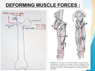
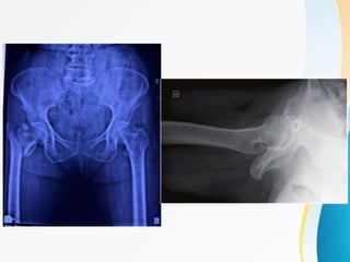
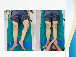
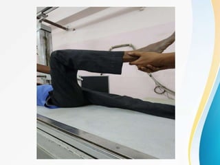
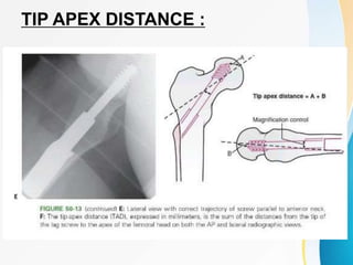
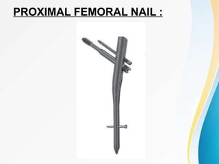
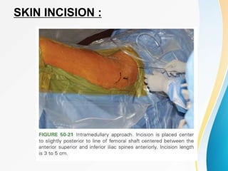
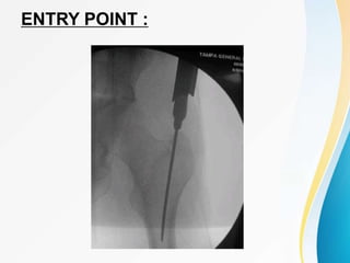
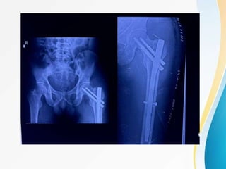
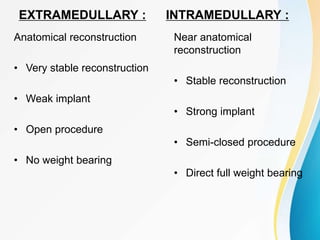
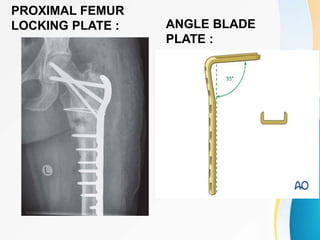
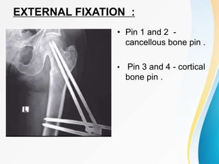
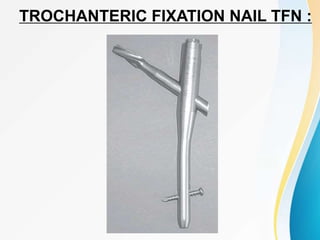
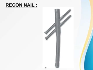
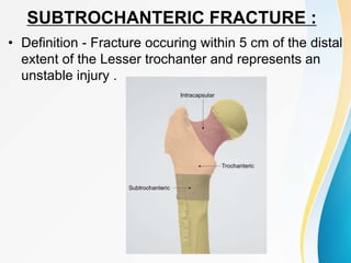
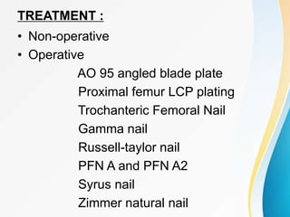
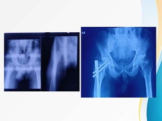
TROCHANTERIC FRACTURE AND
ITS MANAGEMENT
DR.VIGNESHWAR A
TROCHANTERIC FRACTURES :
• Extra-capsular
• occur in the region between Greater
trochanter and lesser trochanter of femur
often extending to the sub-trochanteric
region .
• part of pertrochanteric fracture.
• PERTROCHANTERIC FRACTURE
↓
1. Intertrochanteric fracture
2.Peritrochanteric fracture
• Extending from extracapsilar basilar neck
to LT (proximal to development of
medullary canal) .
HISTORY :
• Before 1930, treatment - conservative by traction until
healing ( ↑ morbidity )
• In 1930, jewett nail was introduced - immediate
stability of fracture fragments and early mobilization of
the patient .
• In 1950, Earnest roll - sliding screw and pugh &
badgley - sliding screw with Trephine tip.
• In 1962, Massie modified sliding screw to allow
collapse and impaction of fragments. Richard
manufacturing co. of USA produced Dynamic hip
screw
• In 1966, Kuntscher and later in 1970 Enders
introduced Condylocephalic intramedullary
devices.
• In 1984, Russel Taylor reconstructed im nail for
pertrochanteric and subtrochanteric fractures.
• In1992, Halder and williams introduced Gamma
nail.
ANATOMY OF PROXIMAL FEMUR :
• Proximal end of femur includes the Head, the
neck , Greater Trochanter, Lesser Trochanter
and intertrochanteric line and intertrochanteric
crest .
• Normal neck shaft angle : 120 to 130 ⁰
• Angle of anteversion : 10 to 30 ⁰
• Intertrochanteric region : Area between GT and
LT represents the zone of transition from the
femur neck to femoral shaft , characterized
primarily by Dense trabecular bone .
AP PA
BLOOD SUPPLY :
Crock described the blood supply to
proximal end of femur , into 3 main groups
1.Extracapsular arterial ring at the base of
femoral neck
2.Ascending cervical branches of the arterial ring
on the surface of femoral neck ( retinacular
vessels)
3.Arteries of the ligamentum teres.
TRABECULAR SYSTEM OF PROXIMAL
FEMUR
• Described by WARD in 1838 .
• In accordance with Wolffs law, trabeculations
arise along the Lines of force to which the bone
is exposed.
• It forms the transition of shaft cortex into
metaphyseal cancellous bone .
• WARD’s triangle .
• non homologous pattern of bone density and
stiffness particularly apparent in osteoporotic
patient
CALCAR FEMORALE :
• Dense vertical
plate of bone .
• posterio-medial
part of femoral
shaft .
DEFORMING MUSCLE FORCES :
MECHANISM OF INJURY :
• 90% IT femur fractures in elderly occurs due to a
trivial fall .
• Fall from standing height .
• most fractures result from a Direct impact to the
greater trochanter area.
• YOUNGER INDIVIDUALS - High energy trauma (
MVA OR FALL FROM HEIGHT )
• ↑ fracture incidence with ↑ Age .
• Hip fractures also results from Cyclic mechanical
stresses like fatigue/pathological fractures
secondary to osteoporosis,osteomalacia, etc.,
CUMMING’s FACTORS :
• The Patient must be oriented to fall or "impact" near
the hip .
• Local Shock absorbers (inadequate soft tissue -
muscle/fat coverage) to prevent fracture .
• Protective responses must be inadequate to reduce
the energy of the fall beyond a certain critical
threshold .
• Residual energy of the fall applied to the proximal
femur must exceed its strength (i.e. Bone strength at
the hip must be insufficient .
ASSOCIATED INJURIES :
• Distal radius / proximal humerus fracture
( most common )
• High energy trauma - ipsilateral lower limb
fractures , pelvic fractures ,.
• Head injury
• Primary neoplastic / metastatic disease -
preceding hip discomfort and subsequent fall .
CLINICAL FEATURES :
• Pain and swelling at the hip
• inability to ambulate after a fall
• limb shortened with External rotation
deformity.
• ecchymosis over the GT .
• undisplaced/ impacted fracture - pt may be
ambulatory .
• Auscultation Lippmann test - sensitive for
detection of occult fractures of the proximal
femur or pelvis .
• Bell of the stethoscope on symphysis pubis and
tapping on the patella of both extremities -
variation in sound conduction determines
discontinuity
• Decreased tone or pitch - Fracture .
RADIOGRAPHIC IMAGING :
• Anteroposterior view - to know fracture
obliquity , quality of the bone .
• cross table lateral view - to assess size,
location and comminution of posterior fracture
fragment and helps to determine the fracture
stability .
• CT and MRI
• Technetium bone scan - suspected hip fracture
but not apparent to standard radiographs , it
requires 2-3 days to become positive .
CLASSIFICATION :
• In 1822, Astley cooper described the first Pre-
radiographic classification of hip fractures ,
- Intra-capsular ( main complication - Non union )
- Extra-capsular ( main complication -Coxa vara )
BOYD AND GRIFFIN CLASSIFICATION :
• TYPE 1 Stable (Two part) Fracture
• TYPE 2 Unstable with posteromedial communition
• TYPE 3 Subtrochanteric extension into lateral shaft,
extension of the fracture distally at or just below the
lesser trochanter (the term Reverse Obliquity was
coined by Wright)
• TYPE 4 Subtrochanteric with intertrochanteric
extension with the fracture lying in atleast two
planes.
EVANS CLASSIFICATION :
Distinguished stable from unstable fractures and also
defined the characteristics of a stable reduction.
• Stable fracture patterns - posteromedial cortex
remains intact OR minimal communition
• Unstable fracture patterns - Disrupted
posteromedial cortex - can be converted into
stable if medial cortical opposition is maintained.
• Reverse Oblique - Inherently unstable due to the
tendency for medial displacement of the femoral
shaft .
OTA/AO CLASSIFICATION : 31 A
DORR’S INDEX :
CANAL-CALCAR RATIO :
DORR’S INDEX :
• Selection of cemented or Non cemented femoral
arthroplasty components .
• Used for implant selection for Hip fracture patients .
• Type A - young patient,narrow metaphysis,Thick
cortex and high constricting isthmus .
↓
Excessive bone removal required for intramedullary
devices either a plate type construct or smaller
diameter reconstruction nail are bone conserving .
Type C - most problematic in geriatric populations
with hip fractures , Wide metephysis , wide medullary
canal and loss of isthmus constriction .
TREATMENT :
NON-OPERATIVE :
• Prolonged bedrest in traction until fracture healing
occurs (usually 10 to 12 weeks), followed by a
lengthy program of ambulation training .
Can be done for :
1. An elderly person with high risk of
mortality from anaesthesia and surgery.
2. Nonambulatory patient who has minimal
discomfort following fracture .
3. Undisplaced/Minimal displaced fracture .
METHODS OF NON-OPERATIVE TREATMENT
• Buck's traction or extension
• Russell skeletal traction
• Balanced traction in Thomas splint
• Plaster spica immobilization
• De-rotation boot
skin traction :
Skeletal traction :
COMPLICATIONS :
• Varus Deformity with Limb shortening .
• Pin-tract infection in skeletal traction
• Decubitus ulcers
• Urinary tract infection
• Pneumonia
• Thromboembolic complications .
OPERATIVE TREATMENT :
• Pertrochanteric fractures are best treated
with Surgical Repair .
• Goal : Strong and stable fixation of fracture
segments .
• OUTLINE :
Position
Approaches
Implant of choice
complications
POSITION :
LATERAL SURGICAL APPROACH :
WATSON-JONES APPROACH :
SLIDING HIP SCREW SHS :
DYNAMIC HIP SCREW :
• PRINCIPLE : Controlled collapse
• Dynamic action reduces the incidence of screw
cut out and penetration of screw into hip joint .
• Used for Stable per-trochanteric fracture .
• Lateral cortical wall of the proximal femur should
be intact ( pre-requisite).
• If not intact , DHS + TSP ( trochanteric
stabilisation plate ) should be used .
SHS :
TIP APEX DISTANCE : TAD
• Sum of distances from the tip of the lag screw
to the apex of the femoral head on both the
anteroposterior and lateral radiographic views.
• The sum should be <25mm to minimize the risk
of lag screw cutout .
• TAD > 25 mm , the surgeon should reassess
the fracture reduction and position of Guide
Pin.
TIP APEX DISTANCE :
PROXIMAL FEMORAL NAIL :
• Better fixation and stability compared to SHS.
• Has two screws- load bearing lag screw and
proximal thinner antirotation screw to counter
rotational tendency.
• Distal locking screws to control rotation and to
increase construct stiffness.
PROXIMAL FEMORAL NAIL :
• The lag screw withstands the bending moment which is
transferred to the intramedullary nail and
counterbalanced by its locking mechanism with the
femoral cortex in the medullary canal.
• Entire load is transferred to the nail and a negligible
portion to the medial femoral cortex.
• PFN acts as an BUTTRESS in preventing medialisation of
the shaft . The entry portal of the PFN through the
trochanter limits the surgical insult to the tendinous hip
abductor muscle only, unlike those nails which require
entry through piriformis fossa .
SKIN INCISION :
ENTRY POINT :
ADVANTAGES OF PFN :
• Superior biomechanics
- Shorter lever arm
- ↓ Tensile strain on the implant
- Controlled shaft medialisation and
collapse
- Less soft tissue dissection
- Early weight bearing
EXTRAMEDULLARY :
Anatomical reconstruction
• Very stable reconstruction
• Weak implant
• Open procedure
• No weight bearing
INTRAMEDULLARY :
Near anatomical
reconstruction
• Stable reconstruction
• Strong implant
• Semi-closed procedure
• Direct full weight bearing
• Lateral migration of the lag screw and the
concomitant medial migration of the
antirotation screw.
• Both the screws come under cyclic loading.
• The proximal thinner screw bends easily, jams,
fails to slide and cuts off the head.
• The distal thicker screw is minimally affected
retaining its sliding properties, permits
impaction of metaphyseal fracture area.
Z EFFECT :
POST-OPERATIVE RADIOGRAPHIC ANALYSIS
• 1. Neck shaft angle
• 2. Neck length
• 3. Horizontal offset
• 4. Cal TAD
• 5. Position of hip screw - Parker's ratio
• 6. Greater trochanter orthogonal line (GTOL)
• 7. Chang's reduction quality criteria(CRQC)
PARKER’S RATIO :
• The Parker's ratio on AP view
is represented by the
percentage AB/ACx100
indicating the distance of the
screw from the inferior border
of the femoral neck (the
superior border is considered
as 100%)
• Cut- out is the most common
mechanical complication of the
osteosynthesis of the
pertrochanteric fracture .
GREATER TROCHANTERIC
ORTHROGONAL LINE :
A) Passing through 1st quadrant - varus NSA
B) Passing through 2nd quadrant - valgus/normal NSA
C) Normal hip GTOL through 2nd quadrant
CHANG’S REDUCTION QUALITY CRITERIA
POSITIVE MEDIAL
CORTEX SUPPORT
NEGATIVE MEDIAL
CORTEX SUPPORT
OTHER IMPLANTS :
Jewett Nail :
SP Nail with
Mclaughlin’s plate :
MEDOFF SLIDING PLATE :
PROXIMAL FEMUR
LOCKING PLATE : ANGLE BLADE
PLATE :
EXTERNAL FIXATION :
• Pin 1 and 2 -
cancellous bone pin .
• Pin 3 and 4 - cortical
bone pin .
TROCHANTERIC FIXATION NAIL TFN :
GAMMA NAIL :
RECON NAIL :
InterTAN :
RECENT ADVANCES :
PFNA 2
• Medio-lateral angle - 5
degrees .
• optimal stress distribution
• lateral flattened cross section
• rotational and angular
stability achieved with one
single element .
• No Z-effect
• Higher cutout resistance .
POSTOPERATIVE CARE :
• Ambulation - under supervision with weight
bearing as tolerated with a walker or crutches .
• Regular strengthening exercises .
• complete weight bearing - Radiographic healing
should be there .
• Radiographic follow up : 6 weeks , 3 months,
6 months, 1 year and 2 year .
• D V T prophylaxis for 6 weeks postoperatively .
• vitamin D3 supplementation
SUBTROCHANTERIC FRACTURE :
• Definition - Fracture occuring within 5 cm of the distal
extent of the Lesser trochanter and represents an
unstable injury .
RUSSELL-TAYLOR CLASSIFICATION :
AO/OTA CLASSIFICATION :
FIELDING AND MAGLIATO’S :
• Type 1 Fracture at the
level of LT
• Type 2 Fracture
2.5cm - 5 cm below
the level of LT
• Type 3 Fracture > 5
cm below the lesser
trochanter .
SEINSHEIMER’S CLASSIFICATION :
• Type 1 Non-displaced / less than 2 mm displacement .
• Type 2 Two part fracture
A.Transverse fracture > 2mm displ.
B.Spiral configuration with LT attached to
proximal fragment
C.Spiral configuration with LT attached to distal
fragment .
• Type 3 Three part fracture
A.Spiral configuration with LT a part of Third
fragment
B.Spiral configuration with third part a butterfly
fragment .
• Type 4 Comminuted fracture with four or more
fragments
• Type 5 Subtrochanteric with intertrochanteric
TREATMENT :
• Non-operative
• Operative
AO 95 angled blade plate
Proximal femur LCP plating
Trochanteric Femoral Nail
Gamma nail
Russell-taylor nail
PFN A and PFN A2
Syrus nail
Zimmer natural nail
COMPLICATIONS :
• Delayed union
• Non-union ( rare in IT fractures )
• Malunion
• Infection
• Implant failure
• Infections
HIP FRACTURES IN CHILDREN :
• classification proposed by Delbet and
popularized by Colonna .
HIP FRACTURES IN CHILDREN :
COMPLICAGTIONS OF HIP FRACTURES IN
CHILDREN :
• Avascular Necrosis
• Coxa Vara
• Non-Union
• Premature epiphyseal closure
MANAGEMENT :
• Rapid union (within 6 to 8 weeks) child’s osteogenic
potential ↑↑
• Initially , skeletal traction for 2-3 weeks to
obtain reduction
↓
Abduction spica cast for 6-12 weeks
If not reduced with TRACTION
↓
closed manipulation + abduction spica cast
Rarely, internal fixation required ( depends on the age
of child ) .
SUBTROCHANTERIC FRACTURES IN
CHILDREN
• WADDELL’S TRIAD :
Fractured femoral shaft
Intra-thoracic/intra-abdominal injury
Contralateral Head injury
MANAGEMENT :
• Infants 0-6 months : pavlik harness
• children 6 months to 5 years : Hip spica cast if
fracture unstable Internal fixation necessary
• children 5-11 years : Flexible IM nails,open plating
or submuscular plating
• children > 11 years : Rigid IM nails using lateral
trochanteric entry .
THANK YOU

2TROCHANTERIC FRACTURES VIGNESH.pptx

  • 1.
    TROCHANTERIC FRACTURE AND ITSMANAGEMENT DR.VIGNESHWAR A
  • 2.
    TROCHANTERIC FRACTURES : •Extra-capsular • occur in the region between Greater trochanter and lesser trochanter of femur often extending to the sub-trochanteric region . • part of pertrochanteric fracture.
  • 3.
    • PERTROCHANTERIC FRACTURE ↓ 1.Intertrochanteric fracture 2.Peritrochanteric fracture • Extending from extracapsilar basilar neck to LT (proximal to development of medullary canal) .
  • 4.
    HISTORY : • Before1930, treatment - conservative by traction until healing ( ↑ morbidity ) • In 1930, jewett nail was introduced - immediate stability of fracture fragments and early mobilization of the patient . • In 1950, Earnest roll - sliding screw and pugh & badgley - sliding screw with Trephine tip. • In 1962, Massie modified sliding screw to allow collapse and impaction of fragments. Richard manufacturing co. of USA produced Dynamic hip screw
  • 5.
    • In 1966,Kuntscher and later in 1970 Enders introduced Condylocephalic intramedullary devices. • In 1984, Russel Taylor reconstructed im nail for pertrochanteric and subtrochanteric fractures. • In1992, Halder and williams introduced Gamma nail.
  • 6.
    ANATOMY OF PROXIMALFEMUR : • Proximal end of femur includes the Head, the neck , Greater Trochanter, Lesser Trochanter and intertrochanteric line and intertrochanteric crest . • Normal neck shaft angle : 120 to 130 ⁰ • Angle of anteversion : 10 to 30 ⁰ • Intertrochanteric region : Area between GT and LT represents the zone of transition from the femur neck to femoral shaft , characterized primarily by Dense trabecular bone .
  • 7.
  • 9.
    BLOOD SUPPLY : Crockdescribed the blood supply to proximal end of femur , into 3 main groups 1.Extracapsular arterial ring at the base of femoral neck 2.Ascending cervical branches of the arterial ring on the surface of femoral neck ( retinacular vessels) 3.Arteries of the ligamentum teres.
  • 13.
    TRABECULAR SYSTEM OFPROXIMAL FEMUR • Described by WARD in 1838 . • In accordance with Wolffs law, trabeculations arise along the Lines of force to which the bone is exposed. • It forms the transition of shaft cortex into metaphyseal cancellous bone . • WARD’s triangle . • non homologous pattern of bone density and stiffness particularly apparent in osteoporotic patient
  • 16.
    CALCAR FEMORALE : •Dense vertical plate of bone . • posterio-medial part of femoral shaft .
  • 17.
  • 18.
    MECHANISM OF INJURY: • 90% IT femur fractures in elderly occurs due to a trivial fall . • Fall from standing height . • most fractures result from a Direct impact to the greater trochanter area. • YOUNGER INDIVIDUALS - High energy trauma ( MVA OR FALL FROM HEIGHT ) • ↑ fracture incidence with ↑ Age . • Hip fractures also results from Cyclic mechanical stresses like fatigue/pathological fractures secondary to osteoporosis,osteomalacia, etc.,
  • 19.
    CUMMING’s FACTORS : •The Patient must be oriented to fall or "impact" near the hip . • Local Shock absorbers (inadequate soft tissue - muscle/fat coverage) to prevent fracture . • Protective responses must be inadequate to reduce the energy of the fall beyond a certain critical threshold . • Residual energy of the fall applied to the proximal femur must exceed its strength (i.e. Bone strength at the hip must be insufficient .
  • 20.
    ASSOCIATED INJURIES : •Distal radius / proximal humerus fracture ( most common ) • High energy trauma - ipsilateral lower limb fractures , pelvic fractures ,. • Head injury • Primary neoplastic / metastatic disease - preceding hip discomfort and subsequent fall .
  • 21.
    CLINICAL FEATURES : •Pain and swelling at the hip • inability to ambulate after a fall • limb shortened with External rotation deformity. • ecchymosis over the GT . • undisplaced/ impacted fracture - pt may be ambulatory .
  • 23.
    • Auscultation Lippmanntest - sensitive for detection of occult fractures of the proximal femur or pelvis . • Bell of the stethoscope on symphysis pubis and tapping on the patella of both extremities - variation in sound conduction determines discontinuity • Decreased tone or pitch - Fracture .
  • 24.
    RADIOGRAPHIC IMAGING : •Anteroposterior view - to know fracture obliquity , quality of the bone . • cross table lateral view - to assess size, location and comminution of posterior fracture fragment and helps to determine the fracture stability . • CT and MRI • Technetium bone scan - suspected hip fracture but not apparent to standard radiographs , it requires 2-3 days to become positive .
  • 28.
    CLASSIFICATION : • In1822, Astley cooper described the first Pre- radiographic classification of hip fractures , - Intra-capsular ( main complication - Non union ) - Extra-capsular ( main complication -Coxa vara )
  • 29.
    BOYD AND GRIFFINCLASSIFICATION : • TYPE 1 Stable (Two part) Fracture • TYPE 2 Unstable with posteromedial communition • TYPE 3 Subtrochanteric extension into lateral shaft, extension of the fracture distally at or just below the lesser trochanter (the term Reverse Obliquity was coined by Wright) • TYPE 4 Subtrochanteric with intertrochanteric extension with the fracture lying in atleast two planes.
  • 31.
  • 32.
    Distinguished stable fromunstable fractures and also defined the characteristics of a stable reduction. • Stable fracture patterns - posteromedial cortex remains intact OR minimal communition • Unstable fracture patterns - Disrupted posteromedial cortex - can be converted into stable if medial cortical opposition is maintained. • Reverse Oblique - Inherently unstable due to the tendency for medial displacement of the femoral shaft .
  • 33.
  • 34.
  • 35.
  • 36.
    DORR’S INDEX : •Selection of cemented or Non cemented femoral arthroplasty components . • Used for implant selection for Hip fracture patients . • Type A - young patient,narrow metaphysis,Thick cortex and high constricting isthmus . ↓ Excessive bone removal required for intramedullary devices either a plate type construct or smaller diameter reconstruction nail are bone conserving . Type C - most problematic in geriatric populations with hip fractures , Wide metephysis , wide medullary canal and loss of isthmus constriction .
  • 37.
    TREATMENT : NON-OPERATIVE : •Prolonged bedrest in traction until fracture healing occurs (usually 10 to 12 weeks), followed by a lengthy program of ambulation training . Can be done for : 1. An elderly person with high risk of mortality from anaesthesia and surgery. 2. Nonambulatory patient who has minimal discomfort following fracture . 3. Undisplaced/Minimal displaced fracture .
  • 38.
    METHODS OF NON-OPERATIVETREATMENT • Buck's traction or extension • Russell skeletal traction • Balanced traction in Thomas splint • Plaster spica immobilization • De-rotation boot
  • 39.
  • 40.
    COMPLICATIONS : • VarusDeformity with Limb shortening . • Pin-tract infection in skeletal traction • Decubitus ulcers • Urinary tract infection • Pneumonia • Thromboembolic complications .
  • 41.
    OPERATIVE TREATMENT : •Pertrochanteric fractures are best treated with Surgical Repair . • Goal : Strong and stable fixation of fracture segments . • OUTLINE : Position Approaches Implant of choice complications
  • 42.
  • 43.
  • 44.
  • 46.
  • 47.
    DYNAMIC HIP SCREW: • PRINCIPLE : Controlled collapse • Dynamic action reduces the incidence of screw cut out and penetration of screw into hip joint . • Used for Stable per-trochanteric fracture . • Lateral cortical wall of the proximal femur should be intact ( pre-requisite). • If not intact , DHS + TSP ( trochanteric stabilisation plate ) should be used .
  • 48.
  • 49.
    TIP APEX DISTANCE: TAD • Sum of distances from the tip of the lag screw to the apex of the femoral head on both the anteroposterior and lateral radiographic views. • The sum should be <25mm to minimize the risk of lag screw cutout . • TAD > 25 mm , the surgeon should reassess the fracture reduction and position of Guide Pin.
  • 50.
  • 51.
  • 52.
    • Better fixationand stability compared to SHS. • Has two screws- load bearing lag screw and proximal thinner antirotation screw to counter rotational tendency. • Distal locking screws to control rotation and to increase construct stiffness. PROXIMAL FEMORAL NAIL :
  • 53.
    • The lagscrew withstands the bending moment which is transferred to the intramedullary nail and counterbalanced by its locking mechanism with the femoral cortex in the medullary canal. • Entire load is transferred to the nail and a negligible portion to the medial femoral cortex. • PFN acts as an BUTTRESS in preventing medialisation of the shaft . The entry portal of the PFN through the trochanter limits the surgical insult to the tendinous hip abductor muscle only, unlike those nails which require entry through piriformis fossa .
  • 54.
  • 55.
  • 57.
    ADVANTAGES OF PFN: • Superior biomechanics - Shorter lever arm - ↓ Tensile strain on the implant - Controlled shaft medialisation and collapse - Less soft tissue dissection - Early weight bearing
  • 58.
    EXTRAMEDULLARY : Anatomical reconstruction •Very stable reconstruction • Weak implant • Open procedure • No weight bearing INTRAMEDULLARY : Near anatomical reconstruction • Stable reconstruction • Strong implant • Semi-closed procedure • Direct full weight bearing
  • 59.
    • Lateral migrationof the lag screw and the concomitant medial migration of the antirotation screw. • Both the screws come under cyclic loading. • The proximal thinner screw bends easily, jams, fails to slide and cuts off the head. • The distal thicker screw is minimally affected retaining its sliding properties, permits impaction of metaphyseal fracture area. Z EFFECT :
  • 61.
    POST-OPERATIVE RADIOGRAPHIC ANALYSIS •1. Neck shaft angle • 2. Neck length • 3. Horizontal offset • 4. Cal TAD • 5. Position of hip screw - Parker's ratio • 6. Greater trochanter orthogonal line (GTOL) • 7. Chang's reduction quality criteria(CRQC)
  • 62.
    PARKER’S RATIO : •The Parker's ratio on AP view is represented by the percentage AB/ACx100 indicating the distance of the screw from the inferior border of the femoral neck (the superior border is considered as 100%) • Cut- out is the most common mechanical complication of the osteosynthesis of the pertrochanteric fracture .
  • 63.
    GREATER TROCHANTERIC ORTHROGONAL LINE: A) Passing through 1st quadrant - varus NSA B) Passing through 2nd quadrant - valgus/normal NSA C) Normal hip GTOL through 2nd quadrant
  • 64.
  • 65.
  • 66.
    OTHER IMPLANTS : JewettNail : SP Nail with Mclaughlin’s plate :
  • 67.
  • 68.
    PROXIMAL FEMUR LOCKING PLATE: ANGLE BLADE PLATE :
  • 69.
    EXTERNAL FIXATION : •Pin 1 and 2 - cancellous bone pin . • Pin 3 and 4 - cortical bone pin .
  • 70.
  • 71.
  • 72.
  • 73.
  • 74.
    RECENT ADVANCES : PFNA2 • Medio-lateral angle - 5 degrees . • optimal stress distribution • lateral flattened cross section • rotational and angular stability achieved with one single element . • No Z-effect • Higher cutout resistance .
  • 75.
    POSTOPERATIVE CARE : •Ambulation - under supervision with weight bearing as tolerated with a walker or crutches . • Regular strengthening exercises . • complete weight bearing - Radiographic healing should be there . • Radiographic follow up : 6 weeks , 3 months, 6 months, 1 year and 2 year . • D V T prophylaxis for 6 weeks postoperatively . • vitamin D3 supplementation
  • 76.
    SUBTROCHANTERIC FRACTURE : •Definition - Fracture occuring within 5 cm of the distal extent of the Lesser trochanter and represents an unstable injury .
  • 77.
  • 78.
  • 79.
    FIELDING AND MAGLIATO’S: • Type 1 Fracture at the level of LT • Type 2 Fracture 2.5cm - 5 cm below the level of LT • Type 3 Fracture > 5 cm below the lesser trochanter .
  • 80.
    SEINSHEIMER’S CLASSIFICATION : •Type 1 Non-displaced / less than 2 mm displacement . • Type 2 Two part fracture A.Transverse fracture > 2mm displ. B.Spiral configuration with LT attached to proximal fragment C.Spiral configuration with LT attached to distal fragment . • Type 3 Three part fracture A.Spiral configuration with LT a part of Third fragment B.Spiral configuration with third part a butterfly fragment . • Type 4 Comminuted fracture with four or more fragments • Type 5 Subtrochanteric with intertrochanteric
  • 81.
    TREATMENT : • Non-operative •Operative AO 95 angled blade plate Proximal femur LCP plating Trochanteric Femoral Nail Gamma nail Russell-taylor nail PFN A and PFN A2 Syrus nail Zimmer natural nail
  • 83.
    COMPLICATIONS : • Delayedunion • Non-union ( rare in IT fractures ) • Malunion • Infection • Implant failure • Infections
  • 84.
    HIP FRACTURES INCHILDREN :
  • 85.
    HIP FRACTURES INCHILDREN : • classification proposed by Delbet and popularized by Colonna .
  • 86.
    HIP FRACTURES INCHILDREN :
  • 87.
    COMPLICAGTIONS OF HIPFRACTURES IN CHILDREN : • Avascular Necrosis • Coxa Vara • Non-Union • Premature epiphyseal closure
  • 88.
    MANAGEMENT : • Rapidunion (within 6 to 8 weeks) child’s osteogenic potential ↑↑ • Initially , skeletal traction for 2-3 weeks to obtain reduction ↓ Abduction spica cast for 6-12 weeks If not reduced with TRACTION ↓ closed manipulation + abduction spica cast Rarely, internal fixation required ( depends on the age of child ) .
  • 89.
    SUBTROCHANTERIC FRACTURES IN CHILDREN •WADDELL’S TRIAD : Fractured femoral shaft Intra-thoracic/intra-abdominal injury Contralateral Head injury
  • 90.
    MANAGEMENT : • Infants0-6 months : pavlik harness • children 6 months to 5 years : Hip spica cast if fracture unstable Internal fixation necessary • children 5-11 years : Flexible IM nails,open plating or submuscular plating • children > 11 years : Rigid IM nails using lateral trochanteric entry .
  • 91.

Editor's Notes

  • #36 type A-champagne and type C - stove-pipe apperance
  • #46 reduction with percutaneous pin using COACH LEVER MANEUVER
  • #47 richards screw pitch 3 thread dia 14 length 22mm core diameter 8mm
  • #63 less than 40%.... CUTOUT - varus collapse of the femoral head neck fragment with extrusion of cephalic screw .
  • #74 LINEAR COMPRESSSION
  • #79 FRACTURE LOCATION GEOMETRY AND PRESENCE OF COMMINUTION