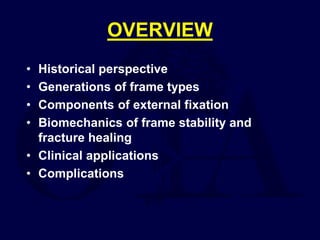
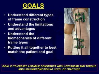
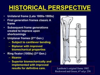
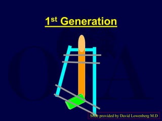
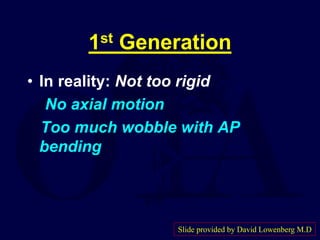
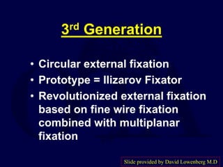
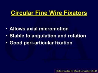
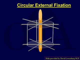
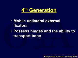
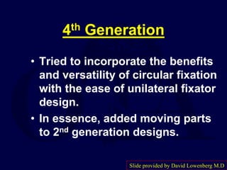
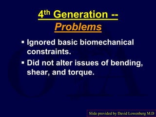
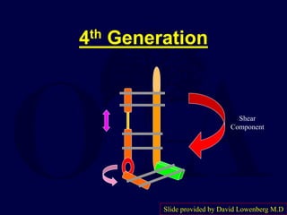
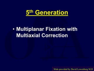
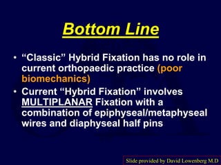
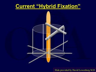
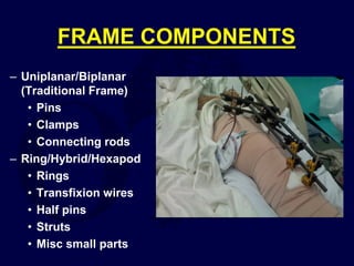
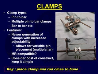
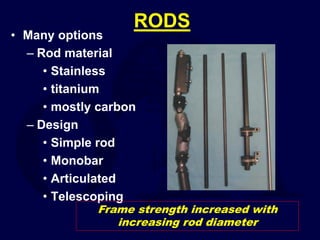
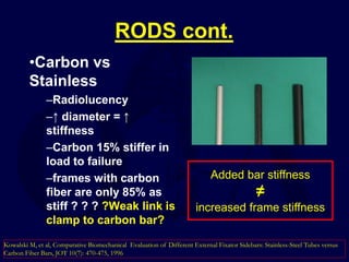
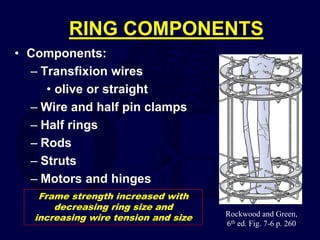
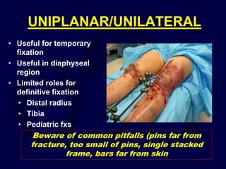
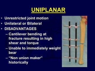
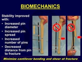
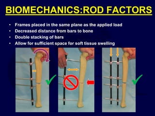
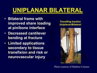
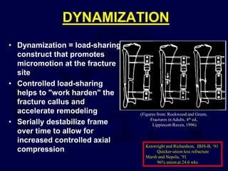
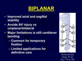
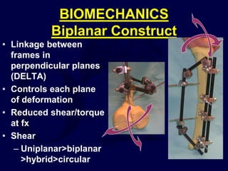
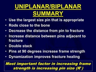
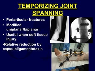
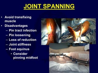
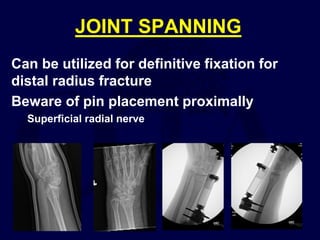
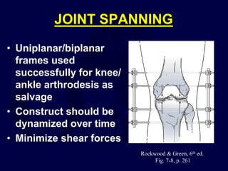
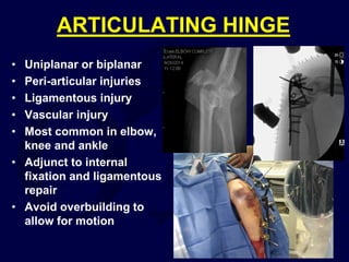
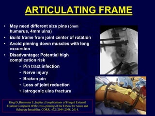
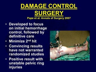
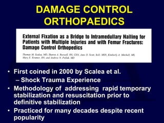
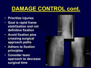
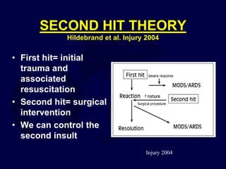
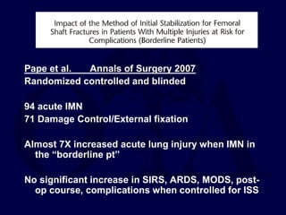
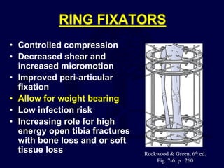
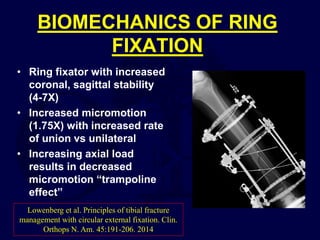
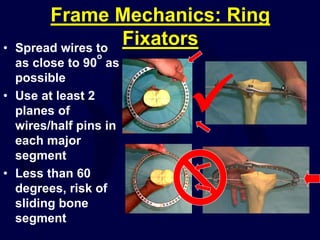
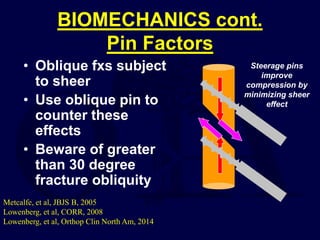
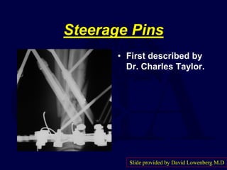
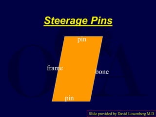
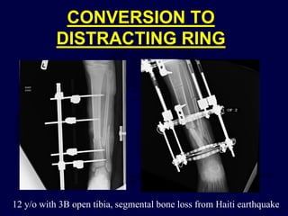
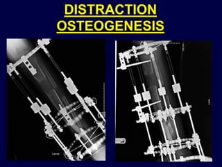
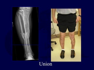
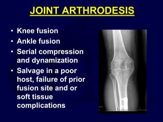
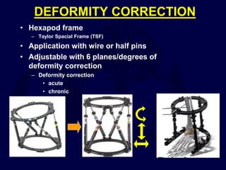
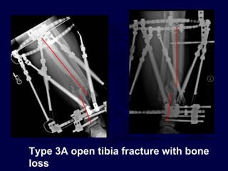
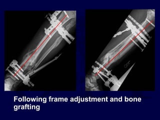
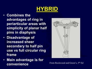
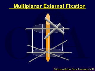
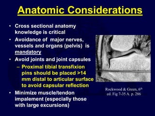
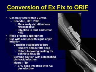
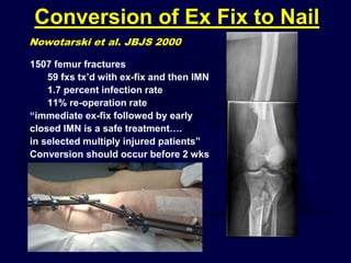
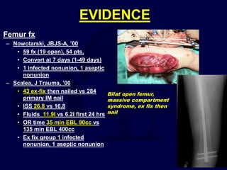
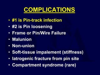
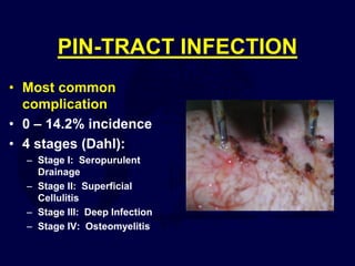
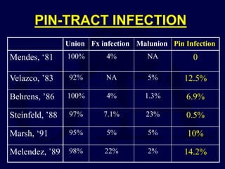
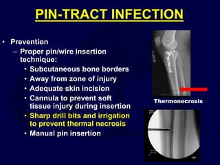
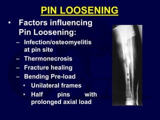
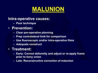
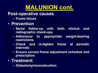
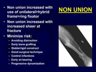
SlideShare a Scribd company logo
1 of 108
Download to read offline
PRINCIPLES OF EXTERNAL
FIXATION
Dan D. Kemper, M.D.
Kaiser Permanente
Orthopaedic Trauma Service
Walnut Creek, California
Revised Oct. 2014
Contributing author:
David W. Lowenberg, M.D.
Clinical Professor
Chief, Orthopaedic Trauma Service
Department of Orthopaedic Surgery
Stanford University School of Medicine
Original Authors: Alvin Ong M.D. & Roman Hayda M.D. 2004
Revised by Roman Hayda M.D. 2008
OVERVIEW
• Historical perspective
• Generations of frame types
• Components of external fixation
• Biomechanics of frame stability and
fracture healing
• Clinical applications
• Complications
GOALS
• Understand different types
of frame construction
• Understand the limitations
and advantages
• Understand the
biomechanics of different
frame types
• Putting it all together to best
match the patient and goal
GOAL IS TO CREATE A STABLE CONSTRUCT WITH LOW SHEAR AND TORQUE
AND HIGH MICROMOTION AT LEVEL OF FRACTURE
HISTORICAL PERSPECTIVE
• Unilateral frame (Late 1800s-1900s)
• First generation frames classic A
frame
• Subsequent frame generations
created to improve upon
shortcomings
• Uniplanar frames (2nd Gen.)
– Subject to cantilever bending
– Biplanar with improved
biomechanical properties
• Ring fixator (1950s) (3rd Gen.)
– Ilizarov
– Superior biomechanically and
implemented with improved
results for definitive care. Lambotte’s original frame 1902
Rockwood and Green, 6th ed p. 258
HISTORICAL PERSPECTIVE
cont.
• Articulated External fixation (4th gen.)
– Allow for joint range of motion
– Modified unilateral frame
• Hexapod (5th gen.)
– Taylor spatial frame (TSF)
– 6 degrees of freedom (6 struts in multi-planar configuration)
– Deformity correction
– Computer software to facilitate correction
• Hybrid ring (6th gen.)
– Improved ease of use
– Mates the advantage of metaphyseal fixation with ease of use
of half pins
– Not biomechanically superior to full ring
1st Generation
• Classic Rigid “A – Frame”
fixation
• “Too Rigid”
• Poor results gave external
fixation a bad name
Slide provided by David Lowenberg M.D.
1st Generation
Slide provided by David Lowenberg M.D
1st Generation
• In reality: Not too rigid
No axial motion
Too much wobble with AP
bending
Slide provided by David Lowenberg M.D
2nd Generation
• The classic “Unilateral Fixator”
• Gained great acceptance
• Became the workhorse of
external fixators
Slide provided by David Lowenberg M.D
2nd Generation (“unilateral”)
Slide provided by David Lowenberg M.D
3rd Generation
• Circular external fixation
• Prototype = Ilizarov Fixator
• Revolutionized external fixation
based on fine wire fixation
combined with multiplanar
fixation
Slide provided by David Lowenberg M.D
Circular Fine Wire Fixators
• Allows axial micromotion
• Stable to angulation and rotation
• Good peri-articular fixation
Slide provided by David Lowenberg M.D
Circular External Fixation
Slide provided by David Lowenberg M.D
4th Generation
• Mobile unilateral external
fixators
• Possess hinges and the ability to
transport bone
Slide provided by David Lowenberg M.D
4th Generation
• Tried to incorporate the benefits
and versatility of circular fixation
with the ease of unilateral fixator
design.
• In essence, added moving parts
to 2nd generation designs.
Slide provided by David Lowenberg M.D
4th Generation --
Problems
 Ignored basic biomechanical
constraints.
 Did not alter issues of bending,
shear, and torque.
Slide provided by David Lowenberg M.D
4th Generation
Shear
Component
Slide provided by David Lowenberg M.D
5th Generation
• Multiplanar Fixation with
Multiaxial Correction
Slide provided by David Lowenberg M.D
Multiplanar External Fixation
Slide provided by David Lowenberg M.D
“Hybrid Fixation”
• Need to understand
biomechanical principles
• Don’t repeat same mistakes
Slide provided by David Lowenberg M.D
Bottom Line
• “Classic” Hybrid Fixation has no role in
current orthopaedic practice (poor
biomechanics)
• Current “Hybrid Fixation” involves
MULTIPLANAR Fixation with a
combination of epiphyseal/metaphyseal
wires and diaphyseal half pins
Slide provided by David Lowenberg M.D
“Classic” Hybrid Fixation
Slide provided by David Lowenberg M.D
Current “Hybrid Fixation”
Slide provided by David Lowenberg M.D
FRAME COMPONENTS
– Uniplanar/Biplanar
(Traditional Frame)
• Pins
• Clamps
• Connecting rods
– Ring/Hybrid/Hexapod
• Rings
• Transfixion wires
• Half pins
• Struts
• Misc small parts
PINS
• Key link
• Pin/bone interface
is critical
• Pin stability
dependent on
radial preload
– Use appropriate size
drill
• Pin loosening is a
common problem
• Loosening brings
Radial pre-load
PINS cont.
• The single most important factor with frame
strength is increasing pin size
• Frame bending stiffness proportional to radius⁴
– Example 5mm pin is 144% stiffer versus
4mm pin
Use the largest size pin that is appropriate
4mm
VS
5mm
PIN OPTIONS
Many options
– 2-6mm sizes
– Self drilling/tapping
– Blunt tip
– Conical
– Fine thread
– Course thread
• Cancellous bone
Material
– Titanium
– Stainless
Coatings
– Non-coated
– Titanium
– Hydroxyapatite
PIN DIAMETERS
GENERAL GUIDELINES
–Femur – 5 or 6 mm
–Tibia – 5 or 6 mm
–Humerus – 5 mm
–Forearm – 4 mm
–Hand, Foot – 2.5-3 mm
Photos courtesy of Matthew Camuso
Use the appropriate pin size for the application
Avoid unicortical pin
SELF DRILLING/SELF TAPPING
• Advantages
– Single stage insertion
– Fast
– OK for short term use
• Disadvantages
– Short drill flutes
resulting in possible
• Thermal necrosis
• Stripping near cortex
– Loss of radial pre-
load
• Decreased torque to
pull out over time
(loosening)
BLUNT PINS
• Multi stage insertion
• Preservation of near
cortex
• Tapered pins
– Improved radial
pre-load
– Beware of
advancing and then
backing up, loss of
radial pre-load with
early loosening
Thermal necrosis possible with any type of pin.
Irrigate and adhere to proper technique with insertion.
PIN COATINGS
• Hydroxyapatite (HA) vs titanium vs
uncoated
– HA with superior retention of extraction torque
– Decreased infection
• 0/50 pts in pertrochanteric region (Moroni
JSBS-A, 05’)
– 13x higher extraction torque vs uncoated
– 2x higher extraction torque vs titanium
– Insertion torque and extraction torque equal with
HA coated pins
– Highly consider HA pins for extended use and or
definitive fracture care.
• Possible future coatings
– Bisphosphonate
– Antibiotic coated
Moroni A, et al, Techniques to Avoid Pin Loosening and Infection in External Fixation. JOT. 16:
189-195, 2002
Moroni A, et al, Dynamic Hip Screw versus External Fixation for Treatment of Osteoporotic
Pertrochanteric Fractures, J Bone Joint Surg Am. 87:753-759, 2005.
PIN INSERTION TECHNIQUE
1. Incise skin
2. Spread soft tissues
to bone
3. Triple sleeve first in
and last out
4. Irrigate while drilling
5. Place appropriate
pin using sleeve
6. Place pin bi-cortical
Avoid soft tissue damage and
bone thermal necrosis
CLAMPS
• Clamp types
– Pin to bar
– Multiple pin to bar clamps
– Bar to bar etc
• Features:
– Newer generation of
clamps with increased
adjustability
• Allows for variable pin
placement (multiplanar)
– MR compatible?
– Consider cost of construct,
keep it simple
Key : place clamp and rod close to bone
RODS
• Many options
– Rod material
• Stainless
• titanium
• mostly carbon
– Design
• Simple rod
• Monobar
• Articulated
• Telescoping
Frame strength increased with
increasing rod diameter
RODS cont.
•Carbon vs
Stainless
–Radiolucency
–↑ diameter = ↑
stiffness
–Carbon 15% stiffer in
load to failure
–frames with carbon
fiber are only 85% as
stiff ? ? ? ?Weak link is
clamp to carbon bar?
Kowalski M, et al, Comparative Biomechanical Evaluation of Different External Fixator Sidebars: Stainless-Steel Tubes versus
Carbon Fiber Bars, JOT 10(7): 470-475, 1996
Added bar stiffness
≠
increased frame stiffness
RING COMPONENTS
• Components:
– Transfixion wires
• olive or straight
– Wire and half pin clamps
– Half rings
– Rods
– Struts
– Motors and hinges
Rockwood and Green,
6th ed. Fig. 7-6 p. 260
Frame strength increased with
decreasing ring size and
increasing wire tension and size
UNIPLANAR/UNILATERAL
• Useful for temporary
fixation
• Useful in diaphyseal
region
• Limited roles for
definitive fixation
• Distal radius
• Tibia
• Pediatric fxs
Beware of common pitfalls (pins far from
fracture, too small of pins, single stacked
frame, bars far from skin
UNIPLANAR
• Unrestricted joint motion
• Unilateral or Bilateral
• DISADVANTAGES
– Cantilever bending at
fracture resulting in high
shear and torque
– Unable to immediately weight
bear
– “Non union maker”
historically
BIOMECHANICS
Stability improved
with:
• Increased pin
diameter
• Increased pin
spread
• Increased
number of pins
• Decreased
distance from pin
to fracture
Minimize cantilever bending and sheer at fracture
BIOMECHANICS:ROD FACTORS
• Frames placed in the same plane as the applied load
• Decreased distance from bars to bone
• Double stacking of bars
• Allow for sufficient space for soft tissue swelling


UNIPLANAR BILATERAL
• Bilateral frame with
improved share loading
at pin/bone interface
• Decreased cantilever
bending at fracture
• Limited applications
secondary to tissue
transfixion and risk or
neurovascular injury
Travelling traction
Uniplanar/Bilateral
Photo courtesy of Matthew Camuso
DYNAMIZATION
• Dynamization = load-sharing
construct that promotes
micromotion at the fracture
site
• Controlled load-sharing
helps to "work harden" the
fracture callus and
accelerate remodeling
• Serially destabilize frame
over time to allow for
increased controlled axial
compression
(Figures from: Rockwood and Green,
Fractures in Adults, 4th ed,
Lippincott-Raven, 1996)
Kenwright and Richardson, JBJS-B, ‘91
Quicker union less refracture
Marsh and Nepola, ’91
96% union at 24.6 wks
BIPLANAR
• Improved axial and sagittal
stability
• Avoids NV injury vs
uniplanar/bilateral
• Major limitations is still cantilever
bending
– Common for temporary
fixation
– Limited applications for
definitive care
Rockwood and
Green, 6th ed.
Fig. 7-14. p. 264
BIOMECHANICS
Biplanar Construct
• Linkage between
frames in
perpendicular planes
(DELTA)
• Controls each plane
of deformation
• Reduced shear/torque
at fx
• Shear
– Uniplanar>biplanar
>hybrid>circular
UNIPLANAR/BIPLANAR
SUMMARY
• Use the largest size pin that is appropriate
• Rods close to the bone
• Decrease the distance from pin to fracture
• Increase distance between pins adjacent to
fracture
• Double stack
• Pins at 90 degrees increase frame strength
• Dynamization improves fracture healing
Most important factor in increasing frame
strength is increasing pin size (R⁴)
TEMPORIZING JOINT
SPANNING
• Periarticular fractures
• Modified
uniplanar/biplanar
• Useful when soft tissue
injury
•Relative reduction by
capsuloligamentotaxis
JOINT SPANNING
• Avoid transfixing
muscle
• Disadvantages
– Pin tract infection
– Pin loosening
– Loss of reduction
– Joint stiffness
– Foot equinus
• Consider
pinning midfoot
JOINT SPANNING
Can be utilized for definitive fixation for
distal radius fracture
Beware of pin placement proximally
Superficial radial nerve
JOINT SPANNING
• Uniplanar/biplanar
frames used
successfully for knee/
ankle arthrodesis as
salvage
• Construct should be
dynamized over time
• Minimize shear forces
Rockwood & Green, 6th ed.
Fig. 7-8, p. 261
ARTICULATING HINGE
• Uniplanar or biplanar
• Peri-articular injuries
• Ligamentous injury
• Vascular injury
• Most common in elbow,
knee and ankle
• Adjunct to internal
fixation and ligamentous
repair
• Avoid overbuilding to
allow for motion
ARTICULATING FRAME
• May need different size pins (5mm
humerus, 4mm ulna)
• Build frame from joint center of rotation
• Avoid pinning down muscles with long
excursion
• Disadvantage: Potential high
complication risk
• Pin tract infection
• Nerve injury
• Broken pin
• Loss of joint reduction
• Iatrogenic ulna fracture
Ring D.,Bruinsma E.,Jupiter.,Complications of Hinged External
Fixation Compared With Cross-pinning of the Elbow for Acute and
Subacute Instability. CORR, 472: 2044-2048, 2014.
DAMAGE CONTROL
SURGERY
Pape et al. Annals of Surgery 2007
• Developed to focus
on initial hemorrhage
control, followed by
definitive care
• Minimize 2nd hit
• Convincing results
have not warranted
randomized studies
• Positive result with
unstable pelvic ring
injuries
DAMAGE CONTROL
ORTHOPAEDICS
• First coined in 2000 by Scalea et al.
– Shock Trauma Experience
• Methodology of addressing rapid temporary
stabilization and resuscitation prior to
definitive stabilization
• Practiced for many decades despite recent
popularity
DAMAGE CONTROL cont.
• Prioritize injuries
• Goal is rapid frame
stabilization and not
definitive fixation
• Avoid fixation pins
crossing surgical
approach paths
• Adhere to fixation
principles
• Consider team
approach to decrease
surgical time
SECOND HIT THEORY
Hildebrand et al. Injury 2004
• First hit= initial
trauma and
associated
resuscitation
• Second hit= surgical
intervention
• We can control the
second insult
Injury 2004
Pape et al. Annals of Surgery 2007
Randomized controlled and blinded
94 acute IMN
71 Damage Control/External fixation
Almost 7X increased acute lung injury when IMN in
the “borderline pt”
No significant increase in SIRS, ARDS, MODS, post-
op course, complications when controlled for ISS
• Retrospective cohort study
– ETC (1981-89)
– INT (1990-92)
– DCO (1993-2000)
• Higher ARDS rate in IMN (15%) vs ETC
(9.1%)
– No change in mortality
• Incidence of MODS/MOF decreased
significantly in all groups from 81-2000
DAMAGE CONTROL
SUMMARY
• Rapidly stabilize pelvis and long bone
injuries
• Peri-articular fxs secondarily
• Avoid crossing surgical planes with pin
fixation
• Goal is stabilization, may require later frame
adjustment
• Don’t confuse speed with carelessness,
adhere to principles
RING FIXATORS
• Controlled compression
• Decreased shear and
increased micromotion
• Improved peri-articular
fixation
• Allow for weight bearing
• Low infection risk
• Increasing role for high
energy open tibia fractures
with bone loss and or soft
tissue loss Rockwood & Green, 6th ed.
Fig. 7-6. p. 260
BIOMECHANICS OF RING
FIXATION
• Ring fixator with increased
coronal, sagittal stability
(4-7X)
• Increased micromotion
(1.75X) with increased rate
of union vs unilateral
• Increasing axial load
results in decreased
micromotion “trampoline
effect”
Lowenberg et al. Principles of tibial fracture
management with circular external fixation. Clin.
Orthops N. Am. 45:191-206. 2014
RING FIXATORS
Key Principles
• Increasing frame strength with:
•Wires as close to 90
o
to each other
•Increasing pin diameter
•Increasing tension (90-130kg)
•Decreased ring size
•Increased number of rings/wires
•Decreasing distance of ring to
fracture
Frame Mechanics: Ring
Fixators
• Spread wires to
as close to 90
o
as
possible
• Use at least 2
planes of
wires/half pins in
each major
segment
• Less than 60
degrees, risk of
sliding bone
segment

BIOMECHANICS cont.
Pin Factors
• Oblique fxs subject
to sheer
• Use oblique pin to
counter these
effects
• Beware of greater
than 30 degree
fracture obliquity
Metcalfe, et al, JBJS B, 2005
Lowenberg, et al, CORR, 2008
Lowenberg, et al, Orthop Clin North Am, 2014
Steerage pins
improve
compression by
minimizing sheer
effect
Steerage Pins
• First described by
Dr. Charles Taylor.
Slide provided by David Lowenberg M.D
Steerage Pins
frame
bone
pin
pin
Slide provided by David Lowenberg M.D
frame
bone
steerage pin
steerage
pin
Structural
Parallelogram
Slide provided by David Lowenberg M.D
0.0
1.0
2.0
3.0
4.0
5.0
6.0
7.0
8.0
9.0
10.0
Mean Fracture
Line Migration
(mm)
0° 15° 30° 40° 50° 60°
Classic Ilizarov Construct
Proximal Half Pin
Double Uniplanar Half Pins
Arced Wires
Single Proximal Steerage Pin
True Steerage Pin Construct
Fracture Obliquity
Mean Fracture Line Migration vs. Fracture Obliquity after
1000 N Load
Slide provided by David Lowenberg M.D
Arced Wires
Slide provided by David Lowenberg M.D
Conclusions
Lowenberg, et al
• Shear becomes a factor in
fracture stability at >30 degrees
of fracture obliquity (“30 – 60
Rule”).
• Arced wires can help, but there
is an inherent need to convert
shear to compression by
creation of a parallelogram at the
fracture site.
Slide provided by David Lowenberg M.D
Conclusions
Lowenberg, et al
• With increasing fracture
obliquity, improve the
frame/fixation geometry.
• Steerage pins are your friend.
Slide provided by David Lowenberg M.D
BIOLOGY
• Relative stability
• Fracture healing by
stable yet less rigid
systems
– Dynamization
– Micromotion
– Uniform compression
• Callus formation
(Figures from: Rockwood and Green,
Fractures in Adults, 4th ed,
Lippincott-Raven, 1996)
Kenwright, CORR, 1998
Larsson, CORR, 2001
SALVAGE
• Ring frame useful as salvage for
segmental bone loss or severe
soft tissue loss
• May acutely shorten limb to allow
soft tissue coverage
• Later conversion to distraction
ring frame, bone transport and
grafting
CONVERSION TO
DISTRACTING RING
12 y/o with 3B open tibia, segmental bone loss from Haiti earthquake
DISTRACTION
OSTEOGENESIS
DOCKING
3B tibia with segmental bone loss, 3A
plateau, temporary spanning ex fix
• Converted to circular
frame, limited open
reduction and internal
fixation of tibial plateau
with screws/wires
• Corticotomy
and transport
Consolidation
*note: docking site bone grafted
Union
JOINT ARTHRODESIS
• Knee fusion
• Ankle fusion
• Serial compression
and dynamization
• Salvage in a poor
host, failure of prior
fusion site and or
soft tissue
complications
DEFORMITY CORRECTION
• Hexapod frame
– Taylor Spacial Frame (TSF)
• Application with wire or half pins
• Adjustable with 6 planes/degrees of
deformity correction
– Deformity correction
• acute
• chronic
Type 3A open tibia fracture with bone
loss
Following frame adjustment and bone
grafting
HYBRID
• Combines the
advantages of ring in
periarticular areas with
simplicity of planar half
pins in diaphysis
• Disadvantage of
increased sheer
secondary to half pin
use vs full circular ring
frame
• Main advantage is for
convenience From Rockwood and Green’s, 5th Ed
HYBRID VS FULL RING
• Ring frames resist axial and bending
deformation better than any hybrid
modification
• Adding 2nd proximal ring and anterior
half pin improves stability of hybrid
frame
Pugh et al, JOT, ‘99
Yilmaz et al, Clin Biomech, 2003
Roberts et al, JOT, 2003
Clinical application: Use full ring fixator for fx with
bone defects or expected long frame time
“Classic” Hybrid Fixation
Slide provided by David Lowenberg M.D
Multiplanar External Fixation
Slide provided by David Lowenberg M.D
Anatomic Considerations
• Cross sectional anatomy
knowledge is critical
• Avoidance of major nerves,
vessels and organs (pelvis) is
mandatory
• Avoid joints and joint capsules
– Proximal tibial transfixion
pins should be placed >14
mm distal to articular surface
to avoid capsular reflection
• Minimize muscle/tendon
impalement (especially those
with large excursions)
Rockwood & Green, 6th
ed. Fig 7-35 A. p. 286
Conversion of Ex Fix to ORIF
• Generally safe within 2-3 wks
– Bhandari, JOT, 2005
• Meta analysis: all but one
retrospective
• Infection in tibia and femur
<4%
• Rods or plates appropriate
• Use with caution with signs of pin
irritation
– Consider staged procedure
• Remove and curette sites
• Return following healing for
definitive fixation
– Extreme caution with established
pin track infection
– Maurer, ’89
• 77% deep infection with h/o
pin infection
Conversion of Ex Fix to Nail
1507 femur fractures
59 fxs tx’d with ex-fix and then IMN
1.7 percent infection rate
11% re-operation rate
“immediate ex-fix followed by early
closed IMN is a safe treatment….
in selected multiply injured patients”
Conversion should occur before 2 wks
Nowotarski et al. JBJS 2000
EVIDENCE
Femur fx
– Nowotarski, JBJS-A, ’00
• 59 fx (19 open), 54 pts,
• Convert at 7 days (1-49 days)
• 1 infected nonunion, 1 aseptic
nonunion
– Scalea, J Trauma, ’00
• 43 ex-fix then nailed vs 284
primary IM nail
• ISS 26.8 vs 16.8
• Fluids 11.9l vs 6.2l first 24 hrs
• OR time 35 min EBL 90cc vs
135 min EBL 400cc
• Ex fix group 1 infected
nonunion, 1 aseptic nonunion
Bilat open femur,
massive compartment
syndrome, ex fix then
nail
COMPLICATIONS
• #1 is Pin-track infection
• #2 is Pin loosening
• Frame or Pin/Wire Failure
• Malunion
• Non-union
• Soft-tissue impalement (stiffness)
• Iatrogenic fracture from pin site
• Compartment syndrome (rare)
PIN-TRACT INFECTION
• Most common
complication
• 0 – 14.2% incidence
• 4 stages (Dahl):
– Stage I: Seropurulent
Drainage
– Stage II: Superficial
Cellulitis
– Stage III: Deep Infection
– Stage IV: Osteomyelitis
DAHL PIN SITE
CLASSIFICATION
• Treatment
– Stage I: aggressive pin-site care and
oral antibiotics
– Stage II: same as Stage I and +/-
Parenteral Abx
– Stage III: Removal/exchange of pin plus
Parenteral Abx
– Stage IV: same as Stage III, culture pin
site for offending organism, specific IV
Abx for 10 to 14 days, surgical
debridement of pin site
PIN-TRACT INFECTION
Union Fx infection Malunion Pin Infection
Mendes, ‘81 100% 4% NA 0
Velazco, ’83 92% NA 5% 12.5%
Behrens, ’86 100% 4% 1.3% 6.9%
Steinfeld, ’88 97% 7.1% 23% 0.5%
Marsh, ‘91 95% 5% 5% 10%
Melendez, ’89 98% 22% 2% 14.2%
PIN-TRACT INFECTION
• Prevention
– Proper pin/wire insertion
technique:
• Subcutaneous bone borders
• Away from zone of injury
• Adequate skin incision
• Cannula to prevent soft
tissue injury during insertion
• Sharp drill bits and irrigation
to prevent thermal necrosis
• Manual pin insertion
Thermonecrosis
PIN LOOSENING
• Factors influencing
Pin Loosening:
– Infection/osteomyelitis
at pin site
– Thermonecrosis
– Fracture healing
– Bending Pre-load
• Unilateral frames
• Half pins with
prolonged axial load
MALUNION
Intra-operative causes:
– Poor technique
• Prevention:
– Clear pre-operative planning
– Prep contralateral limb for comparison
– Use fluoroscopic and/or intra-operative films
– Adequate construct
• Treatment:
– Early: Correct deformity and adjust or re-apply frame
prior to bony union
– Late: Reconstructive correction of malunion
MALUNION cont.
Post-operative causes
– Frame failure
• Prevention
– Serial follow-up with both clinical and
radiographic check-ups
– Adherence to appropriate weight-bearing
restrictions
– Check and re-tighten frame at periodic
intervals
– Ensure correct frame adjustment schedule and
prescription
• Treatment
– Osteotomy/reconstruction
NON UNION
• Non union increased with
use of unilateral>hybrid
frame>ring fixator
• Non union increased with
increased sheer at
fracture
• Minimize risk:
– Avoiding distraction
– Early bone grafting
– Stable/rigid construct
– Good surgical technique
– Control infections
– Early wt bearing
– Progressive dynamization
IATROGENIC FRACTURE
• Rare
• Often related to
placement of pin
• Avoid unicortical
pin and stress riser
• Pin size <1/3
diameter of
diaphysis
67 yo diabetic, charcot contralateral limb
SOFT TISSUE TETHERING
• Entrapment of soft tissues/muscle
– Loss of motion
– Scarring and adhesion
– Neurovascular injury
• Prevention
– Check ROM intra-operatively
– Avoid piercing muscle or tendons
– Position joint in NEUTRAL
– Early stretching and ROM exercises
– Convert to definitive fixation when possible
COMPARTMENT SYNDROME
• Pathophysiology
– Rare
– Initial trauma
– Pin or wire causing
additional bleeding
• Prevention
– Understand anatomy
– Good technique
– Post-operative
vigilance
• Low threshold for
fasciotomy
EXTERNAL FIXATION OF
PELVIC RING
• See: http://ota.org/education/resident-
resources/core-curriculum/pelvis-and-
acetabulum
KEYS TO SUCCESS
• Chose optimal pin diameter
• Use good insertion technique
• Place clamps and frames close to skin
• Build frame in plane of deforming forces
• Double stack frame
• Consider use of ring fixator for definitive
fixation
• Respect the soft tissues and think ahead
about the next step. Don’t burn a bridge
Plan ahead!
SUMMARY
• Multiple applications and
techniques
• Pair technique with clinical
indication(s)
• Appropriate use can lead to
excellent results
• Understand the biomechanics of
each frame type
• Recognize and correct
complications early
Return to
General/Principles
Index
E-mail OTA
about
Questions/Comments
If you would like to volunteer as an author for
the Resident Slide Project or recommend
updates to any of the following slides, please
send an e-mail to ota@ota.org
REFERENCES
1. Bhandari M, Zlowodski M, Tornetta P, Schmidt A, Templeman D. Intramedullary Nailing Following External Fixation in Femoral and Tibial Shaft
Fractures. Evidence-Based Orthopaedic Trauma. JOT, 19(2): 40-144, 2005.
2. Cannada LK, Herzenberg JE, Hughes PM, Belkoff S. Safety and Image Artifact of External Fixators and Magnetic Resonance Imaging. CORR,
317, 206-214:1995.
3. Calhoun JH, Li F, Ledbetter BR, et al. Biomechanics of the Ilizarov Fixator for Fracture Fixation. Clin Orthop Relat Res. 280:15-22. 1992.
4. COTS. Open reduction and Internal Fixation Compared with Circular Fixator Application for Bicondylar Tibial Plateau Fractures. Results of a
Multicenter, Prospective, Randomized Clinical Trial. JBJS-Am. 88(12):2613-23. 2006.
5. Davison BL, Cantu RV, Van Woerkom S. The Magnetic Attraction of Lower Extremity External Fixators in an MRI Suite. JOT, 18 (1): 24-27, 2004.
6. Egol KA, Tejwani NC, Capla EL, Wolinsky PL, Koval KJ., Staged management of high-energy proximal tibia fractures (OTA types 41): the
results of a prospective, standardized protocol. JOT, 19(7):448-55. 2005.
7. Garcia-Cimbrelo E, Marti-Gonzalez JC., Circular External Fixation in Tibial Nonunion. Clin Orthop Relat Res.,419: 65-70. 2004.
8. Giotakis N, Panchani SK, Narayan B,. Segmental Fractures of the Tibia Treated by Circular External Fixation. JBJS-Br. 92B:687-92. 2010.
9. Hildebrand F.,Giannoudis P.,Krettek C.,Pape H.C., Damage Control: Extremiteis. Injury. 35:678-689. 2004
10. Foster PA, Barton SB, Jones SC, Morrison RJ, Britten S. The Treatment of Complex Tibial Shaft Fractures by the Ilizarov Method. JBJS- Br.
12:1678-1683. 2012.
11. Jones CP, Youngblood CS, Waldrop N, Davis WH, Pinaur MS, Tibial Stress Fracture Secondary to Half-Pins in Circular Ring External Fixation
for Charcot Foot. Foot and Ankle Int, 35(6): 572-577. 2014
12. Keating J, Simpson A, Robinson C. The Management of Fracture with Bone Loss. JBJS Br. 87::142-50. 2005.
13. Kenwright J, Richardson JB, Cunningham, et al. Axial Movement and Tibial Fractures. A Controlled Randomized Trial of Treatment, JBJS-Br,
73 (4): 654-650, 1991.
14. Kenwright J , Gardner T. Mechanical Influences on Tibial Fracture Healing. CORR, 355: 179-190,1998.
15. Kowalski, M et al, Comparative Biomechanical Evaluation of Different External Fixator Sidebars: Stainless-Steel Tubes versus Carbon Fiber
Bars, JOT 10(7): 470-475, 1996.
16. Kumar R, Lerski RA, Gandy S, Clift BA, Abboud RJ. Safety of Orthopedic Implants in Magnetic Resonance Imaging: an Experimental
Verification. J Orthop Res, 24 (9): 1799-1802, 2006.
17. Larsson S, Kim W, Caja VL, Egger EL, Inoue N, Chao EY. Effect of Early Axial Dynamization on Tibial Bone Healing: A Study in Dogs. CORR,
388: 240-51, 2001.
18. Lowenberg DW, Nork S, Abruzzo FM. The Correlation of Shearing Force with Fracture Line Migration for Progressive Fracture Obliquities
Stabilized by External Fixation in the Tibial Model. CORR, 466:2947–2954, 2008.
1. Lowenberg DW, Parrett BM, Bunti RF, Et al. Long Term Results and Costs of Muscle Flap Coverage with Ilizarov Bone
Transport in Lower Limb Salvage. JOT. 27(10):576-81. 2013
2. Lowenberg DW, Principles of Tibial Fracture Management with Circular External Fixation. Orthop Clin North Am,
45(2):191-206.
3. Marsh JL. Nepola JV, Wuest TK, Osteen D, Cox K, Oppenheim W. Unilateral External Fixation Until Healing with the
Dynamic Axial Fixator for Severe Open Tibial Fractures. Review of Two Consecutive Series , JOT, 5(3): 341-348, 1991.
4. Maurer DJ, Merkow RL, Gustilo RB. Infection After Intramedullary Nailing of Severe Open Tibial Fractures Initially Treated
with External Fixation. JBJS-A, 71(6), 835-838, 1989.
5. Metcalfe AJ, Saleh M, Yang L. Techniques for Improving Stability in Oblique Fractures Treated by Circular Fixation with
Particular Reference to the Sagittal Plane. JBJS-Br, 87 (6): 868-872, 2005.
6. Moroni A, Faldini C, Marchetti S, Manca M, Consoli V, Giannini S. Improvement of the Bone-Pin Interface Strength in
Osteoporotic Bone with Use of Hydroxyapatite-Coated Tapered External-Fixation Pins: A Prospective, Randomized
Clinical Study of Wrist Fractures . JBJS –A, 83:717-721, 2001.
7. Moroni A, Faldini C. Pegreffi F. Hoang-Kim A. Vannini F. Giannini S. Dynamic Hip Screw versus External Fixation for
Treatment of Osteoporotic Pertrochanteric Fractures, J BJS-Am. 87:753-759, 2005.
8. Moroni A. Faldini C. Rocca M. Stea S. Giannini S. Improvement of the Bone-Screw Interface Strength with Hydroxyapatite-
coated and Titanium-coated AO/ASIF Cortical Screws. J OT. 16(4): 257-63, 2002 .
9. Nowotarski PJ, Turen CH, Brumback RJ, Scarboro JM, Conversion of External Fixation to Intramedullary Nailing for
Fractures of the Shaft of the Femur in Multiply Injured Patients, JBJS-A, 82:781-788, 2000.
10. Pape H.C.,Rixen D., Morley J., et al. Impact of the Method of Initial Stabilization for Femoral Shaft Fractures in Patients
With Multiple Injuries at Risk for Complications (Borderline Patients). Ann Surg. 246 (3):491-501. 2007
11. Patterson MJ, Cole J. Two-Staged Delayed Open Reduction and Internal Fixation of Severe Pilon Fractures. JOT, 13(2): 85-
91, 1999.
12. Pugh K.J, Wolinsky PR, Dawson JM, Stahlman GC. The Biomechanics of Hybrid External Fixation. JOT. 13(1):20-26,
1999.
13. Ring D.,Bruinsma E.,Jupiter.,Complications of Hinged External Fixation Compared With Cross-pinning of the Elbow for
Acute and Subacute Instability. CORR, 472: 2044-2048, 2014.
14. Roberts C, Dodds JC, Perry K, Beck D, Seligson D, Voor M. Hybrid External Fixation of the Proximal Tibia: Strategies to
Improve Frame Stability. JOT, 17(6):415-420, 2003.
15. Scalea TM, Boswell SA, Scott JD, Mitchell KA, Kramer ME, Pollak AN. External Fixation as a Bridge to Intramedullary
Nailing for Patients with Multiple Injuries and with Femur Fractures: Damage Control Orthopedics. J Trauma, 48(4):613-
623, 2000.
16. Sirkin M, Sanders R, DiPasquale T, Herscovici, A Staged Protocol for Soft Tissue Management in the Treatment of
Complex Pilon Fractures. JOT, 13(2): 78-84, 1999.
17. Watson T, Coufal C. Treatment of complex lateral plateau fracture using Ilizarov techniques. Clin Orthop Relat Res.
353:97-106. 1998
18. Yilmaz E, Belhan O, Karakurt L, Arslan N, Serin E. Mechanical performance of hybrid Ilizarov External Fixator in
Comparison with Ilizarov Circular External Fixator. Clin Biomech, 18 (6): 518, 2003.

More Related Content

What's hot

HIPS STEM DESIGN-- Ashish Sharma
HIPS STEM DESIGN-- Ashish SharmaHIPS STEM DESIGN-- Ashish Sharma
HIPS STEM DESIGN-- Ashish Sharmaas747
 
Bio degadable implants used in Orthopaedics by Dr.Vinay
Bio degadable implants used in Orthopaedics by Dr.VinayBio degadable implants used in Orthopaedics by Dr.Vinay
Bio degadable implants used in Orthopaedics by Dr.VinayVenkat Vinay
 
Evolution of Intramedullary Nails
Evolution of Intramedullary NailsEvolution of Intramedullary Nails
Evolution of Intramedullary NailsPrateek Goel
 
Cementless acetabular cups
Cementless acetabular  cupsCementless acetabular  cups
Cementless acetabular cupsIhab El-Desouky
 
Bearing surfaces THR
Bearing surfaces THRBearing surfaces THR
Bearing surfaces THRorthoprince
 
Three column fixation for complex PROXIMAL TIBIA FRACTURES
Three column fixation for complex PROXIMAL TIBIA FRACTURESThree column fixation for complex PROXIMAL TIBIA FRACTURES
Three column fixation for complex PROXIMAL TIBIA FRACTURESLokesh Sharoff
 
Bone cement,Cementing Generations, Complications & Recent advances
Bone cement,Cementing Generations, Complications & Recent advancesBone cement,Cementing Generations, Complications & Recent advances
Bone cement,Cementing Generations, Complications & Recent advancesSameer Ashar
 
Lumbar Disc Replacement
Lumbar Disc ReplacementLumbar Disc Replacement
Lumbar Disc ReplacementPablo Pazmino
 
minimally invasive percutaneous plate osteosynthesis
minimally invasive percutaneous plate osteosynthesisminimally invasive percutaneous plate osteosynthesis
minimally invasive percutaneous plate osteosynthesisSagar Tomar
 
Design and function of surgical screws
Design and function of surgical screwsDesign and function of surgical screws
Design and function of surgical screwsOrthosurg2016
 
Damage Control Orthopaedics (DCO)
 Damage Control Orthopaedics (DCO) Damage Control Orthopaedics (DCO)
Damage Control Orthopaedics (DCO)fathi neana
 
Acl graft fixation options
Acl graft fixation optionsAcl graft fixation options
Acl graft fixation optionsorthoprinciples
 
Inra medullary nailing - basic concepts
Inra medullary nailing - basic conceptsInra medullary nailing - basic concepts
Inra medullary nailing - basic conceptsharivenkat1990
 
Trunnionosis in total hip arthroplasty
Trunnionosis in total hip arthroplastyTrunnionosis in total hip arthroplasty
Trunnionosis in total hip arthroplastySuhail Shaheer
 
Uncemented femoral stem
Uncemented  femoral stemUncemented  femoral stem
Uncemented femoral stemSameer Ashar
 
Masquelet technique ppt
Masquelet technique pptMasquelet technique ppt
Masquelet technique pptApoorv Garg
 
Percutaneous pinning of distal radius (old technique)
Percutaneous pinning of distal radius (old technique)Percutaneous pinning of distal radius (old technique)
Percutaneous pinning of distal radius (old technique)Alberto Mantovani
 
DHS vs PFNA for Intertrochanteric fractures - Dr Chintan N Patel
DHS vs PFNA for Intertrochanteric fractures - Dr Chintan N PatelDHS vs PFNA for Intertrochanteric fractures - Dr Chintan N Patel
DHS vs PFNA for Intertrochanteric fractures - Dr Chintan N PatelDrChintan Patel
 

What's hot (20)

HIPS STEM DESIGN-- Ashish Sharma
HIPS STEM DESIGN-- Ashish SharmaHIPS STEM DESIGN-- Ashish Sharma
HIPS STEM DESIGN-- Ashish Sharma
 
Bio degadable implants used in Orthopaedics by Dr.Vinay
Bio degadable implants used in Orthopaedics by Dr.VinayBio degadable implants used in Orthopaedics by Dr.Vinay
Bio degadable implants used in Orthopaedics by Dr.Vinay
 
Evolution of Intramedullary Nails
Evolution of Intramedullary NailsEvolution of Intramedullary Nails
Evolution of Intramedullary Nails
 
Cementless acetabular cups
Cementless acetabular  cupsCementless acetabular  cups
Cementless acetabular cups
 
Protrusio acetabuli
Protrusio acetabuliProtrusio acetabuli
Protrusio acetabuli
 
Bearing surfaces THR
Bearing surfaces THRBearing surfaces THR
Bearing surfaces THR
 
Three column fixation for complex PROXIMAL TIBIA FRACTURES
Three column fixation for complex PROXIMAL TIBIA FRACTURESThree column fixation for complex PROXIMAL TIBIA FRACTURES
Three column fixation for complex PROXIMAL TIBIA FRACTURES
 
Bone cement,Cementing Generations, Complications & Recent advances
Bone cement,Cementing Generations, Complications & Recent advancesBone cement,Cementing Generations, Complications & Recent advances
Bone cement,Cementing Generations, Complications & Recent advances
 
Lumbar Disc Replacement
Lumbar Disc ReplacementLumbar Disc Replacement
Lumbar Disc Replacement
 
minimally invasive percutaneous plate osteosynthesis
minimally invasive percutaneous plate osteosynthesisminimally invasive percutaneous plate osteosynthesis
minimally invasive percutaneous plate osteosynthesis
 
08 seminar by yash on thr
08 seminar by yash on thr08 seminar by yash on thr
08 seminar by yash on thr
 
Design and function of surgical screws
Design and function of surgical screwsDesign and function of surgical screws
Design and function of surgical screws
 
Damage Control Orthopaedics (DCO)
 Damage Control Orthopaedics (DCO) Damage Control Orthopaedics (DCO)
Damage Control Orthopaedics (DCO)
 
Acl graft fixation options
Acl graft fixation optionsAcl graft fixation options
Acl graft fixation options
 
Inra medullary nailing - basic concepts
Inra medullary nailing - basic conceptsInra medullary nailing - basic concepts
Inra medullary nailing - basic concepts
 
Trunnionosis in total hip arthroplasty
Trunnionosis in total hip arthroplastyTrunnionosis in total hip arthroplasty
Trunnionosis in total hip arthroplasty
 
Uncemented femoral stem
Uncemented  femoral stemUncemented  femoral stem
Uncemented femoral stem
 
Masquelet technique ppt
Masquelet technique pptMasquelet technique ppt
Masquelet technique ppt
 
Percutaneous pinning of distal radius (old technique)
Percutaneous pinning of distal radius (old technique)Percutaneous pinning of distal radius (old technique)
Percutaneous pinning of distal radius (old technique)
 
DHS vs PFNA for Intertrochanteric fractures - Dr Chintan N Patel
DHS vs PFNA for Intertrochanteric fractures - Dr Chintan N PatelDHS vs PFNA for Intertrochanteric fractures - Dr Chintan N Patel
DHS vs PFNA for Intertrochanteric fractures - Dr Chintan N Patel
 

Similar to G11-Principles of External Fixation.pdf

External fixator
External fixatorExternal fixator
External fixatorAkshay Shah
 
G11-Ex-Fix-Principles-JTG-rev-2-4-10.ppt
G11-Ex-Fix-Principles-JTG-rev-2-4-10.pptG11-Ex-Fix-Principles-JTG-rev-2-4-10.ppt
G11-Ex-Fix-Principles-JTG-rev-2-4-10.pptmvuyekule1
 
Materials used in Orthodontics _ Dr. Nabil Al-Zubair
Materials used in Orthodontics _ Dr. Nabil Al-ZubairMaterials used in Orthodontics _ Dr. Nabil Al-Zubair
Materials used in Orthodontics _ Dr. Nabil Al-ZubairNabil Al-Zubair
 
MAXILLARY MAJOR CONNECTORS
MAXILLARY MAJOR CONNECTORSMAXILLARY MAJOR CONNECTORS
MAXILLARY MAJOR CONNECTORSDr.Richa Sahai
 
Dental implant introduction
Dental implant introductionDental implant introduction
Dental implant introductionmarwan mohamed
 
Principles of external fixator
Principles of external fixatorPrinciples of external fixator
Principles of external fixatorDR. D. P. SWAMI
 
Endodontic instruments /certified fixed orthodontic courses by Indian dental...
Endodontic instruments  /certified fixed orthodontic courses by Indian dental...Endodontic instruments  /certified fixed orthodontic courses by Indian dental...
Endodontic instruments /certified fixed orthodontic courses by Indian dental...Indian dental academy
 
external fixation re
external fixation reexternal fixation re
external fixation reReza Fahlevi
 
Orthodontics archwaire & treatment plan
Orthodontics archwaire & treatment plan Orthodontics archwaire & treatment plan
Orthodontics archwaire & treatment plan jojo smn
 
Custom made post & Core in endodontics
Custom made post & Core in endodonticsCustom made post & Core in endodontics
Custom made post & Core in endodonticsDr. Arpit Viradiya
 
Plating principles in Orthopaedics
Plating principles in OrthopaedicsPlating principles in Orthopaedics
Plating principles in OrthopaedicsHimashis Medhi
 
Principles of locking compression plates
Principles of locking compression platesPrinciples of locking compression plates
Principles of locking compression platesDr Souvik Paul
 
Principles of fixation and osteosynthesis in trauma
Principles of fixation and osteosynthesis in traumaPrinciples of fixation and osteosynthesis in trauma
Principles of fixation and osteosynthesis in traumaDr Bhavik Miyani
 
Post endodontic restoration /certified fixed orthodontic courses by Indian de...
Post endodontic restoration /certified fixed orthodontic courses by Indian de...Post endodontic restoration /certified fixed orthodontic courses by Indian de...
Post endodontic restoration /certified fixed orthodontic courses by Indian de...Indian dental academy
 
prp.pptxbiomechanics
prp.pptxbiomechanicsprp.pptxbiomechanics
prp.pptxbiomechanicsAbdallahOmer9
 
Intramedullary nailing.pptx
Intramedullary nailing.pptxIntramedullary nailing.pptx
Intramedullary nailing.pptxAbdallahOmer9
 

Similar to G11-Principles of External Fixation.pdf (20)

External fixator
External fixatorExternal fixator
External fixator
 
G11-Ex-Fix-Principles-JTG-rev-2-4-10.ppt
G11-Ex-Fix-Principles-JTG-rev-2-4-10.pptG11-Ex-Fix-Principles-JTG-rev-2-4-10.ppt
G11-Ex-Fix-Principles-JTG-rev-2-4-10.ppt
 
Materials used in Orthodontics _ Dr. Nabil Al-Zubair
Materials used in Orthodontics _ Dr. Nabil Al-ZubairMaterials used in Orthodontics _ Dr. Nabil Al-Zubair
Materials used in Orthodontics _ Dr. Nabil Al-Zubair
 
MAXILLARY MAJOR CONNECTORS
MAXILLARY MAJOR CONNECTORSMAXILLARY MAJOR CONNECTORS
MAXILLARY MAJOR CONNECTORS
 
Dental implant introduction
Dental implant introductionDental implant introduction
Dental implant introduction
 
Principles of external fixator
Principles of external fixatorPrinciples of external fixator
Principles of external fixator
 
Ilizarov ring fixator
Ilizarov ring fixatorIlizarov ring fixator
Ilizarov ring fixator
 
Endodontic instruments /certified fixed orthodontic courses by Indian dental...
Endodontic instruments  /certified fixed orthodontic courses by Indian dental...Endodontic instruments  /certified fixed orthodontic courses by Indian dental...
Endodontic instruments /certified fixed orthodontic courses by Indian dental...
 
external fixation re
external fixation reexternal fixation re
external fixation re
 
Orthodontics archwaire & treatment plan
Orthodontics archwaire & treatment plan Orthodontics archwaire & treatment plan
Orthodontics archwaire & treatment plan
 
Custom made post & Core in endodontics
Custom made post & Core in endodonticsCustom made post & Core in endodontics
Custom made post & Core in endodontics
 
Leveling and Aligning
Leveling and AligningLeveling and Aligning
Leveling and Aligning
 
Clasp Designs - Dr. devi
Clasp Designs - Dr. deviClasp Designs - Dr. devi
Clasp Designs - Dr. devi
 
Plating principles in Orthopaedics
Plating principles in OrthopaedicsPlating principles in Orthopaedics
Plating principles in Orthopaedics
 
Principles of locking compression plates
Principles of locking compression platesPrinciples of locking compression plates
Principles of locking compression plates
 
External fixation
External fixation External fixation
External fixation
 
Principles of fixation and osteosynthesis in trauma
Principles of fixation and osteosynthesis in traumaPrinciples of fixation and osteosynthesis in trauma
Principles of fixation and osteosynthesis in trauma
 
Post endodontic restoration /certified fixed orthodontic courses by Indian de...
Post endodontic restoration /certified fixed orthodontic courses by Indian de...Post endodontic restoration /certified fixed orthodontic courses by Indian de...
Post endodontic restoration /certified fixed orthodontic courses by Indian de...
 
prp.pptxbiomechanics
prp.pptxbiomechanicsprp.pptxbiomechanics
prp.pptxbiomechanics
 
Intramedullary nailing.pptx
Intramedullary nailing.pptxIntramedullary nailing.pptx
Intramedullary nailing.pptx
 

More from Universitas Kristen Krida Wacana (Ukrida)

BIMBINGAN 7 - CTS, TTS, Neuropati, Peroneal Palsy, Neurogenic Bladder, HNP, R...
BIMBINGAN 7 - CTS, TTS, Neuropati, Peroneal Palsy, Neurogenic Bladder, HNP, R...BIMBINGAN 7 - CTS, TTS, Neuropati, Peroneal Palsy, Neurogenic Bladder, HNP, R...
BIMBINGAN 7 - CTS, TTS, Neuropati, Peroneal Palsy, Neurogenic Bladder, HNP, R...Universitas Kristen Krida Wacana (Ukrida)
 

More from Universitas Kristen Krida Wacana (Ukrida) (20)

anscanjcs - jncjkanbsnjsjakbncjhakbscjka.pptx
anscanjcs - jncjkanbsnjsjakbncjhakbscjka.pptxanscanjcs - jncjkanbsnjsjakbncjhakbscjka.pptx
anscanjcs - jncjkanbsnjsjakbncjhakbscjka.pptx
 
RIB FRACTURE.pptx
 RIB FRACTURE.pptx RIB FRACTURE.pptx
RIB FRACTURE.pptx
 
Compartment_Syndrome - COK ver 02112022.ppt
Compartment_Syndrome - COK ver 02112022.pptCompartment_Syndrome - COK ver 02112022.ppt
Compartment_Syndrome - COK ver 02112022.ppt
 
COMPARTMENT SYNDROME-DR COKORDA.pptx
COMPARTMENT SYNDROME-DR COKORDA.pptxCOMPARTMENT SYNDROME-DR COKORDA.pptx
COMPARTMENT SYNDROME-DR COKORDA.pptx
 
BIMBINGAN 7 - CTS, TTS, Neuropati, Peroneal Palsy, Neurogenic Bladder, HNP, R...
BIMBINGAN 7 - CTS, TTS, Neuropati, Peroneal Palsy, Neurogenic Bladder, HNP, R...BIMBINGAN 7 - CTS, TTS, Neuropati, Peroneal Palsy, Neurogenic Bladder, HNP, R...
BIMBINGAN 7 - CTS, TTS, Neuropati, Peroneal Palsy, Neurogenic Bladder, HNP, R...
 
malignantbonetumours-170608150210.pdf
malignantbonetumours-170608150210.pdfmalignantbonetumours-170608150210.pdf
malignantbonetumours-170608150210.pdf
 
Osteosarcoma & Ewing Sarcoma.pptx
Osteosarcoma & Ewing Sarcoma.pptxOsteosarcoma & Ewing Sarcoma.pptx
Osteosarcoma & Ewing Sarcoma.pptx
 
INTRO.pptx
INTRO.pptxINTRO.pptx
INTRO.pptx
 
DISCUSSION KVN.pptx
DISCUSSION KVN.pptxDISCUSSION KVN.pptx
DISCUSSION KVN.pptx
 
TATALAKSANA SARPUS ASY.pptx
TATALAKSANA SARPUS ASY.pptxTATALAKSANA SARPUS ASY.pptx
TATALAKSANA SARPUS ASY.pptx
 
FEMUR.pptx
FEMUR.pptxFEMUR.pptx
FEMUR.pptx
 
Compartment Syndrome.pptx
Compartment Syndrome.pptxCompartment Syndrome.pptx
Compartment Syndrome.pptx
 
Proposal.pptx
Proposal.pptxProposal.pptx
Proposal.pptx
 
4 Juli 2022.pptx
4 Juli 2022.pptx4 Juli 2022.pptx
4 Juli 2022.pptx
 
Kegawatan Muskuloskeletal Non Trauma.ppt
Kegawatan Muskuloskeletal Non Trauma.pptKegawatan Muskuloskeletal Non Trauma.ppt
Kegawatan Muskuloskeletal Non Trauma.ppt
 
LBP-HNP DR LAN.pptx
LBP-HNP DR LAN.pptxLBP-HNP DR LAN.pptx
LBP-HNP DR LAN.pptx
 
PPT.pptx
PPT.pptxPPT.pptx
PPT.pptx
 
Perlengkapan VISITASI AKREDITASI LAM-PTKES 2022.pptx
Perlengkapan VISITASI AKREDITASI LAM-PTKES 2022.pptxPerlengkapan VISITASI AKREDITASI LAM-PTKES 2022.pptx
Perlengkapan VISITASI AKREDITASI LAM-PTKES 2022.pptx
 
KONAS Update 1 .pptx
KONAS Update 1 .pptxKONAS Update 1 .pptx
KONAS Update 1 .pptx
 
Skoliosis (1).pptx
Skoliosis (1).pptxSkoliosis (1).pptx
Skoliosis (1).pptx
 

Recently uploaded

Call Girls Bangalore - 450+ Call Girl Cash Payment 💯Call Us 🔝 6378878445 🔝 💃 ...
Call Girls Bangalore - 450+ Call Girl Cash Payment 💯Call Us 🔝 6378878445 🔝 💃 ...Call Girls Bangalore - 450+ Call Girl Cash Payment 💯Call Us 🔝 6378878445 🔝 💃 ...
Call Girls Bangalore - 450+ Call Girl Cash Payment 💯Call Us 🔝 6378878445 🔝 💃 ...gragneelam30
 
Cheap Rate Call Girls Bangalore {9179660964} ❤️VVIP BEBO Call Girls in Bangal...
Cheap Rate Call Girls Bangalore {9179660964} ❤️VVIP BEBO Call Girls in Bangal...Cheap Rate Call Girls Bangalore {9179660964} ❤️VVIP BEBO Call Girls in Bangal...
Cheap Rate Call Girls Bangalore {9179660964} ❤️VVIP BEBO Call Girls in Bangal...Sheetaleventcompany
 
Call Girl in Chennai | Whatsapp No 📞 7427069034 📞 VIP Escorts Service Availab...
Call Girl in Chennai | Whatsapp No 📞 7427069034 📞 VIP Escorts Service Availab...Call Girl in Chennai | Whatsapp No 📞 7427069034 📞 VIP Escorts Service Availab...
Call Girl in Chennai | Whatsapp No 📞 7427069034 📞 VIP Escorts Service Availab...amritaverma53
 
Low Cost Call Girls Bangalore {9179660964} ❤️VVIP NISHA Call Girls in Bangalo...
Low Cost Call Girls Bangalore {9179660964} ❤️VVIP NISHA Call Girls in Bangalo...Low Cost Call Girls Bangalore {9179660964} ❤️VVIP NISHA Call Girls in Bangalo...
Low Cost Call Girls Bangalore {9179660964} ❤️VVIP NISHA Call Girls in Bangalo...Sheetaleventcompany
 
💰Call Girl In Bangalore☎️63788-78445💰 Call Girl service in Bangalore☎️Bangalo...
💰Call Girl In Bangalore☎️63788-78445💰 Call Girl service in Bangalore☎️Bangalo...💰Call Girl In Bangalore☎️63788-78445💰 Call Girl service in Bangalore☎️Bangalo...
💰Call Girl In Bangalore☎️63788-78445💰 Call Girl service in Bangalore☎️Bangalo...gragneelam30
 
💚Reliable Call Girls Chandigarh 💯Niamh 📲🔝8868886958🔝Call Girl In Chandigarh N...
💚Reliable Call Girls Chandigarh 💯Niamh 📲🔝8868886958🔝Call Girl In Chandigarh N...💚Reliable Call Girls Chandigarh 💯Niamh 📲🔝8868886958🔝Call Girl In Chandigarh N...
💚Reliable Call Girls Chandigarh 💯Niamh 📲🔝8868886958🔝Call Girl In Chandigarh N...Sheetaleventcompany
 
7 steps How to prevent Thalassemia : Dr Sharda Jain & Vandana Gupta
7 steps How to prevent Thalassemia : Dr Sharda Jain & Vandana Gupta7 steps How to prevent Thalassemia : Dr Sharda Jain & Vandana Gupta
7 steps How to prevent Thalassemia : Dr Sharda Jain & Vandana GuptaLifecare Centre
 
Premium Call Girls Nagpur {9xx000xx09} ❤️VVIP POOJA Call Girls in Nagpur Maha...
Premium Call Girls Nagpur {9xx000xx09} ❤️VVIP POOJA Call Girls in Nagpur Maha...Premium Call Girls Nagpur {9xx000xx09} ❤️VVIP POOJA Call Girls in Nagpur Maha...
Premium Call Girls Nagpur {9xx000xx09} ❤️VVIP POOJA Call Girls in Nagpur Maha...Sheetaleventcompany
 
Difference Between Skeletal Smooth and Cardiac Muscles
Difference Between Skeletal Smooth and Cardiac MusclesDifference Between Skeletal Smooth and Cardiac Muscles
Difference Between Skeletal Smooth and Cardiac MusclesMedicoseAcademics
 
Call 8250092165 Patna Call Girls ₹4.5k Cash Payment With Room Delivery
Call 8250092165 Patna Call Girls ₹4.5k Cash Payment With Room DeliveryCall 8250092165 Patna Call Girls ₹4.5k Cash Payment With Room Delivery
Call 8250092165 Patna Call Girls ₹4.5k Cash Payment With Room DeliveryJyoti singh
 
Cardiac Output, Venous Return, and Their Regulation
Cardiac Output, Venous Return, and Their RegulationCardiac Output, Venous Return, and Their Regulation
Cardiac Output, Venous Return, and Their RegulationMedicoseAcademics
 
Chandigarh Call Girls Service ❤️🍑 9809698092 👄🫦Independent Escort Service Cha...
Chandigarh Call Girls Service ❤️🍑 9809698092 👄🫦Independent Escort Service Cha...Chandigarh Call Girls Service ❤️🍑 9809698092 👄🫦Independent Escort Service Cha...
Chandigarh Call Girls Service ❤️🍑 9809698092 👄🫦Independent Escort Service Cha...Sheetaleventcompany
 
tongue disease lecture Dr Assadawy legacy
tongue disease lecture Dr Assadawy legacytongue disease lecture Dr Assadawy legacy
tongue disease lecture Dr Assadawy legacyDrMohamed Assadawy
 
Call Girl In Indore 📞9235973566📞 Just📲 Call Inaaya Indore Call Girls Service ...
Call Girl In Indore 📞9235973566📞 Just📲 Call Inaaya Indore Call Girls Service ...Call Girl In Indore 📞9235973566📞 Just📲 Call Inaaya Indore Call Girls Service ...
Call Girl In Indore 📞9235973566📞 Just📲 Call Inaaya Indore Call Girls Service ...Sheetaleventcompany
 
Call Girls Rishikesh Just Call 9667172968 Top Class Call Girl Service Available
Call Girls Rishikesh Just Call 9667172968 Top Class Call Girl Service AvailableCall Girls Rishikesh Just Call 9667172968 Top Class Call Girl Service Available
Call Girls Rishikesh Just Call 9667172968 Top Class Call Girl Service Availableperfect solution
 
Call Girls in Lucknow Just Call 👉👉8630512678 Top Class Call Girl Service Avai...
Call Girls in Lucknow Just Call 👉👉8630512678 Top Class Call Girl Service Avai...Call Girls in Lucknow Just Call 👉👉8630512678 Top Class Call Girl Service Avai...
Call Girls in Lucknow Just Call 👉👉8630512678 Top Class Call Girl Service Avai...soniyagrag336
 
💚Chandigarh Call Girls Service 💯Piya 📲🔝8868886958🔝Call Girls In Chandigarh No...
💚Chandigarh Call Girls Service 💯Piya 📲🔝8868886958🔝Call Girls In Chandigarh No...💚Chandigarh Call Girls Service 💯Piya 📲🔝8868886958🔝Call Girls In Chandigarh No...
💚Chandigarh Call Girls Service 💯Piya 📲🔝8868886958🔝Call Girls In Chandigarh No...Sheetaleventcompany
 
Ahmedabad Call Girls Book Now 9630942363 Top Class Ahmedabad Escort Service A...
Ahmedabad Call Girls Book Now 9630942363 Top Class Ahmedabad Escort Service A...Ahmedabad Call Girls Book Now 9630942363 Top Class Ahmedabad Escort Service A...
Ahmedabad Call Girls Book Now 9630942363 Top Class Ahmedabad Escort Service A...Genuine Call Girls
 
Chennai ❣️ Call Girl 6378878445 Call Girls in Chennai Escort service book now
Chennai ❣️ Call Girl 6378878445 Call Girls in Chennai Escort service book nowChennai ❣️ Call Girl 6378878445 Call Girls in Chennai Escort service book now
Chennai ❣️ Call Girl 6378878445 Call Girls in Chennai Escort service book nowtanudubay92
 
Call Girls Kathua Just Call 8250077686 Top Class Call Girl Service Available
Call Girls Kathua Just Call 8250077686 Top Class Call Girl Service AvailableCall Girls Kathua Just Call 8250077686 Top Class Call Girl Service Available
Call Girls Kathua Just Call 8250077686 Top Class Call Girl Service AvailableDipal Arora
 

Recently uploaded (20)

Call Girls Bangalore - 450+ Call Girl Cash Payment 💯Call Us 🔝 6378878445 🔝 💃 ...
Call Girls Bangalore - 450+ Call Girl Cash Payment 💯Call Us 🔝 6378878445 🔝 💃 ...Call Girls Bangalore - 450+ Call Girl Cash Payment 💯Call Us 🔝 6378878445 🔝 💃 ...
Call Girls Bangalore - 450+ Call Girl Cash Payment 💯Call Us 🔝 6378878445 🔝 💃 ...
 
Cheap Rate Call Girls Bangalore {9179660964} ❤️VVIP BEBO Call Girls in Bangal...
Cheap Rate Call Girls Bangalore {9179660964} ❤️VVIP BEBO Call Girls in Bangal...Cheap Rate Call Girls Bangalore {9179660964} ❤️VVIP BEBO Call Girls in Bangal...
Cheap Rate Call Girls Bangalore {9179660964} ❤️VVIP BEBO Call Girls in Bangal...
 
Call Girl in Chennai | Whatsapp No 📞 7427069034 📞 VIP Escorts Service Availab...
Call Girl in Chennai | Whatsapp No 📞 7427069034 📞 VIP Escorts Service Availab...Call Girl in Chennai | Whatsapp No 📞 7427069034 📞 VIP Escorts Service Availab...
Call Girl in Chennai | Whatsapp No 📞 7427069034 📞 VIP Escorts Service Availab...
 
Low Cost Call Girls Bangalore {9179660964} ❤️VVIP NISHA Call Girls in Bangalo...
Low Cost Call Girls Bangalore {9179660964} ❤️VVIP NISHA Call Girls in Bangalo...Low Cost Call Girls Bangalore {9179660964} ❤️VVIP NISHA Call Girls in Bangalo...
Low Cost Call Girls Bangalore {9179660964} ❤️VVIP NISHA Call Girls in Bangalo...
 
💰Call Girl In Bangalore☎️63788-78445💰 Call Girl service in Bangalore☎️Bangalo...
💰Call Girl In Bangalore☎️63788-78445💰 Call Girl service in Bangalore☎️Bangalo...💰Call Girl In Bangalore☎️63788-78445💰 Call Girl service in Bangalore☎️Bangalo...
💰Call Girl In Bangalore☎️63788-78445💰 Call Girl service in Bangalore☎️Bangalo...
 
💚Reliable Call Girls Chandigarh 💯Niamh 📲🔝8868886958🔝Call Girl In Chandigarh N...
💚Reliable Call Girls Chandigarh 💯Niamh 📲🔝8868886958🔝Call Girl In Chandigarh N...💚Reliable Call Girls Chandigarh 💯Niamh 📲🔝8868886958🔝Call Girl In Chandigarh N...
💚Reliable Call Girls Chandigarh 💯Niamh 📲🔝8868886958🔝Call Girl In Chandigarh N...
 
7 steps How to prevent Thalassemia : Dr Sharda Jain & Vandana Gupta
7 steps How to prevent Thalassemia : Dr Sharda Jain & Vandana Gupta7 steps How to prevent Thalassemia : Dr Sharda Jain & Vandana Gupta
7 steps How to prevent Thalassemia : Dr Sharda Jain & Vandana Gupta
 
Premium Call Girls Nagpur {9xx000xx09} ❤️VVIP POOJA Call Girls in Nagpur Maha...
Premium Call Girls Nagpur {9xx000xx09} ❤️VVIP POOJA Call Girls in Nagpur Maha...Premium Call Girls Nagpur {9xx000xx09} ❤️VVIP POOJA Call Girls in Nagpur Maha...
Premium Call Girls Nagpur {9xx000xx09} ❤️VVIP POOJA Call Girls in Nagpur Maha...
 
Difference Between Skeletal Smooth and Cardiac Muscles
Difference Between Skeletal Smooth and Cardiac MusclesDifference Between Skeletal Smooth and Cardiac Muscles
Difference Between Skeletal Smooth and Cardiac Muscles
 
Call 8250092165 Patna Call Girls ₹4.5k Cash Payment With Room Delivery
Call 8250092165 Patna Call Girls ₹4.5k Cash Payment With Room DeliveryCall 8250092165 Patna Call Girls ₹4.5k Cash Payment With Room Delivery
Call 8250092165 Patna Call Girls ₹4.5k Cash Payment With Room Delivery
 
Cardiac Output, Venous Return, and Their Regulation
Cardiac Output, Venous Return, and Their RegulationCardiac Output, Venous Return, and Their Regulation
Cardiac Output, Venous Return, and Their Regulation
 
Chandigarh Call Girls Service ❤️🍑 9809698092 👄🫦Independent Escort Service Cha...
Chandigarh Call Girls Service ❤️🍑 9809698092 👄🫦Independent Escort Service Cha...Chandigarh Call Girls Service ❤️🍑 9809698092 👄🫦Independent Escort Service Cha...
Chandigarh Call Girls Service ❤️🍑 9809698092 👄🫦Independent Escort Service Cha...
 
tongue disease lecture Dr Assadawy legacy
tongue disease lecture Dr Assadawy legacytongue disease lecture Dr Assadawy legacy
tongue disease lecture Dr Assadawy legacy
 
Call Girl In Indore 📞9235973566📞 Just📲 Call Inaaya Indore Call Girls Service ...
Call Girl In Indore 📞9235973566📞 Just📲 Call Inaaya Indore Call Girls Service ...Call Girl In Indore 📞9235973566📞 Just📲 Call Inaaya Indore Call Girls Service ...
Call Girl In Indore 📞9235973566📞 Just📲 Call Inaaya Indore Call Girls Service ...
 
Call Girls Rishikesh Just Call 9667172968 Top Class Call Girl Service Available
Call Girls Rishikesh Just Call 9667172968 Top Class Call Girl Service AvailableCall Girls Rishikesh Just Call 9667172968 Top Class Call Girl Service Available
Call Girls Rishikesh Just Call 9667172968 Top Class Call Girl Service Available
 
Call Girls in Lucknow Just Call 👉👉8630512678 Top Class Call Girl Service Avai...
Call Girls in Lucknow Just Call 👉👉8630512678 Top Class Call Girl Service Avai...Call Girls in Lucknow Just Call 👉👉8630512678 Top Class Call Girl Service Avai...
Call Girls in Lucknow Just Call 👉👉8630512678 Top Class Call Girl Service Avai...
 
💚Chandigarh Call Girls Service 💯Piya 📲🔝8868886958🔝Call Girls In Chandigarh No...
💚Chandigarh Call Girls Service 💯Piya 📲🔝8868886958🔝Call Girls In Chandigarh No...💚Chandigarh Call Girls Service 💯Piya 📲🔝8868886958🔝Call Girls In Chandigarh No...
💚Chandigarh Call Girls Service 💯Piya 📲🔝8868886958🔝Call Girls In Chandigarh No...
 
Ahmedabad Call Girls Book Now 9630942363 Top Class Ahmedabad Escort Service A...
Ahmedabad Call Girls Book Now 9630942363 Top Class Ahmedabad Escort Service A...Ahmedabad Call Girls Book Now 9630942363 Top Class Ahmedabad Escort Service A...
Ahmedabad Call Girls Book Now 9630942363 Top Class Ahmedabad Escort Service A...
 
Chennai ❣️ Call Girl 6378878445 Call Girls in Chennai Escort service book now
Chennai ❣️ Call Girl 6378878445 Call Girls in Chennai Escort service book nowChennai ❣️ Call Girl 6378878445 Call Girls in Chennai Escort service book now
Chennai ❣️ Call Girl 6378878445 Call Girls in Chennai Escort service book now
 
Call Girls Kathua Just Call 8250077686 Top Class Call Girl Service Available
Call Girls Kathua Just Call 8250077686 Top Class Call Girl Service AvailableCall Girls Kathua Just Call 8250077686 Top Class Call Girl Service Available
Call Girls Kathua Just Call 8250077686 Top Class Call Girl Service Available
 

G11-Principles of External Fixation.pdf

  • 1. PRINCIPLES OF EXTERNAL FIXATION Dan D. Kemper, M.D. Kaiser Permanente Orthopaedic Trauma Service Walnut Creek, California Revised Oct. 2014 Contributing author: David W. Lowenberg, M.D. Clinical Professor Chief, Orthopaedic Trauma Service Department of Orthopaedic Surgery Stanford University School of Medicine Original Authors: Alvin Ong M.D. & Roman Hayda M.D. 2004 Revised by Roman Hayda M.D. 2008
  • 2. OVERVIEW • Historical perspective • Generations of frame types • Components of external fixation • Biomechanics of frame stability and fracture healing • Clinical applications • Complications
  • 3. GOALS • Understand different types of frame construction • Understand the limitations and advantages • Understand the biomechanics of different frame types • Putting it all together to best match the patient and goal GOAL IS TO CREATE A STABLE CONSTRUCT WITH LOW SHEAR AND TORQUE AND HIGH MICROMOTION AT LEVEL OF FRACTURE
  • 4. HISTORICAL PERSPECTIVE • Unilateral frame (Late 1800s-1900s) • First generation frames classic A frame • Subsequent frame generations created to improve upon shortcomings • Uniplanar frames (2nd Gen.) – Subject to cantilever bending – Biplanar with improved biomechanical properties • Ring fixator (1950s) (3rd Gen.) – Ilizarov – Superior biomechanically and implemented with improved results for definitive care. Lambotte’s original frame 1902 Rockwood and Green, 6th ed p. 258
  • 5. HISTORICAL PERSPECTIVE cont. • Articulated External fixation (4th gen.) – Allow for joint range of motion – Modified unilateral frame • Hexapod (5th gen.) – Taylor spatial frame (TSF) – 6 degrees of freedom (6 struts in multi-planar configuration) – Deformity correction – Computer software to facilitate correction • Hybrid ring (6th gen.) – Improved ease of use – Mates the advantage of metaphyseal fixation with ease of use of half pins – Not biomechanically superior to full ring
  • 6. 1st Generation • Classic Rigid “A – Frame” fixation • “Too Rigid” • Poor results gave external fixation a bad name Slide provided by David Lowenberg M.D.
  • 7. 1st Generation Slide provided by David Lowenberg M.D
  • 8. 1st Generation • In reality: Not too rigid No axial motion Too much wobble with AP bending Slide provided by David Lowenberg M.D
  • 9. 2nd Generation • The classic “Unilateral Fixator” • Gained great acceptance • Became the workhorse of external fixators Slide provided by David Lowenberg M.D
  • 10. 2nd Generation (“unilateral”) Slide provided by David Lowenberg M.D
  • 11. 3rd Generation • Circular external fixation • Prototype = Ilizarov Fixator • Revolutionized external fixation based on fine wire fixation combined with multiplanar fixation Slide provided by David Lowenberg M.D
  • 12. Circular Fine Wire Fixators • Allows axial micromotion • Stable to angulation and rotation • Good peri-articular fixation Slide provided by David Lowenberg M.D
  • 13. Circular External Fixation Slide provided by David Lowenberg M.D
  • 14. 4th Generation • Mobile unilateral external fixators • Possess hinges and the ability to transport bone Slide provided by David Lowenberg M.D
  • 15. 4th Generation • Tried to incorporate the benefits and versatility of circular fixation with the ease of unilateral fixator design. • In essence, added moving parts to 2nd generation designs. Slide provided by David Lowenberg M.D
  • 16. 4th Generation -- Problems  Ignored basic biomechanical constraints.  Did not alter issues of bending, shear, and torque. Slide provided by David Lowenberg M.D
  • 18. 5th Generation • Multiplanar Fixation with Multiaxial Correction Slide provided by David Lowenberg M.D
  • 19. Multiplanar External Fixation Slide provided by David Lowenberg M.D
  • 20. “Hybrid Fixation” • Need to understand biomechanical principles • Don’t repeat same mistakes Slide provided by David Lowenberg M.D
  • 21. Bottom Line • “Classic” Hybrid Fixation has no role in current orthopaedic practice (poor biomechanics) • Current “Hybrid Fixation” involves MULTIPLANAR Fixation with a combination of epiphyseal/metaphyseal wires and diaphyseal half pins Slide provided by David Lowenberg M.D
  • 22. “Classic” Hybrid Fixation Slide provided by David Lowenberg M.D
  • 23. Current “Hybrid Fixation” Slide provided by David Lowenberg M.D
  • 24. FRAME COMPONENTS – Uniplanar/Biplanar (Traditional Frame) • Pins • Clamps • Connecting rods – Ring/Hybrid/Hexapod • Rings • Transfixion wires • Half pins • Struts • Misc small parts
  • 25. PINS • Key link • Pin/bone interface is critical • Pin stability dependent on radial preload – Use appropriate size drill • Pin loosening is a common problem • Loosening brings Radial pre-load
  • 26. PINS cont. • The single most important factor with frame strength is increasing pin size • Frame bending stiffness proportional to radius⁴ – Example 5mm pin is 144% stiffer versus 4mm pin Use the largest size pin that is appropriate 4mm VS 5mm
  • 27. PIN OPTIONS Many options – 2-6mm sizes – Self drilling/tapping – Blunt tip – Conical – Fine thread – Course thread • Cancellous bone Material – Titanium – Stainless Coatings – Non-coated – Titanium – Hydroxyapatite
  • 28. PIN DIAMETERS GENERAL GUIDELINES –Femur – 5 or 6 mm –Tibia – 5 or 6 mm –Humerus – 5 mm –Forearm – 4 mm –Hand, Foot – 2.5-3 mm Photos courtesy of Matthew Camuso Use the appropriate pin size for the application Avoid unicortical pin
  • 29. SELF DRILLING/SELF TAPPING • Advantages – Single stage insertion – Fast – OK for short term use • Disadvantages – Short drill flutes resulting in possible • Thermal necrosis • Stripping near cortex – Loss of radial pre- load • Decreased torque to pull out over time (loosening)
  • 30. BLUNT PINS • Multi stage insertion • Preservation of near cortex • Tapered pins – Improved radial pre-load – Beware of advancing and then backing up, loss of radial pre-load with early loosening Thermal necrosis possible with any type of pin. Irrigate and adhere to proper technique with insertion.
  • 31. PIN COATINGS • Hydroxyapatite (HA) vs titanium vs uncoated – HA with superior retention of extraction torque – Decreased infection • 0/50 pts in pertrochanteric region (Moroni JSBS-A, 05’) – 13x higher extraction torque vs uncoated – 2x higher extraction torque vs titanium – Insertion torque and extraction torque equal with HA coated pins – Highly consider HA pins for extended use and or definitive fracture care. • Possible future coatings – Bisphosphonate – Antibiotic coated Moroni A, et al, Techniques to Avoid Pin Loosening and Infection in External Fixation. JOT. 16: 189-195, 2002 Moroni A, et al, Dynamic Hip Screw versus External Fixation for Treatment of Osteoporotic Pertrochanteric Fractures, J Bone Joint Surg Am. 87:753-759, 2005.
  • 32. PIN INSERTION TECHNIQUE 1. Incise skin 2. Spread soft tissues to bone 3. Triple sleeve first in and last out 4. Irrigate while drilling 5. Place appropriate pin using sleeve 6. Place pin bi-cortical Avoid soft tissue damage and bone thermal necrosis
  • 33. CLAMPS • Clamp types – Pin to bar – Multiple pin to bar clamps – Bar to bar etc • Features: – Newer generation of clamps with increased adjustability • Allows for variable pin placement (multiplanar) – MR compatible? – Consider cost of construct, keep it simple Key : place clamp and rod close to bone
  • 34. RODS • Many options – Rod material • Stainless • titanium • mostly carbon – Design • Simple rod • Monobar • Articulated • Telescoping Frame strength increased with increasing rod diameter
  • 35. RODS cont. •Carbon vs Stainless –Radiolucency –↑ diameter = ↑ stiffness –Carbon 15% stiffer in load to failure –frames with carbon fiber are only 85% as stiff ? ? ? ?Weak link is clamp to carbon bar? Kowalski M, et al, Comparative Biomechanical Evaluation of Different External Fixator Sidebars: Stainless-Steel Tubes versus Carbon Fiber Bars, JOT 10(7): 470-475, 1996 Added bar stiffness ≠ increased frame stiffness
  • 36. RING COMPONENTS • Components: – Transfixion wires • olive or straight – Wire and half pin clamps – Half rings – Rods – Struts – Motors and hinges Rockwood and Green, 6th ed. Fig. 7-6 p. 260 Frame strength increased with decreasing ring size and increasing wire tension and size
  • 37. UNIPLANAR/UNILATERAL • Useful for temporary fixation • Useful in diaphyseal region • Limited roles for definitive fixation • Distal radius • Tibia • Pediatric fxs Beware of common pitfalls (pins far from fracture, too small of pins, single stacked frame, bars far from skin
  • 38. UNIPLANAR • Unrestricted joint motion • Unilateral or Bilateral • DISADVANTAGES – Cantilever bending at fracture resulting in high shear and torque – Unable to immediately weight bear – “Non union maker” historically
  • 39. BIOMECHANICS Stability improved with: • Increased pin diameter • Increased pin spread • Increased number of pins • Decreased distance from pin to fracture Minimize cantilever bending and sheer at fracture
  • 40. BIOMECHANICS:ROD FACTORS • Frames placed in the same plane as the applied load • Decreased distance from bars to bone • Double stacking of bars • Allow for sufficient space for soft tissue swelling  
  • 41. UNIPLANAR BILATERAL • Bilateral frame with improved share loading at pin/bone interface • Decreased cantilever bending at fracture • Limited applications secondary to tissue transfixion and risk or neurovascular injury Travelling traction Uniplanar/Bilateral Photo courtesy of Matthew Camuso
  • 42. DYNAMIZATION • Dynamization = load-sharing construct that promotes micromotion at the fracture site • Controlled load-sharing helps to "work harden" the fracture callus and accelerate remodeling • Serially destabilize frame over time to allow for increased controlled axial compression (Figures from: Rockwood and Green, Fractures in Adults, 4th ed, Lippincott-Raven, 1996) Kenwright and Richardson, JBJS-B, ‘91 Quicker union less refracture Marsh and Nepola, ’91 96% union at 24.6 wks
  • 43. BIPLANAR • Improved axial and sagittal stability • Avoids NV injury vs uniplanar/bilateral • Major limitations is still cantilever bending – Common for temporary fixation – Limited applications for definitive care Rockwood and Green, 6th ed. Fig. 7-14. p. 264
  • 44. BIOMECHANICS Biplanar Construct • Linkage between frames in perpendicular planes (DELTA) • Controls each plane of deformation • Reduced shear/torque at fx • Shear – Uniplanar>biplanar >hybrid>circular
  • 45. UNIPLANAR/BIPLANAR SUMMARY • Use the largest size pin that is appropriate • Rods close to the bone • Decrease the distance from pin to fracture • Increase distance between pins adjacent to fracture • Double stack • Pins at 90 degrees increase frame strength • Dynamization improves fracture healing Most important factor in increasing frame strength is increasing pin size (R⁴)
  • 46. TEMPORIZING JOINT SPANNING • Periarticular fractures • Modified uniplanar/biplanar • Useful when soft tissue injury •Relative reduction by capsuloligamentotaxis
  • 47. JOINT SPANNING • Avoid transfixing muscle • Disadvantages – Pin tract infection – Pin loosening – Loss of reduction – Joint stiffness – Foot equinus • Consider pinning midfoot
  • 48. JOINT SPANNING Can be utilized for definitive fixation for distal radius fracture Beware of pin placement proximally Superficial radial nerve
  • 49. JOINT SPANNING • Uniplanar/biplanar frames used successfully for knee/ ankle arthrodesis as salvage • Construct should be dynamized over time • Minimize shear forces Rockwood & Green, 6th ed. Fig. 7-8, p. 261
  • 50. ARTICULATING HINGE • Uniplanar or biplanar • Peri-articular injuries • Ligamentous injury • Vascular injury • Most common in elbow, knee and ankle • Adjunct to internal fixation and ligamentous repair • Avoid overbuilding to allow for motion
  • 51. ARTICULATING FRAME • May need different size pins (5mm humerus, 4mm ulna) • Build frame from joint center of rotation • Avoid pinning down muscles with long excursion • Disadvantage: Potential high complication risk • Pin tract infection • Nerve injury • Broken pin • Loss of joint reduction • Iatrogenic ulna fracture Ring D.,Bruinsma E.,Jupiter.,Complications of Hinged External Fixation Compared With Cross-pinning of the Elbow for Acute and Subacute Instability. CORR, 472: 2044-2048, 2014.
  • 52. DAMAGE CONTROL SURGERY Pape et al. Annals of Surgery 2007 • Developed to focus on initial hemorrhage control, followed by definitive care • Minimize 2nd hit • Convincing results have not warranted randomized studies • Positive result with unstable pelvic ring injuries
  • 53. DAMAGE CONTROL ORTHOPAEDICS • First coined in 2000 by Scalea et al. – Shock Trauma Experience • Methodology of addressing rapid temporary stabilization and resuscitation prior to definitive stabilization • Practiced for many decades despite recent popularity
  • 54. DAMAGE CONTROL cont. • Prioritize injuries • Goal is rapid frame stabilization and not definitive fixation • Avoid fixation pins crossing surgical approach paths • Adhere to fixation principles • Consider team approach to decrease surgical time
  • 55. SECOND HIT THEORY Hildebrand et al. Injury 2004 • First hit= initial trauma and associated resuscitation • Second hit= surgical intervention • We can control the second insult Injury 2004
  • 56. Pape et al. Annals of Surgery 2007 Randomized controlled and blinded 94 acute IMN 71 Damage Control/External fixation Almost 7X increased acute lung injury when IMN in the “borderline pt” No significant increase in SIRS, ARDS, MODS, post- op course, complications when controlled for ISS
  • 57. • Retrospective cohort study – ETC (1981-89) – INT (1990-92) – DCO (1993-2000) • Higher ARDS rate in IMN (15%) vs ETC (9.1%) – No change in mortality • Incidence of MODS/MOF decreased significantly in all groups from 81-2000
  • 58. DAMAGE CONTROL SUMMARY • Rapidly stabilize pelvis and long bone injuries • Peri-articular fxs secondarily • Avoid crossing surgical planes with pin fixation • Goal is stabilization, may require later frame adjustment • Don’t confuse speed with carelessness, adhere to principles
  • 59. RING FIXATORS • Controlled compression • Decreased shear and increased micromotion • Improved peri-articular fixation • Allow for weight bearing • Low infection risk • Increasing role for high energy open tibia fractures with bone loss and or soft tissue loss Rockwood & Green, 6th ed. Fig. 7-6. p. 260
  • 60. BIOMECHANICS OF RING FIXATION • Ring fixator with increased coronal, sagittal stability (4-7X) • Increased micromotion (1.75X) with increased rate of union vs unilateral • Increasing axial load results in decreased micromotion “trampoline effect” Lowenberg et al. Principles of tibial fracture management with circular external fixation. Clin. Orthops N. Am. 45:191-206. 2014
  • 61. RING FIXATORS Key Principles • Increasing frame strength with: •Wires as close to 90 o to each other •Increasing pin diameter •Increasing tension (90-130kg) •Decreased ring size •Increased number of rings/wires •Decreasing distance of ring to fracture
  • 62. Frame Mechanics: Ring Fixators • Spread wires to as close to 90 o as possible • Use at least 2 planes of wires/half pins in each major segment • Less than 60 degrees, risk of sliding bone segment 
  • 63. BIOMECHANICS cont. Pin Factors • Oblique fxs subject to sheer • Use oblique pin to counter these effects • Beware of greater than 30 degree fracture obliquity Metcalfe, et al, JBJS B, 2005 Lowenberg, et al, CORR, 2008 Lowenberg, et al, Orthop Clin North Am, 2014 Steerage pins improve compression by minimizing sheer effect
  • 64. Steerage Pins • First described by Dr. Charles Taylor. Slide provided by David Lowenberg M.D
  • 67. 0.0 1.0 2.0 3.0 4.0 5.0 6.0 7.0 8.0 9.0 10.0 Mean Fracture Line Migration (mm) 0° 15° 30° 40° 50° 60° Classic Ilizarov Construct Proximal Half Pin Double Uniplanar Half Pins Arced Wires Single Proximal Steerage Pin True Steerage Pin Construct Fracture Obliquity Mean Fracture Line Migration vs. Fracture Obliquity after 1000 N Load Slide provided by David Lowenberg M.D
  • 68. Arced Wires Slide provided by David Lowenberg M.D
  • 69. Conclusions Lowenberg, et al • Shear becomes a factor in fracture stability at >30 degrees of fracture obliquity (“30 – 60 Rule”). • Arced wires can help, but there is an inherent need to convert shear to compression by creation of a parallelogram at the fracture site. Slide provided by David Lowenberg M.D
  • 70. Conclusions Lowenberg, et al • With increasing fracture obliquity, improve the frame/fixation geometry. • Steerage pins are your friend. Slide provided by David Lowenberg M.D
  • 71. BIOLOGY • Relative stability • Fracture healing by stable yet less rigid systems – Dynamization – Micromotion – Uniform compression • Callus formation (Figures from: Rockwood and Green, Fractures in Adults, 4th ed, Lippincott-Raven, 1996) Kenwright, CORR, 1998 Larsson, CORR, 2001
  • 72. SALVAGE • Ring frame useful as salvage for segmental bone loss or severe soft tissue loss • May acutely shorten limb to allow soft tissue coverage • Later conversion to distraction ring frame, bone transport and grafting
  • 73. CONVERSION TO DISTRACTING RING 12 y/o with 3B open tibia, segmental bone loss from Haiti earthquake
  • 76. 3B tibia with segmental bone loss, 3A plateau, temporary spanning ex fix
  • 77. • Converted to circular frame, limited open reduction and internal fixation of tibial plateau with screws/wires • Corticotomy and transport
  • 79. Union
  • 80. JOINT ARTHRODESIS • Knee fusion • Ankle fusion • Serial compression and dynamization • Salvage in a poor host, failure of prior fusion site and or soft tissue complications
  • 81. DEFORMITY CORRECTION • Hexapod frame – Taylor Spacial Frame (TSF) • Application with wire or half pins • Adjustable with 6 planes/degrees of deformity correction – Deformity correction • acute • chronic
  • 82. Type 3A open tibia fracture with bone loss
  • 83. Following frame adjustment and bone grafting
  • 84. HYBRID • Combines the advantages of ring in periarticular areas with simplicity of planar half pins in diaphysis • Disadvantage of increased sheer secondary to half pin use vs full circular ring frame • Main advantage is for convenience From Rockwood and Green’s, 5th Ed
  • 85. HYBRID VS FULL RING • Ring frames resist axial and bending deformation better than any hybrid modification • Adding 2nd proximal ring and anterior half pin improves stability of hybrid frame Pugh et al, JOT, ‘99 Yilmaz et al, Clin Biomech, 2003 Roberts et al, JOT, 2003 Clinical application: Use full ring fixator for fx with bone defects or expected long frame time
  • 86. “Classic” Hybrid Fixation Slide provided by David Lowenberg M.D
  • 87. Multiplanar External Fixation Slide provided by David Lowenberg M.D
  • 88. Anatomic Considerations • Cross sectional anatomy knowledge is critical • Avoidance of major nerves, vessels and organs (pelvis) is mandatory • Avoid joints and joint capsules – Proximal tibial transfixion pins should be placed >14 mm distal to articular surface to avoid capsular reflection • Minimize muscle/tendon impalement (especially those with large excursions) Rockwood & Green, 6th ed. Fig 7-35 A. p. 286
  • 89. Conversion of Ex Fix to ORIF • Generally safe within 2-3 wks – Bhandari, JOT, 2005 • Meta analysis: all but one retrospective • Infection in tibia and femur <4% • Rods or plates appropriate • Use with caution with signs of pin irritation – Consider staged procedure • Remove and curette sites • Return following healing for definitive fixation – Extreme caution with established pin track infection – Maurer, ’89 • 77% deep infection with h/o pin infection
  • 90. Conversion of Ex Fix to Nail 1507 femur fractures 59 fxs tx’d with ex-fix and then IMN 1.7 percent infection rate 11% re-operation rate “immediate ex-fix followed by early closed IMN is a safe treatment…. in selected multiply injured patients” Conversion should occur before 2 wks Nowotarski et al. JBJS 2000
  • 91. EVIDENCE Femur fx – Nowotarski, JBJS-A, ’00 • 59 fx (19 open), 54 pts, • Convert at 7 days (1-49 days) • 1 infected nonunion, 1 aseptic nonunion – Scalea, J Trauma, ’00 • 43 ex-fix then nailed vs 284 primary IM nail • ISS 26.8 vs 16.8 • Fluids 11.9l vs 6.2l first 24 hrs • OR time 35 min EBL 90cc vs 135 min EBL 400cc • Ex fix group 1 infected nonunion, 1 aseptic nonunion Bilat open femur, massive compartment syndrome, ex fix then nail
  • 92. COMPLICATIONS • #1 is Pin-track infection • #2 is Pin loosening • Frame or Pin/Wire Failure • Malunion • Non-union • Soft-tissue impalement (stiffness) • Iatrogenic fracture from pin site • Compartment syndrome (rare)
  • 93. PIN-TRACT INFECTION • Most common complication • 0 – 14.2% incidence • 4 stages (Dahl): – Stage I: Seropurulent Drainage – Stage II: Superficial Cellulitis – Stage III: Deep Infection – Stage IV: Osteomyelitis
  • 94. DAHL PIN SITE CLASSIFICATION • Treatment – Stage I: aggressive pin-site care and oral antibiotics – Stage II: same as Stage I and +/- Parenteral Abx – Stage III: Removal/exchange of pin plus Parenteral Abx – Stage IV: same as Stage III, culture pin site for offending organism, specific IV Abx for 10 to 14 days, surgical debridement of pin site
  • 95. PIN-TRACT INFECTION Union Fx infection Malunion Pin Infection Mendes, ‘81 100% 4% NA 0 Velazco, ’83 92% NA 5% 12.5% Behrens, ’86 100% 4% 1.3% 6.9% Steinfeld, ’88 97% 7.1% 23% 0.5% Marsh, ‘91 95% 5% 5% 10% Melendez, ’89 98% 22% 2% 14.2%
  • 96. PIN-TRACT INFECTION • Prevention – Proper pin/wire insertion technique: • Subcutaneous bone borders • Away from zone of injury • Adequate skin incision • Cannula to prevent soft tissue injury during insertion • Sharp drill bits and irrigation to prevent thermal necrosis • Manual pin insertion Thermonecrosis
  • 97. PIN LOOSENING • Factors influencing Pin Loosening: – Infection/osteomyelitis at pin site – Thermonecrosis – Fracture healing – Bending Pre-load • Unilateral frames • Half pins with prolonged axial load
  • 98. MALUNION Intra-operative causes: – Poor technique • Prevention: – Clear pre-operative planning – Prep contralateral limb for comparison – Use fluoroscopic and/or intra-operative films – Adequate construct • Treatment: – Early: Correct deformity and adjust or re-apply frame prior to bony union – Late: Reconstructive correction of malunion
  • 99. MALUNION cont. Post-operative causes – Frame failure • Prevention – Serial follow-up with both clinical and radiographic check-ups – Adherence to appropriate weight-bearing restrictions – Check and re-tighten frame at periodic intervals – Ensure correct frame adjustment schedule and prescription • Treatment – Osteotomy/reconstruction
  • 100. NON UNION • Non union increased with use of unilateral>hybrid frame>ring fixator • Non union increased with increased sheer at fracture • Minimize risk: – Avoiding distraction – Early bone grafting – Stable/rigid construct – Good surgical technique – Control infections – Early wt bearing – Progressive dynamization
  • 101. IATROGENIC FRACTURE • Rare • Often related to placement of pin • Avoid unicortical pin and stress riser • Pin size <1/3 diameter of diaphysis 67 yo diabetic, charcot contralateral limb
  • 102. SOFT TISSUE TETHERING • Entrapment of soft tissues/muscle – Loss of motion – Scarring and adhesion – Neurovascular injury • Prevention – Check ROM intra-operatively – Avoid piercing muscle or tendons – Position joint in NEUTRAL – Early stretching and ROM exercises – Convert to definitive fixation when possible
  • 103. COMPARTMENT SYNDROME • Pathophysiology – Rare – Initial trauma – Pin or wire causing additional bleeding • Prevention – Understand anatomy – Good technique – Post-operative vigilance • Low threshold for fasciotomy
  • 104. EXTERNAL FIXATION OF PELVIC RING • See: http://ota.org/education/resident- resources/core-curriculum/pelvis-and- acetabulum
  • 105. KEYS TO SUCCESS • Chose optimal pin diameter • Use good insertion technique • Place clamps and frames close to skin • Build frame in plane of deforming forces • Double stack frame • Consider use of ring fixator for definitive fixation • Respect the soft tissues and think ahead about the next step. Don’t burn a bridge Plan ahead!
  • 106. SUMMARY • Multiple applications and techniques • Pair technique with clinical indication(s) • Appropriate use can lead to excellent results • Understand the biomechanics of each frame type • Recognize and correct complications early Return to General/Principles Index E-mail OTA about Questions/Comments If you would like to volunteer as an author for the Resident Slide Project or recommend updates to any of the following slides, please send an e-mail to ota@ota.org
  • 107. REFERENCES 1. Bhandari M, Zlowodski M, Tornetta P, Schmidt A, Templeman D. Intramedullary Nailing Following External Fixation in Femoral and Tibial Shaft Fractures. Evidence-Based Orthopaedic Trauma. JOT, 19(2): 40-144, 2005. 2. Cannada LK, Herzenberg JE, Hughes PM, Belkoff S. Safety and Image Artifact of External Fixators and Magnetic Resonance Imaging. CORR, 317, 206-214:1995. 3. Calhoun JH, Li F, Ledbetter BR, et al. Biomechanics of the Ilizarov Fixator for Fracture Fixation. Clin Orthop Relat Res. 280:15-22. 1992. 4. COTS. Open reduction and Internal Fixation Compared with Circular Fixator Application for Bicondylar Tibial Plateau Fractures. Results of a Multicenter, Prospective, Randomized Clinical Trial. JBJS-Am. 88(12):2613-23. 2006. 5. Davison BL, Cantu RV, Van Woerkom S. The Magnetic Attraction of Lower Extremity External Fixators in an MRI Suite. JOT, 18 (1): 24-27, 2004. 6. Egol KA, Tejwani NC, Capla EL, Wolinsky PL, Koval KJ., Staged management of high-energy proximal tibia fractures (OTA types 41): the results of a prospective, standardized protocol. JOT, 19(7):448-55. 2005. 7. Garcia-Cimbrelo E, Marti-Gonzalez JC., Circular External Fixation in Tibial Nonunion. Clin Orthop Relat Res.,419: 65-70. 2004. 8. Giotakis N, Panchani SK, Narayan B,. Segmental Fractures of the Tibia Treated by Circular External Fixation. JBJS-Br. 92B:687-92. 2010. 9. Hildebrand F.,Giannoudis P.,Krettek C.,Pape H.C., Damage Control: Extremiteis. Injury. 35:678-689. 2004 10. Foster PA, Barton SB, Jones SC, Morrison RJ, Britten S. The Treatment of Complex Tibial Shaft Fractures by the Ilizarov Method. JBJS- Br. 12:1678-1683. 2012. 11. Jones CP, Youngblood CS, Waldrop N, Davis WH, Pinaur MS, Tibial Stress Fracture Secondary to Half-Pins in Circular Ring External Fixation for Charcot Foot. Foot and Ankle Int, 35(6): 572-577. 2014 12. Keating J, Simpson A, Robinson C. The Management of Fracture with Bone Loss. JBJS Br. 87::142-50. 2005. 13. Kenwright J, Richardson JB, Cunningham, et al. Axial Movement and Tibial Fractures. A Controlled Randomized Trial of Treatment, JBJS-Br, 73 (4): 654-650, 1991. 14. Kenwright J , Gardner T. Mechanical Influences on Tibial Fracture Healing. CORR, 355: 179-190,1998. 15. Kowalski, M et al, Comparative Biomechanical Evaluation of Different External Fixator Sidebars: Stainless-Steel Tubes versus Carbon Fiber Bars, JOT 10(7): 470-475, 1996. 16. Kumar R, Lerski RA, Gandy S, Clift BA, Abboud RJ. Safety of Orthopedic Implants in Magnetic Resonance Imaging: an Experimental Verification. J Orthop Res, 24 (9): 1799-1802, 2006. 17. Larsson S, Kim W, Caja VL, Egger EL, Inoue N, Chao EY. Effect of Early Axial Dynamization on Tibial Bone Healing: A Study in Dogs. CORR, 388: 240-51, 2001. 18. Lowenberg DW, Nork S, Abruzzo FM. The Correlation of Shearing Force with Fracture Line Migration for Progressive Fracture Obliquities Stabilized by External Fixation in the Tibial Model. CORR, 466:2947–2954, 2008.
  • 108. 1. Lowenberg DW, Parrett BM, Bunti RF, Et al. Long Term Results and Costs of Muscle Flap Coverage with Ilizarov Bone Transport in Lower Limb Salvage. JOT. 27(10):576-81. 2013 2. Lowenberg DW, Principles of Tibial Fracture Management with Circular External Fixation. Orthop Clin North Am, 45(2):191-206. 3. Marsh JL. Nepola JV, Wuest TK, Osteen D, Cox K, Oppenheim W. Unilateral External Fixation Until Healing with the Dynamic Axial Fixator for Severe Open Tibial Fractures. Review of Two Consecutive Series , JOT, 5(3): 341-348, 1991. 4. Maurer DJ, Merkow RL, Gustilo RB. Infection After Intramedullary Nailing of Severe Open Tibial Fractures Initially Treated with External Fixation. JBJS-A, 71(6), 835-838, 1989. 5. Metcalfe AJ, Saleh M, Yang L. Techniques for Improving Stability in Oblique Fractures Treated by Circular Fixation with Particular Reference to the Sagittal Plane. JBJS-Br, 87 (6): 868-872, 2005. 6. Moroni A, Faldini C, Marchetti S, Manca M, Consoli V, Giannini S. Improvement of the Bone-Pin Interface Strength in Osteoporotic Bone with Use of Hydroxyapatite-Coated Tapered External-Fixation Pins: A Prospective, Randomized Clinical Study of Wrist Fractures . JBJS –A, 83:717-721, 2001. 7. Moroni A, Faldini C. Pegreffi F. Hoang-Kim A. Vannini F. Giannini S. Dynamic Hip Screw versus External Fixation for Treatment of Osteoporotic Pertrochanteric Fractures, J BJS-Am. 87:753-759, 2005. 8. Moroni A. Faldini C. Rocca M. Stea S. Giannini S. Improvement of the Bone-Screw Interface Strength with Hydroxyapatite- coated and Titanium-coated AO/ASIF Cortical Screws. J OT. 16(4): 257-63, 2002 . 9. Nowotarski PJ, Turen CH, Brumback RJ, Scarboro JM, Conversion of External Fixation to Intramedullary Nailing for Fractures of the Shaft of the Femur in Multiply Injured Patients, JBJS-A, 82:781-788, 2000. 10. Pape H.C.,Rixen D., Morley J., et al. Impact of the Method of Initial Stabilization for Femoral Shaft Fractures in Patients With Multiple Injuries at Risk for Complications (Borderline Patients). Ann Surg. 246 (3):491-501. 2007 11. Patterson MJ, Cole J. Two-Staged Delayed Open Reduction and Internal Fixation of Severe Pilon Fractures. JOT, 13(2): 85- 91, 1999. 12. Pugh K.J, Wolinsky PR, Dawson JM, Stahlman GC. The Biomechanics of Hybrid External Fixation. JOT. 13(1):20-26, 1999. 13. Ring D.,Bruinsma E.,Jupiter.,Complications of Hinged External Fixation Compared With Cross-pinning of the Elbow for Acute and Subacute Instability. CORR, 472: 2044-2048, 2014. 14. Roberts C, Dodds JC, Perry K, Beck D, Seligson D, Voor M. Hybrid External Fixation of the Proximal Tibia: Strategies to Improve Frame Stability. JOT, 17(6):415-420, 2003. 15. Scalea TM, Boswell SA, Scott JD, Mitchell KA, Kramer ME, Pollak AN. External Fixation as a Bridge to Intramedullary Nailing for Patients with Multiple Injuries and with Femur Fractures: Damage Control Orthopedics. J Trauma, 48(4):613- 623, 2000. 16. Sirkin M, Sanders R, DiPasquale T, Herscovici, A Staged Protocol for Soft Tissue Management in the Treatment of Complex Pilon Fractures. JOT, 13(2): 78-84, 1999. 17. Watson T, Coufal C. Treatment of complex lateral plateau fracture using Ilizarov techniques. Clin Orthop Relat Res. 353:97-106. 1998 18. Yilmaz E, Belhan O, Karakurt L, Arslan N, Serin E. Mechanical performance of hybrid Ilizarov External Fixator in Comparison with Ilizarov Circular External Fixator. Clin Biomech, 18 (6): 518, 2003.