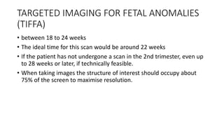
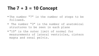
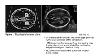
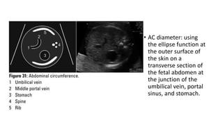
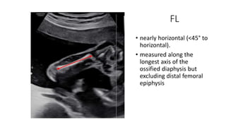
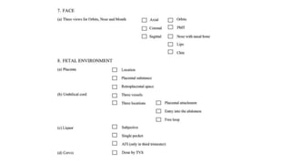
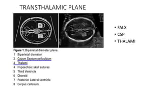
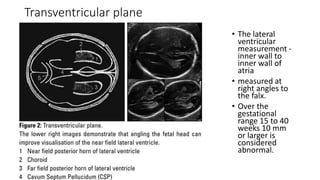
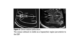
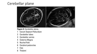
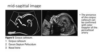
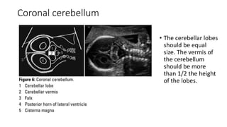
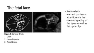
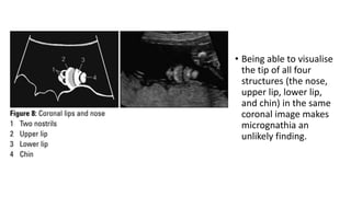
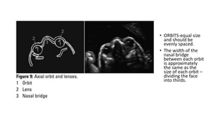
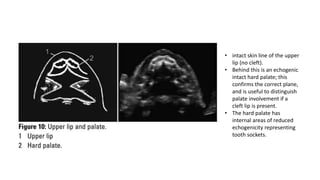
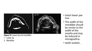
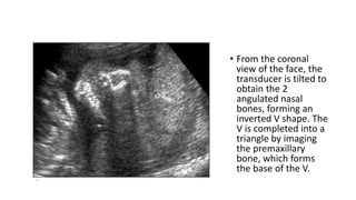
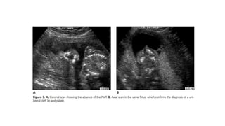
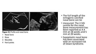
ANOMALY SCAN
 INTRODUCTION
 TARGETED SCAN OF FETAL BRAIN AND FACE
Dr Leslie
JR RD
TARGETED IMAGING FOR FETAL ANOMALIES
(TIFFA)
• between 18 to 24 weeks
• The ideal time for this scan would be around 22 weeks
• If the patient has not undergone a scan in the 2nd trimester, even up
to 28 weeks or later, if technically feasible.
• When taking images the structure of interest should occupy about
75% of the screen to maximise resolution.
The 7 + 3 = 10 Concept
• The number “7” is the number of steps to be
followed.
• The number “3” is the number of anatomical
structures to be seen in each plane
• “10” is the outer limit of normal for
measurements of lateral ventricles, cistern
magna and renal pelvis.
seven step process
• History
• Survey
• Biometry
• Targeted imaging
• Fetal activity
• Fetal environment
• Reporting
• at the level of the thalami and cavum septi pellucidi
without visualization of the cerebellum.
• BPD is commonly measured from the leading edge
(outer edge of the proximal skull) to the leading
edge (inner edge of the distal skull),
• HC is measured around the outside of the skull
bone echoes.
BPD AND HC
• AC diameter: using
the ellipse function at
the outer surface of
the skin on a
transverse section of
the fetal abdomen at
the junction of the
umbilical vein, portal
sinus, and stomach.
FL
• nearly horizontal (<45° to
horizontal).
• measured along the
longest axis of the
ossified diaphysis but
excluding distal femoral
epiphysis
Checklist for Targeted Scan—Rule of Three
TRANSTHALAMIC PLANE
• FALX
• CSP
• THALAMI
Transventricular plane
• The lateral
ventricular
measurement -
inner wall to
inner wall of
atria
• measured at
right angles to
the falx.
• Over the
gestational
range 15 to 40
weeks 10 mm
or larger is
considered
abnormal.
Cerebellar plane
mid-sagittal image
• The presence
of the corpus
callosum can
be confirmed
with color
Doppler of the
pericallosal
artery
Coronal cerebellum
• The cerebellar lobes
should be equal
size. The vermis of
the cerebellum
should be more
than 1/2 the height
of the lobes.
The fetal face
• Areas which
warrant particular
attention are the
size and spacing of
the eyes as well as
the upper lip
• Being able to visualise
the tip of all four
structures (the nose,
upper lip, lower lip,
and chin) in the same
coronal image makes
micrognathia an
unlikely finding.
• ORBITS-equal size
and should be
evenly spaced.
• The width of the
nasal bridge
between each orbit
is approximately
the same as the
size of each orbit –
dividing the face
into thirds.
• intact skin line of the upper
lip (no cleft).
• Behind this is an echogenic
intact hard palate; this
confirms the correct plane,
and is useful to distinguish
palate involvement if a
cleft lip is present.
• The hard palate has
internal areas of reduced
echogenicity representing
tooth sockets.
• intact lower jaw
line.
• The width of the
mandible should
be similar to the
width of the
maxilla and may
be reduced in
micrognathia.
• tooth sockets .
• From the coronal
view of the face, the
transducer is tilted to
obtain the 2
angulated nasal
bones, forming an
inverted V shape. The
V is completed into a
triangle by imaging
the premaxillary
bone, which forms
the base of the V.
• The full length of the
echogenic calcified
nasal bone can be
• measured. The 2.5th
centile for the nasal
bone measurement has
been reported as 4.4
mm at 18 weeks and 5
mm at 20 weeks.
• hypoplastic nasal bone
has been associated
with an increased risk
of Down Syndrome.
REFERENCES
• ICRI GUIDELINES 2022 (ICRI SUBSPECIALITY GROUP ON FETAL RADIOLOGY)
• Suresh, S., Suresh, I. The Second Trimester Obstetric Scan (7 + 3 = 10): A Rational
Approach (Including the “Rule of Three”). J. Fetal Med. 1, 59–73 (2014).
https://doi.org/10.1007/s40556-014-0019-6
• Bethune M, Alibrahim E, Davies B, Yong E. A pictorial guide for the second trimester ultrasound. Australas J
Ultrasound Med. 2013 Aug;16(3):98-113. doi: 10.1002/j.2205-0140.2013.tb00106.x. Epub 2015 Dec 31.
PMID: 28191183; PMCID: PMC5029995.
THANK YOU

anomaly scan. fetal anomaly scan. second trimester scan

  • 1.
    ANOMALY SCAN  INTRODUCTION TARGETED SCAN OF FETAL BRAIN AND FACE Dr Leslie JR RD
  • 2.
    TARGETED IMAGING FORFETAL ANOMALIES (TIFFA) • between 18 to 24 weeks • The ideal time for this scan would be around 22 weeks • If the patient has not undergone a scan in the 2nd trimester, even up to 28 weeks or later, if technically feasible. • When taking images the structure of interest should occupy about 75% of the screen to maximise resolution.
  • 3.
    The 7 +3 = 10 Concept • The number “7” is the number of steps to be followed. • The number “3” is the number of anatomical structures to be seen in each plane • “10” is the outer limit of normal for measurements of lateral ventricles, cistern magna and renal pelvis.
  • 4.
    seven step process •History • Survey • Biometry • Targeted imaging • Fetal activity • Fetal environment • Reporting
  • 5.
    • at thelevel of the thalami and cavum septi pellucidi without visualization of the cerebellum. • BPD is commonly measured from the leading edge (outer edge of the proximal skull) to the leading edge (inner edge of the distal skull), • HC is measured around the outside of the skull bone echoes. BPD AND HC
  • 6.
    • AC diameter:using the ellipse function at the outer surface of the skin on a transverse section of the fetal abdomen at the junction of the umbilical vein, portal sinus, and stomach.
  • 7.
    FL • nearly horizontal(<45° to horizontal). • measured along the longest axis of the ossified diaphysis but excluding distal femoral epiphysis
  • 8.
    Checklist for TargetedScan—Rule of Three
  • 10.
  • 11.
    Transventricular plane • Thelateral ventricular measurement - inner wall to inner wall of atria • measured at right angles to the falx. • Over the gestational range 15 to 40 weeks 10 mm or larger is considered abnormal.
  • 13.
  • 14.
    mid-sagittal image • Thepresence of the corpus callosum can be confirmed with color Doppler of the pericallosal artery
  • 15.
    Coronal cerebellum • Thecerebellar lobes should be equal size. The vermis of the cerebellum should be more than 1/2 the height of the lobes.
  • 16.
    The fetal face •Areas which warrant particular attention are the size and spacing of the eyes as well as the upper lip
  • 17.
    • Being ableto visualise the tip of all four structures (the nose, upper lip, lower lip, and chin) in the same coronal image makes micrognathia an unlikely finding.
  • 18.
    • ORBITS-equal size andshould be evenly spaced. • The width of the nasal bridge between each orbit is approximately the same as the size of each orbit – dividing the face into thirds.
  • 19.
    • intact skinline of the upper lip (no cleft). • Behind this is an echogenic intact hard palate; this confirms the correct plane, and is useful to distinguish palate involvement if a cleft lip is present. • The hard palate has internal areas of reduced echogenicity representing tooth sockets.
  • 20.
    • intact lowerjaw line. • The width of the mandible should be similar to the width of the maxilla and may be reduced in micrognathia. • tooth sockets .
  • 21.
    • From thecoronal view of the face, the transducer is tilted to obtain the 2 angulated nasal bones, forming an inverted V shape. The V is completed into a triangle by imaging the premaxillary bone, which forms the base of the V.
  • 23.
    • The fulllength of the echogenic calcified nasal bone can be • measured. The 2.5th centile for the nasal bone measurement has been reported as 4.4 mm at 18 weeks and 5 mm at 20 weeks. • hypoplastic nasal bone has been associated with an increased risk of Down Syndrome.
  • 25.
    REFERENCES • ICRI GUIDELINES2022 (ICRI SUBSPECIALITY GROUP ON FETAL RADIOLOGY) • Suresh, S., Suresh, I. The Second Trimester Obstetric Scan (7 + 3 = 10): A Rational Approach (Including the “Rule of Three”). J. Fetal Med. 1, 59–73 (2014). https://doi.org/10.1007/s40556-014-0019-6 • Bethune M, Alibrahim E, Davies B, Yong E. A pictorial guide for the second trimester ultrasound. Australas J Ultrasound Med. 2013 Aug;16(3):98-113. doi: 10.1002/j.2205-0140.2013.tb00106.x. Epub 2015 Dec 31. PMID: 28191183; PMCID: PMC5029995.
  • 26.

Editor's Notes

  • #3 terminations are allowed up to 24 weeks, it would be logical to suggest 22 weeks since the fetal organs would be bigger, brain better developed
  • #4 By following the “rule of three”, it is possible to say that all aspects of the fetus that are expected to be seen have indeed been imaged. This ensures that the examination has been done satisfactorily.
  • #5 “high risk” or “low risk”.. whether we need to extend our examination beyond the “rule of three”. For example, if the previous child was diagnosed as a harlequin fetus, measurement of the foot length in this pregnancy would be needed in addition to routine biometry. Number of fetuses Lie and position of the spine The viability The location of placenta The available space around the baby amniotic fluid long axis of the spine of the baby will help us to locate all the major anatomical fetal parts. Fetal activity can be observed while performing the targeted scan. In this step, we specifically look for flexion, extension movements of the limbs including opening and closing of fingers. An actively moving fetus is reassuring. This is the sixth step and includes the imaging of the 1.Placenta 2.Liquor 3.Umbilical cord
  • #6 In the second trimester a difference of >10 days between last menstrual period (LMP) GAand USG GA between 14 and 21 weeks and >14 days between 20 and 28 weeks. The EDD has to be recalculated. If a first trimester scan has been done, it would be appropriate to assign the gestational age by the first trimester parameters and use the second trimester biometry to predict the interval growth and size of the baby. humerus length (HL), nasal bone length (NB), nuchal fold (NF), cerebellar diameter (TCD), cisterna magna and cervical length.
  • #7 more than 50% of the image should be the fetal abdomen. The ellipse should be placed at the outer edge of the soft tissue (soft tissue is included in the ellipse).
  • #8 A measurement less than thie 2.5th centile may increase the risk of aneuploidy and raise concerns about skeletal dysplasia. more than 50% of the image should be the fetal thigh.
  • #11 The normal bone density of the skull should be more echogenic than the falx.
  • #13 just above the BPD plane, at the level of the atrium of the lateral ventricles but still with the CSP in view. The lateral ventricular measurement can be taken from inner wall to inner wall at the level of the glomus of the choroid plexus. The lateral ventricle should be measured at right angles to the falx. Over the gestational range 15 to 40 weeks 10 mm or larger is considered abnormal.
  • #15 inferior to the BPD plane with the probe tilted backward into the posterior fossa. The plane is correct when one can visualise the thalami and cavum septum pellucidum in the same plane as the cerebellum. The trans-cerebellar diameter is the widest measurement across the cerebellum, perpendicular to the falx. Cerebellar size in millimetres correlates with gestational age up to 20 weeks and is larger than gestational age after this time. A cerebellum measuring 2 mm less than gestational age is a concerning finding.16 The cisterna magna can be measured from the posterior margin of the cerebellar vermis to the inside of occipital bone in the midline (following an imaginary continuation of the falx). A measurement of 2–10 mm is normal in the second and third trimesters.17 The nuchal fold is a measurement taken from outer skin. line to outer bone in the midline (following an imaginary continuation of the falx). Less than 6 mm is considered normal up to 22 weeks.
  • #16 It sits superior to the cavum septum pellucidum and extends backwards as a hypoechoic line
  • #17 The cerebellar lobes should be equal size. The vermis of the cerebellum should be more than 1/2 the height of the lobes.
  • #18 The orbits should be equal size with the gap between each orbit approximately the same as the width of each orbit (dividing the face into thirds). The lenses can be seen as central circles that should not have internal echogenicity. Lens opacity may indicate congenital cataracts
  • #20 face needs to be looking up
  • #21 This is a transverse image used to obtain a cross section through the upper lip and hard palate
  • #22 This is a transverse image obtaining a cross section through the lower lip and jaw.
  • #25 This is a mid-sagittal image best taken with the angle of the face at about 45° Shortening (hypoplasia) of the nasal bone has been associated with Down syndrome