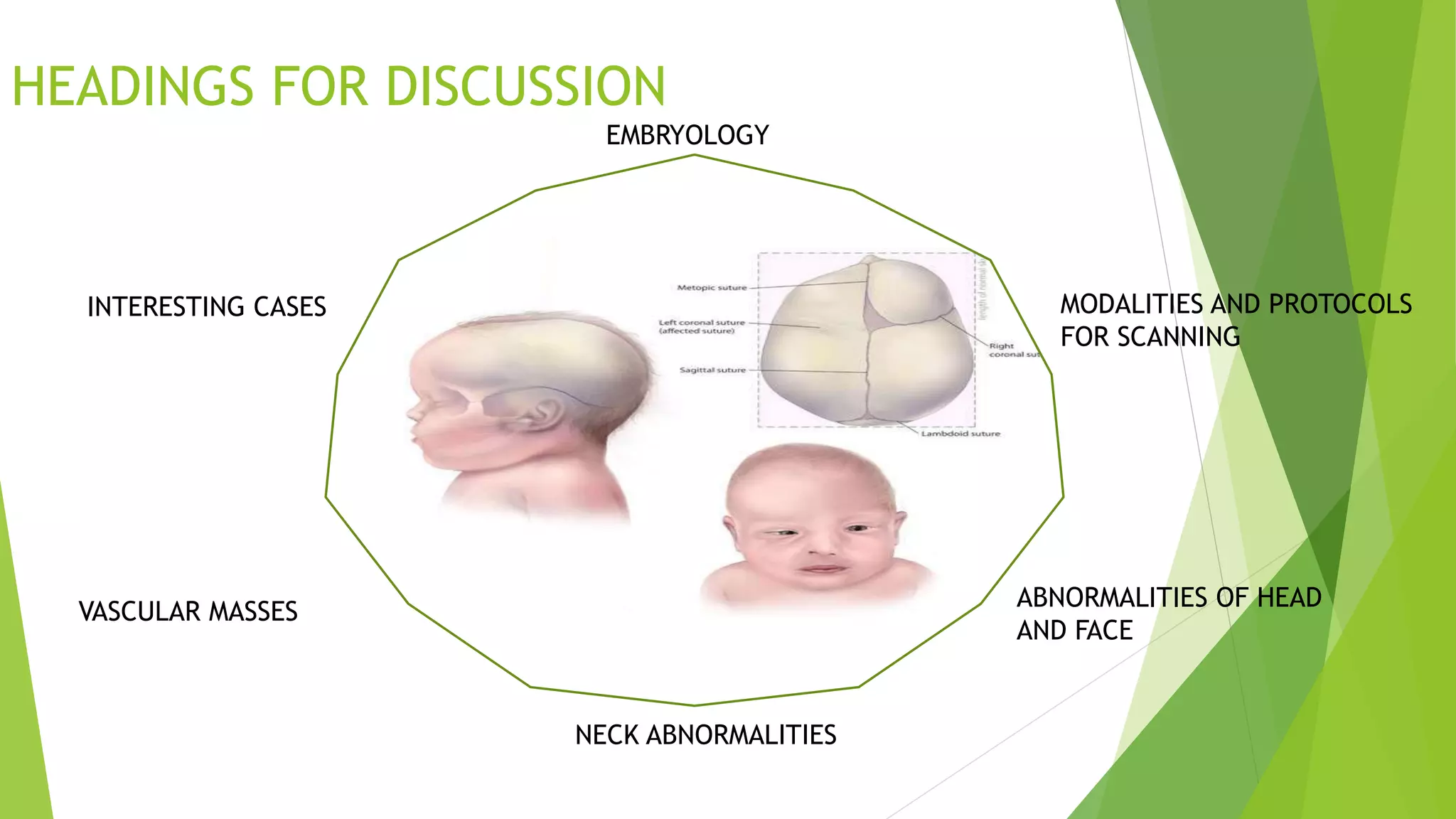
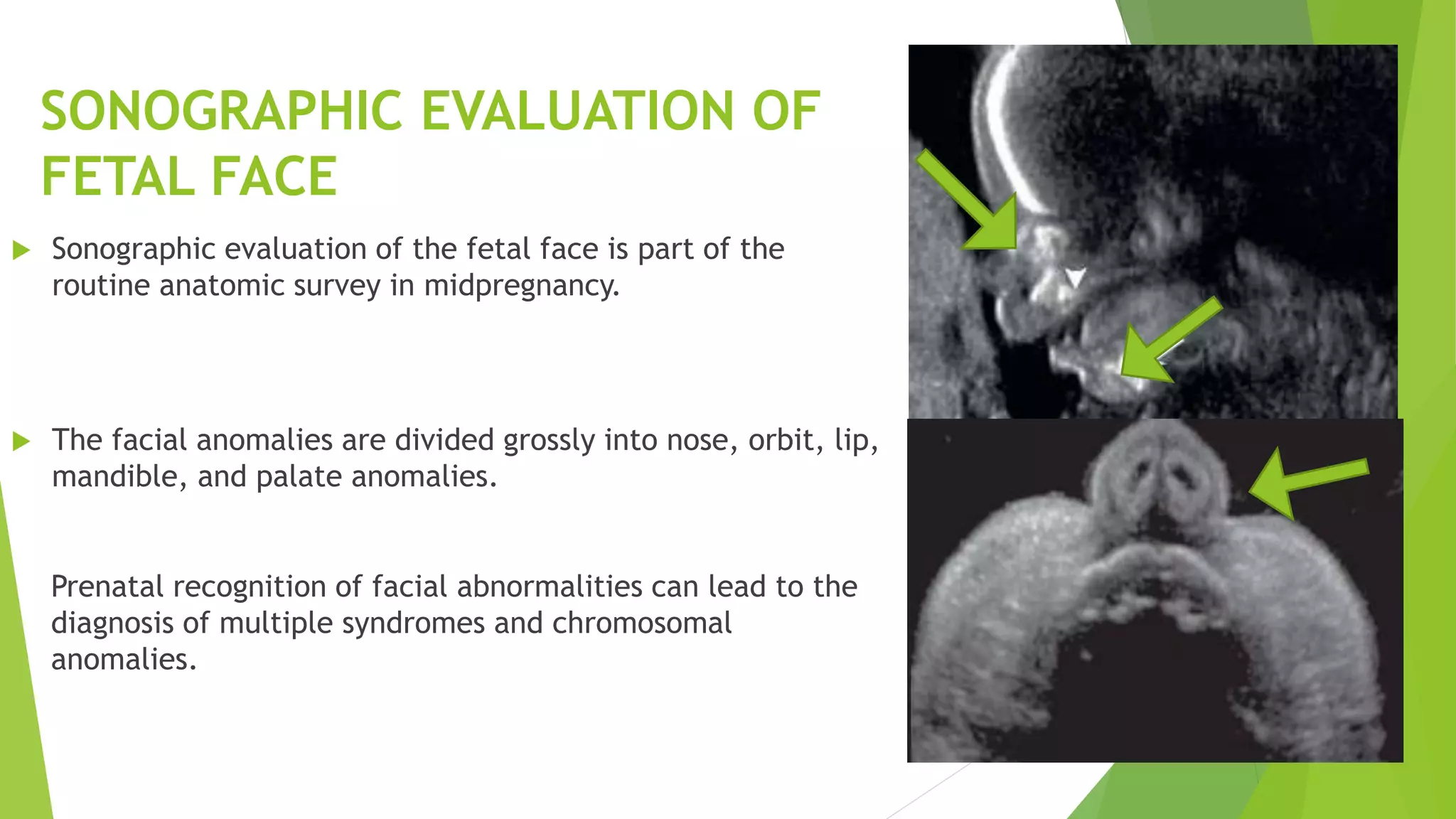
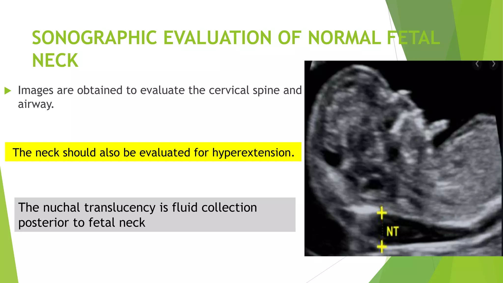
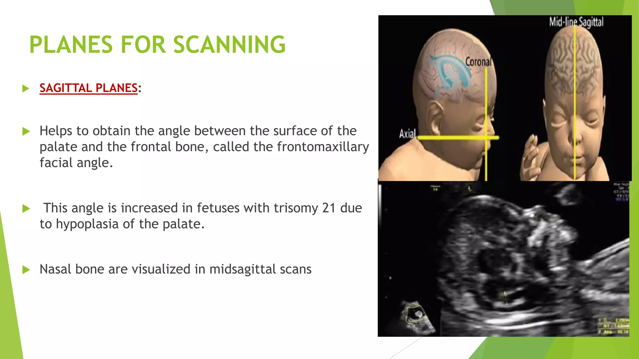
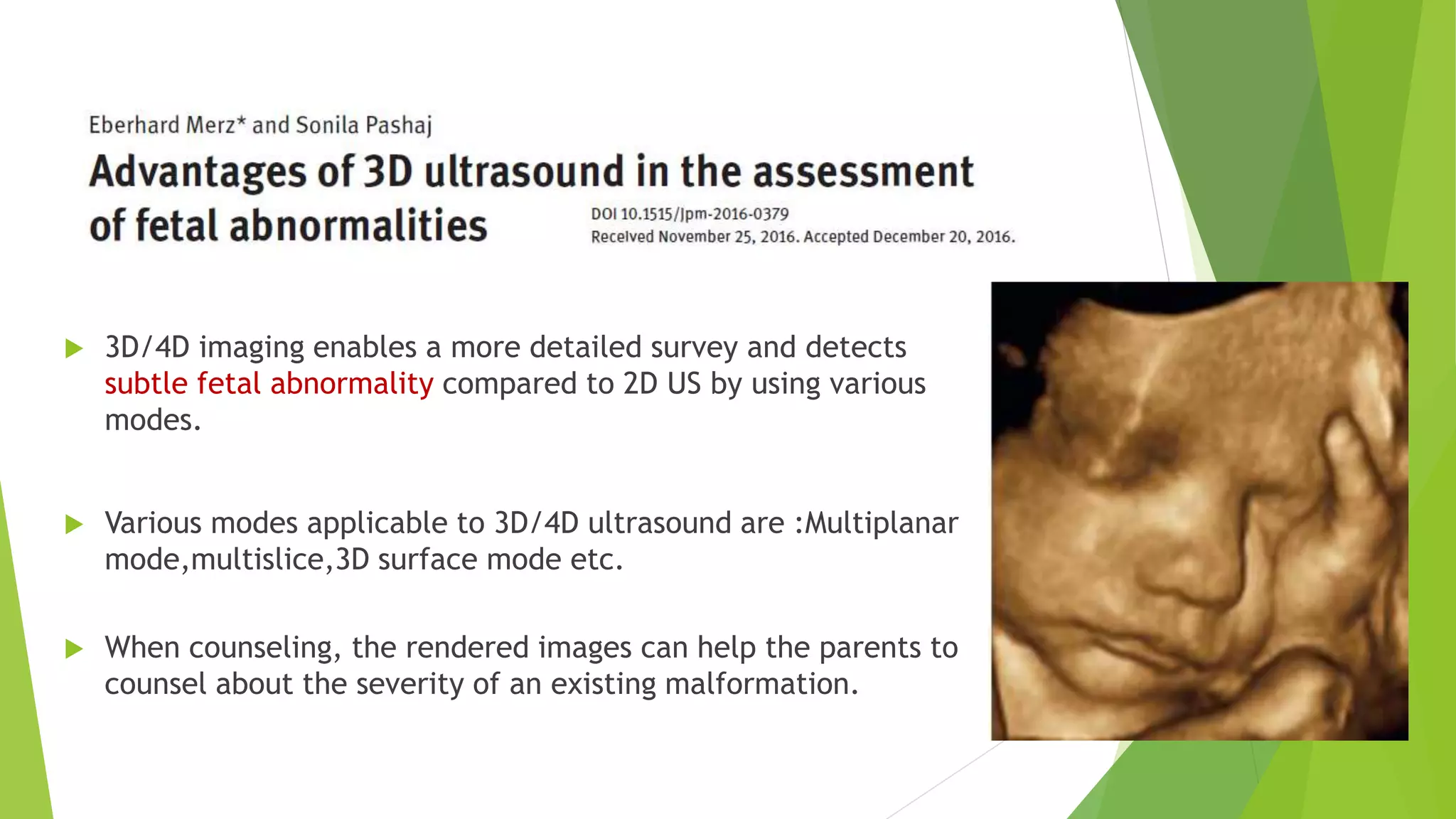
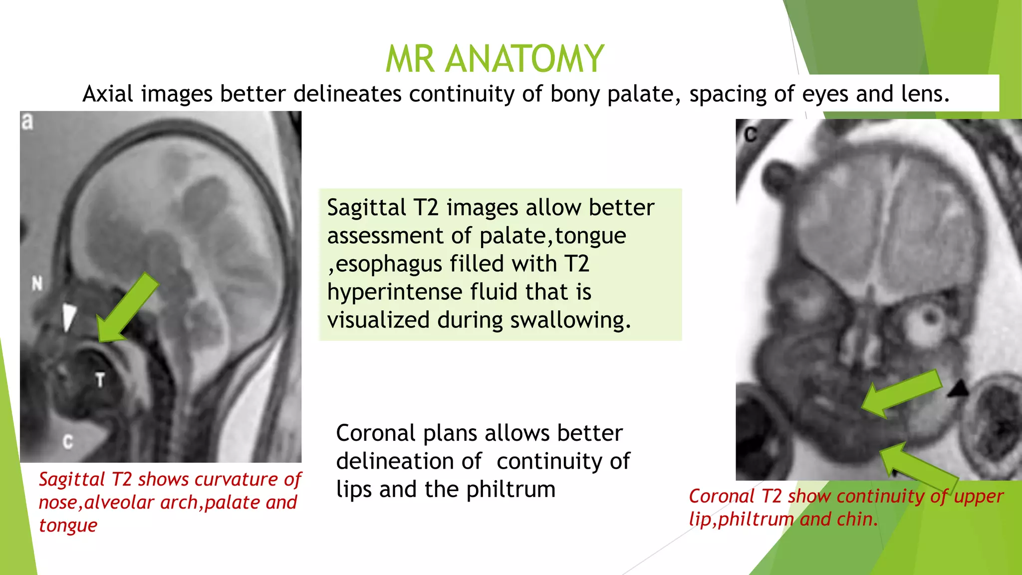
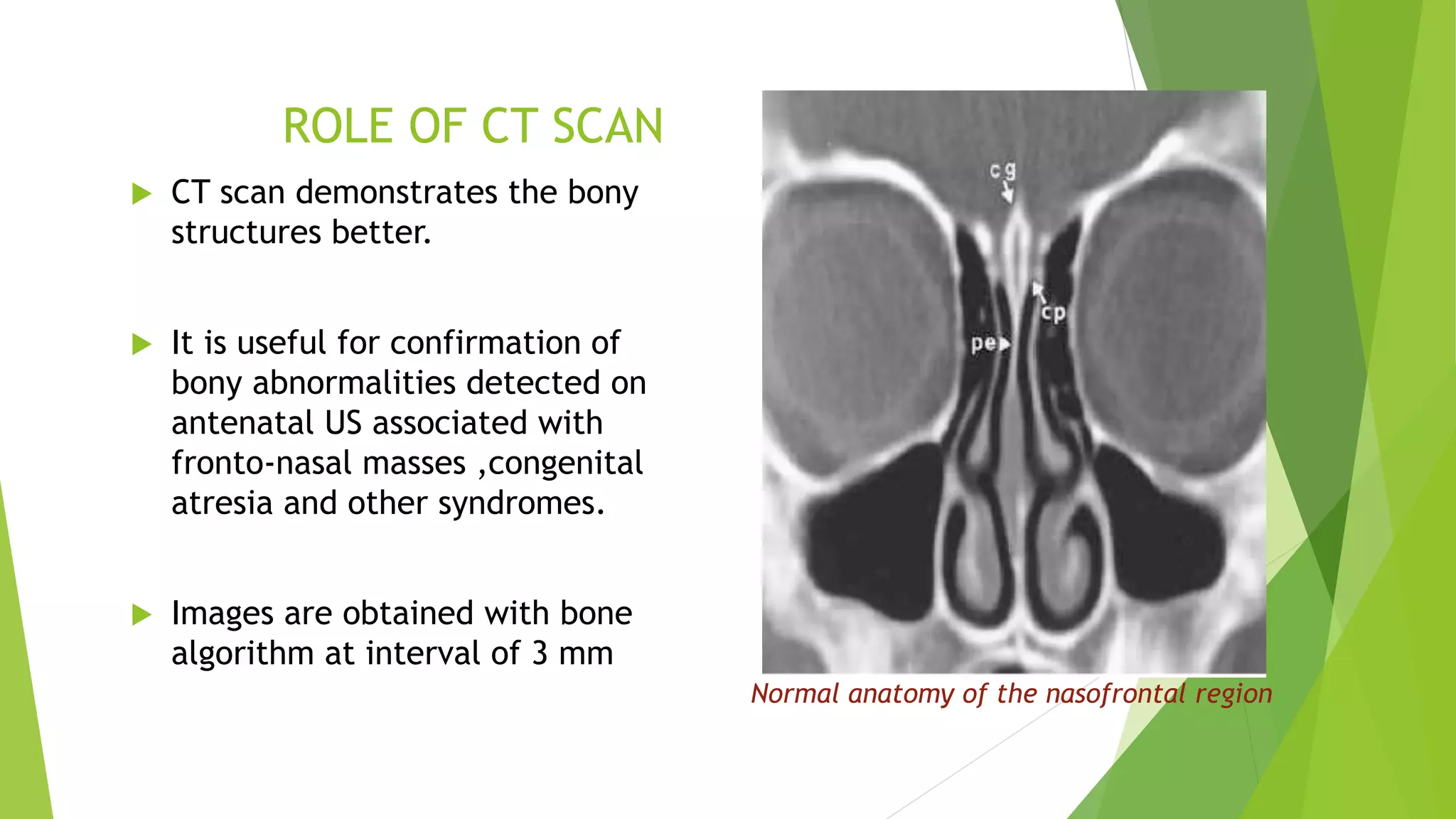
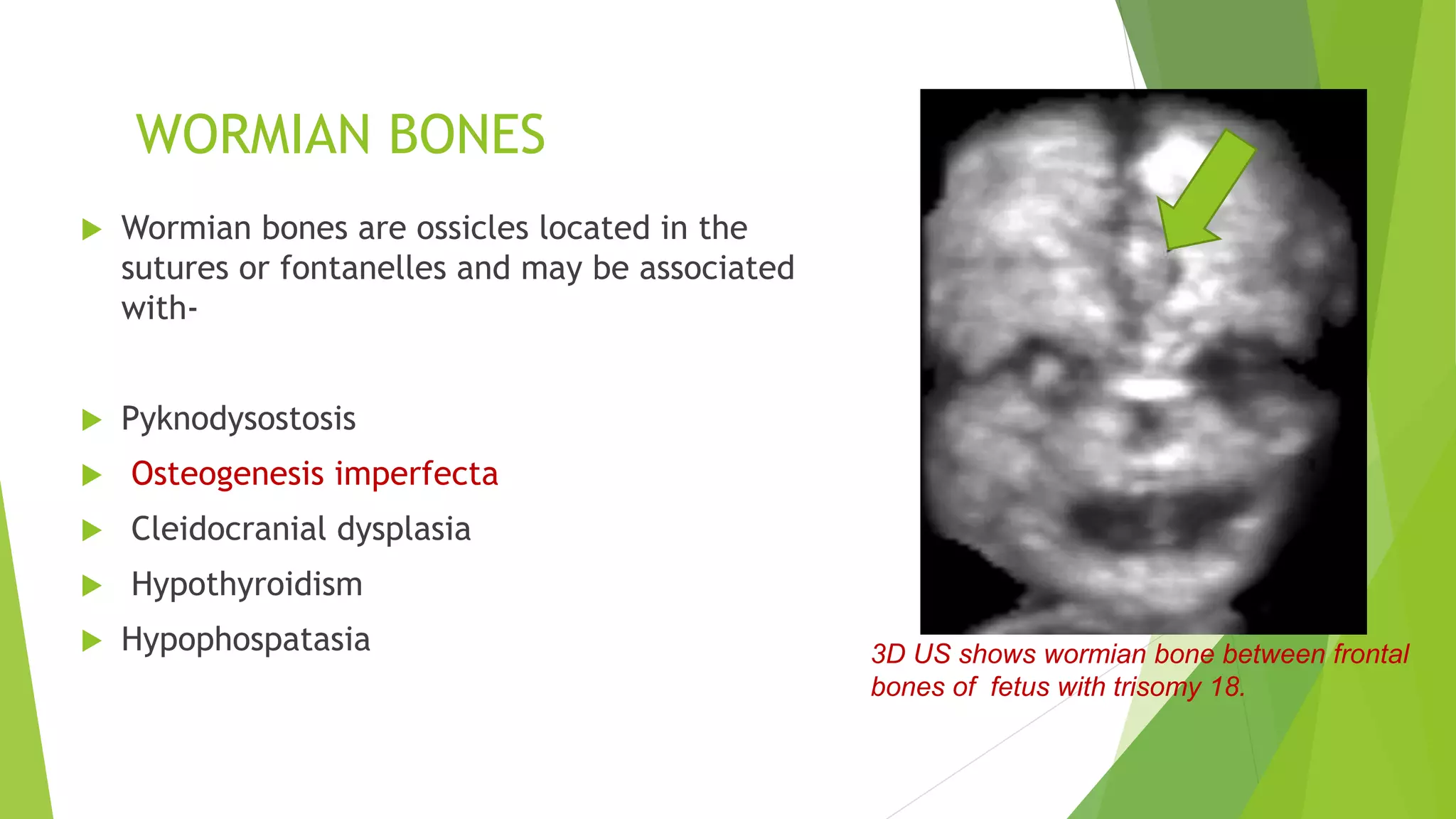
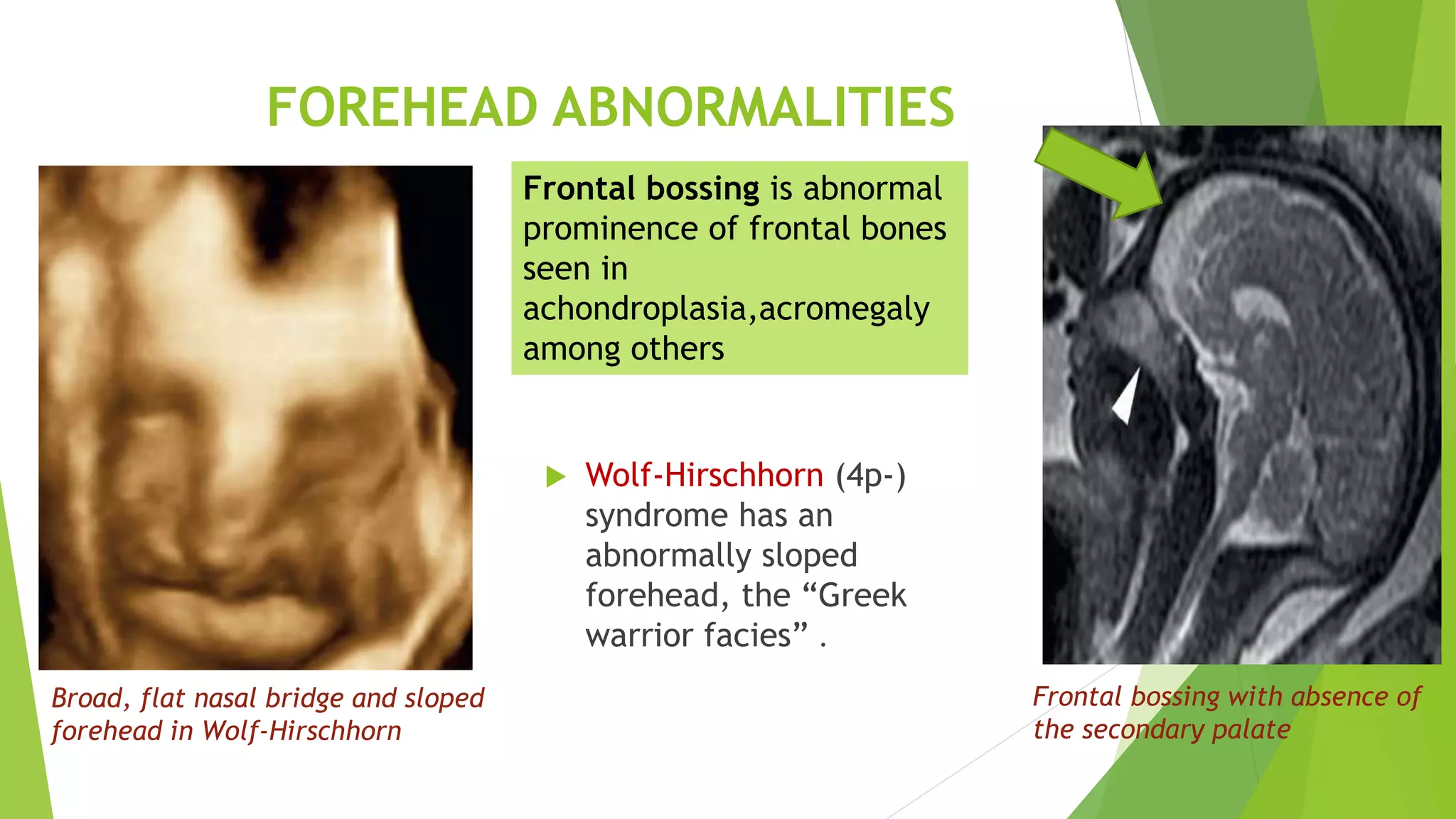
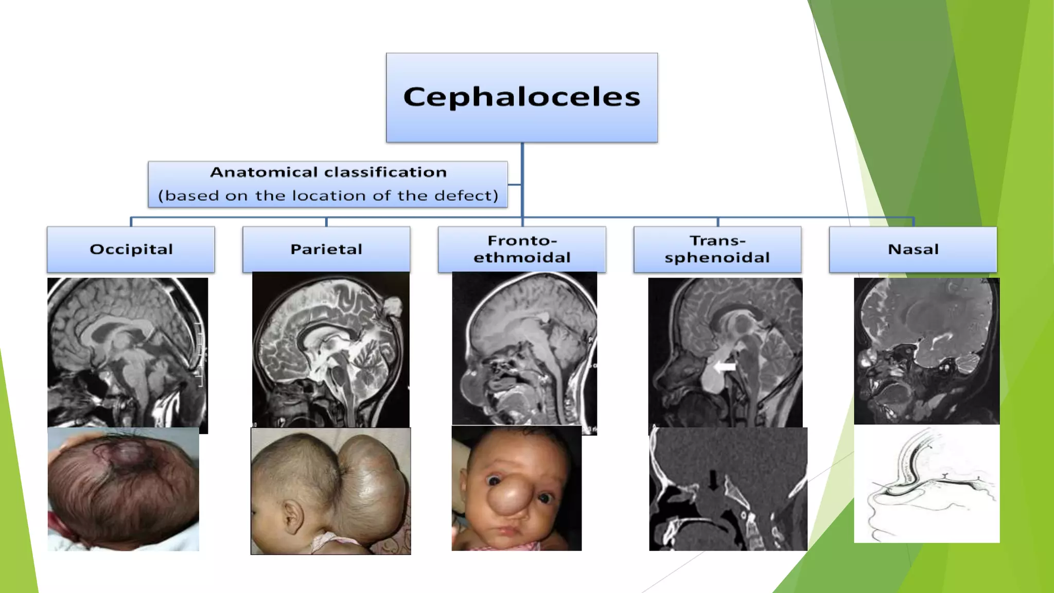
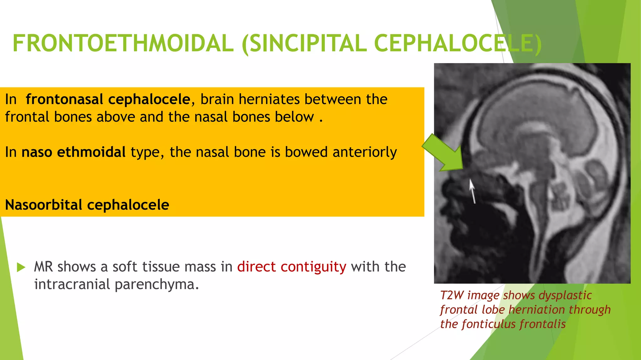
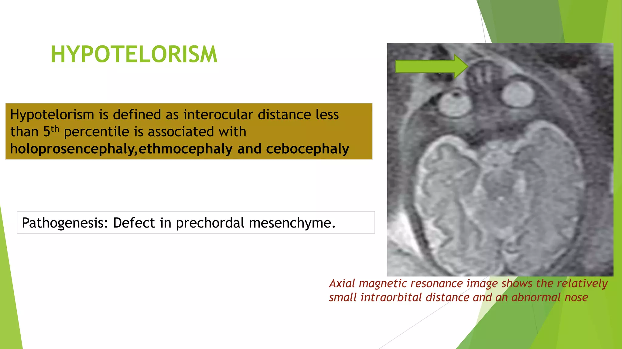
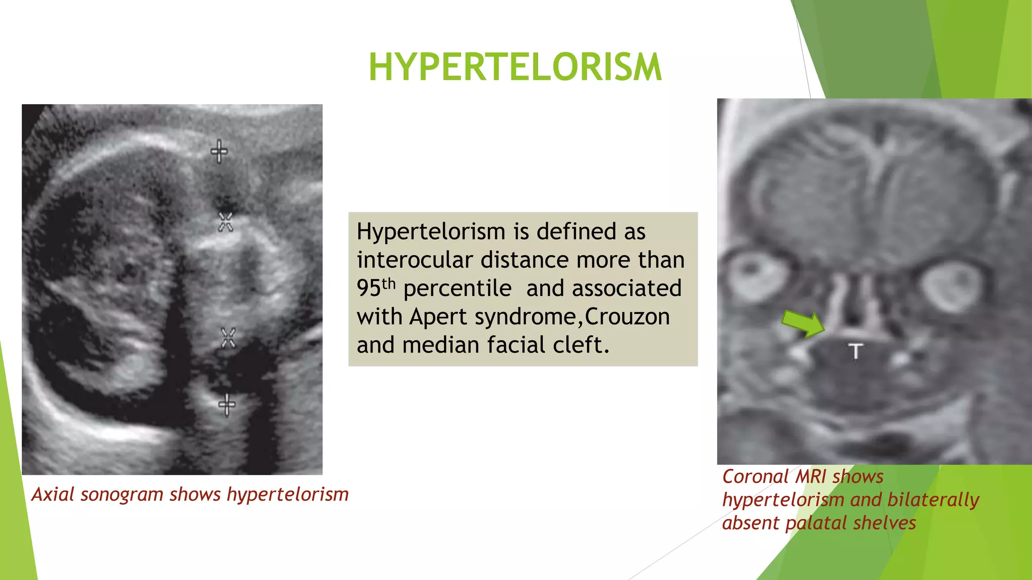
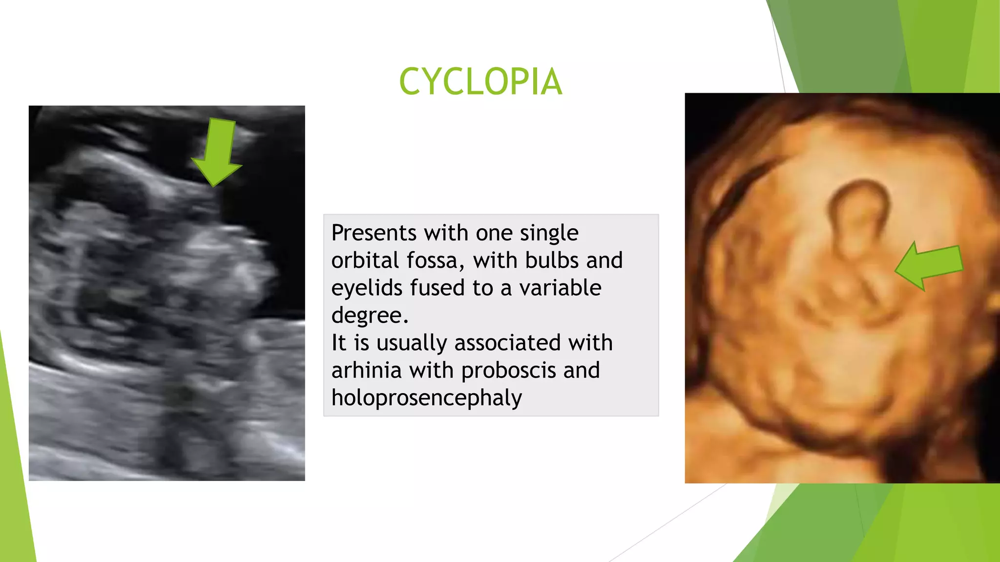
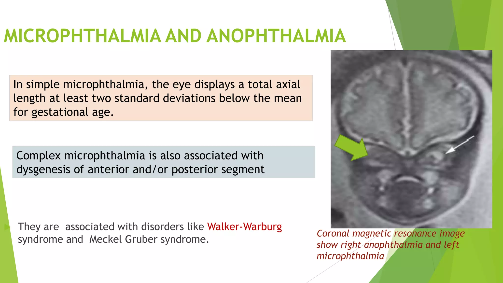
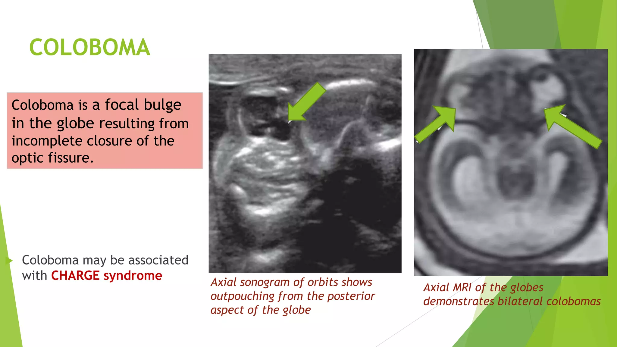
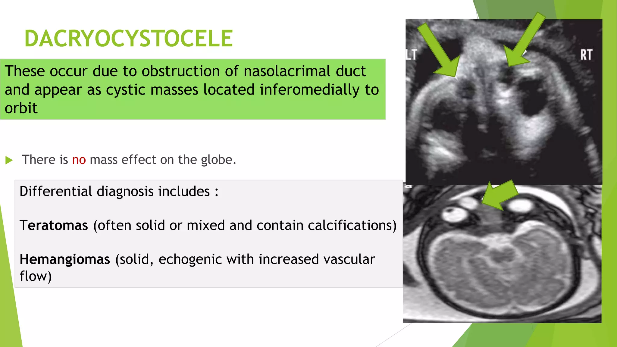
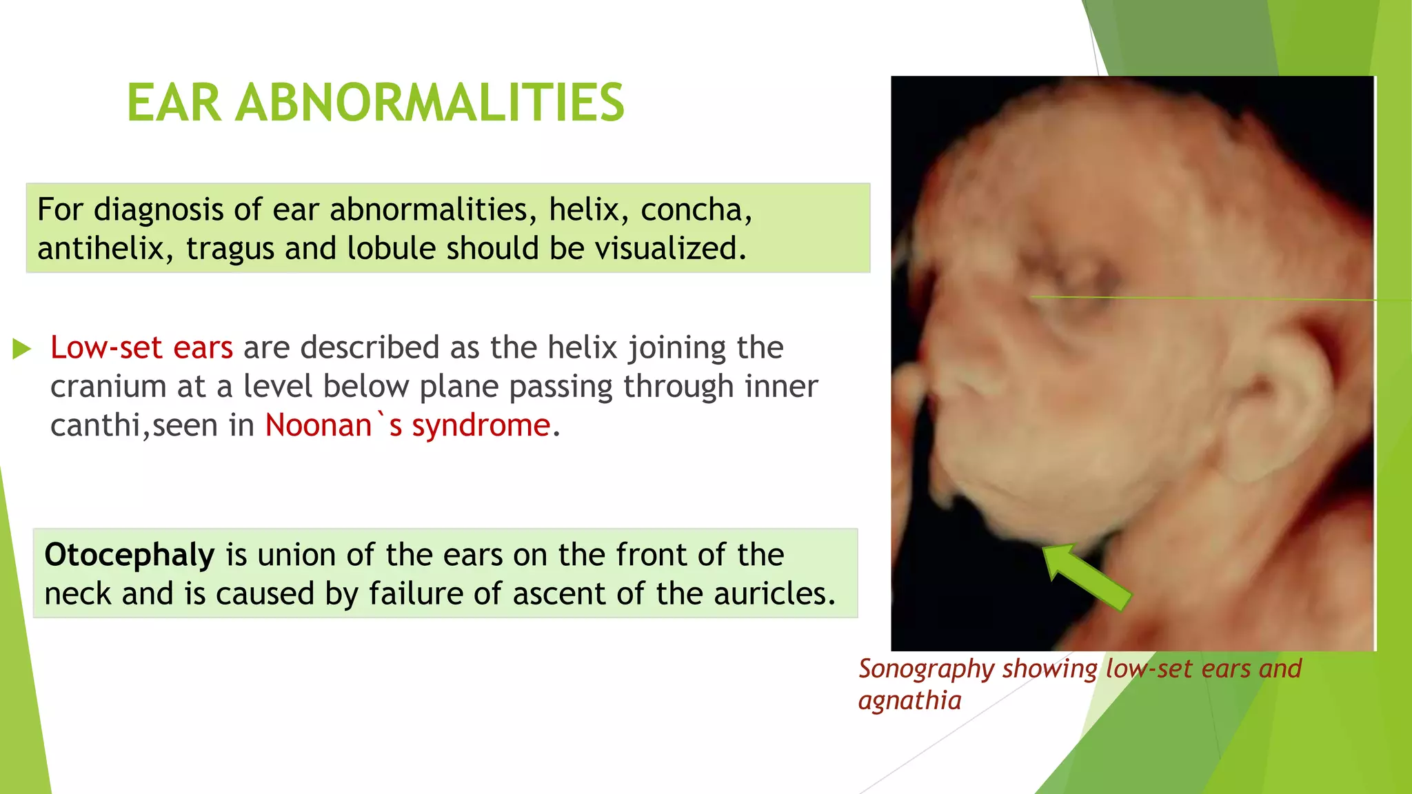
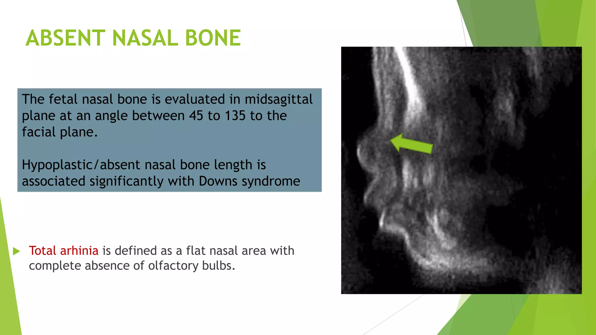
SlideShare a Scribd company logo
1 of 111
CONGENITAL ANOMALIES
OF HEAD,NECK AND
FACE
PRESENTER:DR.SHAURYA AGARWAL
MODERATOR:DR.NABANITA DEKA,M.D
ASSOCIATE PROFESSOR OF RADIOLOGY,GMCH
HEADINGS FOR DISCUSSION
EMBRYOLOGY
MODALITIES AND PROTOCOLS
FOR SCANNING
ABNORMALITIES OF HEAD
AND FACE
NECK ABNORMALITIES
VASCULAR MASSES
INTERESTING CASES
EMBRYOLOGY AND DEVELOPMENT
 Mesenchyme for formation of head region is derived from paraxial and lateral plate
mesoderm, neural crest, and thickened regions of ectoderm known as ectodermal
placodes.
 Paraxial mesoderm (somites and somitomeres) forms a large portion of the membranous
and cartilaginous components of skull and all voluntary muscles of the craniofacial region
 Neural crest cells originate in the neuroectoderm of forebrain, midbrain, and hindbrain
regions and migrate ventrally into the pharyngeal arches, forming the entire face.
Mesenchyme for these structures is derived from
neural crest(blue), paraxial mesoderm (red)and
lateral plate mesoderm (yellow)
Migration pathways of neural crest cells from forebrain,
midbrain, and hindbrain regions into their final locations
in the pharyngeal arches.
EMBRYOGENESIS OF THE BRANCHIAL APPARATUS
The anatomic structures of the face and neck predominantly derive from the branchial apparatus,
that develops between the second and seventh weeks of gestation
 The branchial apparatus consists of six paired arches.
 The cervical sinus of His appears by mesodermal growth cranially of the first arch and a portion
of the second arch and caudally by growth of mesoderm lateral to the fifth-sixth arch.
 Branchial abnormalities develop from incomplete obliteration of the branchial apparatus
Pharyngeal pouches shown as outpocketings of the
foregut and thyroid primordium
Each arch consists of a mesenchymal core
lined internally by endoderm and externally by ectoderm.
Each arch also contains an artery
When the embryo is 42 days old, five mesenchymal prominences can be recognized:
 Frontonasal prominence cranial to the stomodeum
 Median and lateral nasal prominences.
 Maxillary and Mandibular prominences
FACIAL DEVELOPMENT
GESTATIONAL AGE WISE FACIAL DEVELOPMENT
 4th Week: The optic discs are present posterolaterally.
 5th week: The lens vesicles invaginate within optic discs.
 6th week: The hyaloid vessels are present in the optic stalk.
 Six auricular hillocks forms pinna of the ears.
 Nasolacrimal duct forms from the medial corner of eye to the inferior meatus of nasal
cavity
 7th week: The central axis of the nose and philtrum are formed
DEVELOPMENT OF TONGUE AND THYROID
 The tongue develops from two lateral lingual swellings and one
medial lingual swelling,the tuberculum impar,originating from
1st branchial arch.
 Hypobranchial eminence is formed by the mesoderm of the
2nd,3rd,4th pharngeal clefts.
 The thyroid appears as proliferation between tuberculum impar
and hypobranchial eminence indicated by foramen cecum.
 The thyroid then migrates infront of trachea and remains
connected by tongue by thyroglossal duct.
DEVELOPMENT OF NASAL CAVITIES
INTERMAXILLARY SEGMENT
 As a result of growth of the maxillary prominences, the
two medial nasal prominences merges and forms inter
maxillary segment.
 It is composed of (1) labial component, which forms
the philtrum; (2) upper jaw component, which carries
the four incisors; and (3) palatal component, which
forms the triangular primary palate
DEVELOPMENT OF SECONDARY PALATE
 The main part of the definitive palate is formed by two
shelf-like outgrowths from maxillary prominences.
 These palatine shelves appear in the 6th week and
directed downward.
 In the 7th week the palatine shelves ascend to attain a
horizontal position above the tongue and fuse forming
the secondary palate.
 Anteriorly, the shelves fuse with the triangular primary
palate, and incisive foramen is the midline landmark
between the primary and secondary palates.
DEVELOPMENT OF THE NECK
 The laryngotracheal diverticulum forms during the 4th week of gestation
along the floor of the primitive mouth.
 The distal end forms the lung bud. The endoderm of this diverticulum
forms the epithelium of the larynx and trachea.
 The 4th and 6th pharyngeal arches form surrounding cartilage and
muscles.
SONOGRAPHIC EVALUATION OF
FETAL FACE
 Sonographic evaluation of the fetal face is part of the
routine anatomic survey in midpregnancy.
 The facial anomalies are divided grossly into nose, orbit, lip,
mandible, and palate anomalies.
Prenatal recognition of facial abnormalities can lead to the
diagnosis of multiple syndromes and chromosomal
anomalies.
SONOGRAPHIC EVALUATION OF NORMAL FETAL
NECK
 Images are obtained to evaluate the cervical spine and
airway.
The nuchal translucency is fluid collection
posterior to fetal neck
The neck should also be evaluated for hyperextension.
PLANES FOR SCANNING
 SAGITTAL PLANES:
 Helps to obtain the angle between the surface of the
palate and the frontal bone, called the frontomaxillary
facial angle.
 This angle is increased in fetuses with trisomy 21 due
to hypoplasia of the palate.
 Nasal bone are visualized in midsagittal scans
AXIAL PLANES
 Orbits may be visualized simultaneously,
by means of an axial plane.
CORONAL PLANES
Coronal planes better visualizes the eyelids,lips,
forehead, and nose(nostrils appear as two little
anechoic areas)
 3D/4D imaging enables a more detailed survey and detects
subtle fetal abnormality compared to 2D US by using various
modes.
 Various modes applicable to 3D/4D ultrasound are :Multiplanar
mode,multislice,3D surface mode etc.
 When counseling, the rendered images can help the parents to
counsel about the severity of an existing malformation.
FETAL MRI
 Ultrasound is limited by factors such as oligohydramnios, maternal body
habitus, complex fetal anomalies, and operator dependency.
 MRI has emerged as a valuable adjunct because of development of ultrafast
sequences with few motion artefacts ,excellent soft tissue contrast,less
operator dependency and excellent spatial resolution.
 Fetal MRI is optimal at 18-22 weeks.
 Static Magnetic Field Exposure:FDA has limits on magnetic field exposure of 4T for
patients less than one month of age.
 Specific Absorption Rate:Less than 3.2 W/kg over 10 minutes exposure in head
 Acoustic Noise and Sound Pressure Levels :should not exceed 140 db
TECHNICAL PARAMETERS
 T1-weighted sequences are obtained in order to detect mucine or blood.
 T2-weighted images consist of ultra-fast-spin-echo sequences and steady-state free precession
sequences
 Dynamic sequences (cine T2 true sequence) may be used to improve tracheal or laryngeal
obstruction by showing a fluid T2-hyperintense pouch during swallowing.
 3D FIESTA help clarify small lesions as a cleft lip by decreasing the slice thickness to 1.6 mm.
 Diffusion Weighted Imaging (DWI) is obtained to detect brain malformation associated.
MR ANATOMY
Sagittal T2 images allow better
assessment of palate,tongue
,esophagus filled with T2
hyperintense fluid that is
visualized during swallowing.
Coronal plans allows better
delineation of continuity of
lips and the philtrum
Axial images better delineates continuity of bony palate, spacing of eyes and lens.
Sagittal T2 shows curvature of
nose,alveolar arch,palate and
tongue
Coronal T2 show continuity of upper
lip,philtrum and chin.
ROLE OF CT SCAN
 CT scan demonstrates the bony
structures better.
 It is useful for confirmation of
bony abnormalities detected on
antenatal US associated with
fronto-nasal masses ,congenital
atresia and other syndromes.
 Images are obtained with bone
algorithm at interval of 3 mm
Normal anatomy of the nasofrontal region
ABNORMALITIES OF THE HEAD
 The HC gives proper estimates of gestational age.
Abnormal Size
However,if HC is three SD below the mean, microcephaly is diagnosed,seen in
trisomies and holoprosencephaly.
If HC is greater than 3 SDs, macrocephaly is detected leading to cephalo-pelvic
disproportion and difficulty in delivery.
Anencephaly is the most severe form characterized by a defective calvarium
and scalp whereas ineincepahly is occiput defect with cervical vertebrae
absence.
Sagittal T2-weighted image showing
anencephaly and absent brainstem
ABNORMAL SHAPE
 Craniosynostosis:Craniosynostosis describes a group of disorders in which there is premature
fusion of one or several of the cranial sutures.
Craniosynostosis can be isolated or associated with syndromes including Apert, Crouzon,
Pfeiffer, Carpenter and thanatophoric dysplasia.
Imaging plays an essential role in recognition of craniosynostoses, identification of
coexisting brain anomalies,preoperative treatment planning, and postoperative followup.
Lemon Shaped Skull
strawberry
shape
cloverleaf skullDolicocepahly
Turribrachycephaly
SYNDROMES ASSOCIATED WITH CRANIOSYNOSTOSIS
 Shallow orbits with hypertelorism are characteristic ocular findings.
The airways may be compromised by hypoplasia of the posterior choanae.
Spinal fusion anomalies and midfacial hypoplasia are also common.
Acrocephalosyndactyly type I includes brachycephaly and symmetric syndactyly associated
with a mitten like deformity of the hand.
APERT’S SYNDROME
Postnatal CT scan shows midface hypoplasia ,narrowing of
pyriform aperture and posterior choanal stenosis
Sonogram showing prominent forehead and
midfacial hypoplasia
CROUZON’S SYNDROME
Sagittal T1 MR image reveals steep clivus, vertically
oriented brainstem and Chiari I malformation
Crouzon syndrome is characterized
by brachycephaly,mid facial
hypoplasia with vertically oriented
brainstem, and cleft palate.
Cranial abnormalities include
Chiari malformation, hypoplastic
transverse sinus
Brachycephaly
PFEIFFER’S SYNDROME
CARPENTER’S SYNDROME
It is associated with broad, medially rotated thumbs and varus deformity of the great toes.
It is associated with Craniosynostosis, polydactyly,
syndactyly and cardiac defects
Polydactyly
WORMIAN BONES
 Wormian bones are ossicles located in the
sutures or fontanelles and may be associated
with-
 Pyknodysostosis
 Osteogenesis imperfecta
 Cleidocranial dysplasia
 Hypothyroidism
 Hypophospatasia 3D US shows wormian bone between frontal
bones of fetus with trisomy 18.
FOREHEAD ABNORMALITIES
 Wolf-Hirschhorn (4p-)
syndrome has an
abnormally sloped
forehead, the “Greek
warrior facies” .
Broad, flat nasal bridge and sloped
forehead in Wolf-Hirschhorn
Frontal bossing with absence of
the secondary palate
Frontal bossing is abnormal
prominence of frontal bones
seen in
achondroplasia,acromegaly
among others
OCCIPITAL CEPHALOCELES
High occipital defect
Saggital T2 image showing the
high occipital defect in the skull
Associated abnormalities
such as Chiari 2 and Dandy-
Walker spectrum disorders
Occipital cephalocele
are classified into
occipitocervical , low
occipital and high
occipital (involving
occipital bone only)
FRONTOETHMOIDAL (SINCIPITAL CEPHALOCELE)
 MR shows a soft tissue mass in direct contiguity with the
intracranial parenchyma.
T2W image shows dysplastic
frontal lobe herniation through
the fonticulus frontalis
In frontonasal cephalocele, brain herniates between the
frontal bones above and the nasal bones below .
In naso ethmoidal type, the nasal bone is bowed anteriorly
Nasoorbital cephalocele
PARIETAL CEPHALOCELES
T2-weighted images show a fluid-filled lesion between
parietal bones and the falcine sinus
Parietal cephaloceles (atretic cephaloceles )
represent as lesions present as scalp masses near
the posterior vertex.
Associated abnormalities include persistent falcine
sinus and partial absence of the straight sinus.
ORBIT ABNORMALITIES
Axial planes evaluates the presence of both orbits,their
sizes and shapes and the measuring the BOD and IOD.
BOD is measured between the malar margins of
vitreous, while IOD is obtained between the two
ethmoidal margins of vitreous.
The sagittal view help to evaluate proptosis.
HYPOTELORISM
Axial magnetic resonance image shows the relatively
small intraorbital distance and an abnormal nose
Hypotelorism is defined as interocular distance less
than 5th percentile is associated with
holoprosencephaly,ethmocephaly and cebocephaly
Pathogenesis: Defect in prechordal mesenchyme.
HYPERTELORISM
Axial sonogram shows hypertelorism
Coronal MRI shows
hypertelorism and bilaterally
absent palatal shelves
Hypertelorism is defined as
interocular distance more than
95th percentile and associated
with Apert syndrome,Crouzon
and median facial cleft.
CYCLOPIA
Presents with one single
orbital fossa, with bulbs and
eyelids fused to a variable
degree.
It is usually associated with
arhinia with proboscis and
holoprosencephaly
MICROPHTHALMIA AND ANOPHTHALMIA
 They are associated with disorders like Walker-Warburg
syndrome and Meckel Gruber syndrome.
Coronal magnetic resonance image
show right anophthalmia and left
microphthalmia
In simple microphthalmia, the eye displays a total axial
length at least two standard deviations below the mean
for gestational age.
Complex microphthalmia is also associated with
dysgenesis of anterior and/or posterior segment
PERSISTENT HYPERPLASTIC PRIMARY VITREOUS (PHPV)
Axial and sagittal T2W images show linear T2 hypointense structure
corresponding to Cloquet’s canal with retinal detachment
MRI demonstrate an abnormal
low T2 signal line through the
central vitreous corresponding
to Cloquet’s canal.
There is association with Warburg
syndrome and retinal dysplasia.
COLOBOMA
 Coloboma may be associated
with CHARGE syndrome Axial sonogram of orbits shows
outpouching from the posterior
aspect of the globe
Axial MRI of the globes
demonstrates bilateral colobomas
Coloboma is a focal bulge
in the globe resulting from
incomplete closure of the
optic fissure.
CONGENITAL CATARACTS
 Congenital cataracts show a rounded
echogenic mass in the anterior portion of the
globe.
Echogenic lens
Causes includes:
Arthrogryposis Chondrodysplasia punctata
Chromosomal abnormalities (21, 18, 13)
Infection(Rubella,Toxoplasmosis)
DACRYOCYSTOCELE
 There is no mass effect on the globe.
These occur due to obstruction of nasolacrimal duct
and appear as cystic masses located inferomedially to
orbit
Differential diagnosis includes :
Teratomas (often solid or mixed and contain calcifications)
Hemangiomas (solid, echogenic with increased vascular
flow)
EAR ABNORMALITIES
 Low-set ears are described as the helix joining the
cranium at a level below plane passing through inner
canthi,seen in Noonan`s syndrome.
Sonography showing low-set ears and
agnathia
For diagnosis of ear abnormalities, helix, concha,
antihelix, tragus and lobule should be visualized.
Otocephaly is union of the ears on the front of the
neck and is caused by failure of ascent of the auricles.
MIDFACE ABNORMALITIES
 The midface is the area between the upper lip and
forehead.
HYPOPLASIA
Midface hypoplasia occurs in syndromes such as Apert,
Crouzon and trisomy 21.It is associated with
micropthalmia.
3D view showing midface
hypoplasia with microphthalmia
ABSENT NASAL BONE
 Total arhinia is defined as a flat nasal area with
complete absence of olfactory bulbs.
The fetal nasal bone is evaluated in midsagittal
plane at an angle between 45 to 135 to the
facial plane.
Hypoplastic/absent nasal bone length is
associated significantly with Downs syndrome
CONGENITAL MIDLINE NASOFRONTAL MASSES
 These are the result of faulty regression of embryologic dural diverticulum from prenasal space.
 The mass may be intranasal, extranasal, or combination.
 These includes-
Findings that suggest intracranial extension include widening of foramen cecum and a bifid crista galli
EncephalocelesNasal gliomas HemangiomasDermoid
(midline glabellar fatty
masses)
Epidermoid (fluid
attenuation)
NASAL GLIOMA
 Nasal gliomas result from sequestering herniated glial tissue appearing isointense to the gray
matter on T1 and T2 sequences.
 Most of them are extranasal(60%)occuring at the root of the nose
 MRI allows differentiation of nasal glioma having mixed-signal-intensity from encephalocele.
Desquamation of lining cells of dermal sinus tract results in formation of
dermoid/epidermoid tumors along tract.
T2 W reveals a round and well-limited mass of the nasal
region displaying a signal similar to
gray matter signal on MR
Saggital T2 W images show bright signal intensity
in glabella suggestive of dermoid cyst
accompanied by a dermal sinus tract extending
through foramen cecum.
NASAL ENCEPHALOCELE
 Frontonasal encephaloceles result from herniation of dura mater through foramen cecum and
the fonticulus frontalis into glabella whereas nasoethmoidal encephaloceles occur due to
herniation through foramen cecum into nasal cavity.
 Differential diagnosis includes dermoid (midline) opposed to encephaloceles (lateral)to crista.
MRI demonstrates continuity of brain tissue via enlarged foramen cecum and intrathecal injection of
contrast material may demonstrate a subarachnoid connection
Sagittal T1W image shows brain parenchyma within a
glabellar mass with outward bowing of the inferior
frontal bone and associated absence of the corpus
callosum suggesting frontonasal encephalocele
Nasoethmoidal encephalocele
demonstrating a nasal mass containing
cerebrospinal fluid and brain parenchyma
CLEFT LIP AND PALATE
 They present with difficulty with feeding,otitis media, and difficulty with hearing and speech.
 Central clefting includes Pierre Robin anomalad, whereas lateral clefting includes Treacher Collins.
Cleft lip usually result from failure of fusion of medial nasal prominences and maxillary prominence.
Palatal clefts usually are located between incisors and canines and are result of failure of fusion
of one side of primary palatal process with secondary process
Associated Signs of Cleft Palate With Cleft Lip includes on axial and coronal views
Lips: cleft
Nares: flattened or deformed
Vomer: deviated away from side of cleft
Maxilla: wide gap
Orbits: hypertelorism
sagittal views: Profile: Micrognathia, Tongue: high position.
Complete cleft lip is defined as a cleft that fully divides the lip ,extends into nostril and
associated with a cleft of the alveolus
An incomplete cleft lip involves a portion of the lip, but a band of soft tissue spans the
cleft.
NORMAL ISOLATED CLEFT LIP
UNILATERAL CLEFT LIP AND
ANTERIOR PALATE
BILATERAL CLEFT LIP AND
ANTERIOR PALATE ISOLATED CLEFT PALATE
BILATERAL CLEFT LIP AND
PALATE
Nyberg et al proposed a ultrasonographic classification of CL with or without
CP.
CL alone as type 1
Unilateral CL and CP as type 2
Bilateral CL and CP as type 3
Midline CL and CP as type 4
Facial defects associated with amniotic bands as type 5.
UNILATERAL CLEFT LIP OR PALATE
In a unilateral cleft lip or
palate, there is often an
offset between the two
sides of the cleft,
described as the “greater
segment” on the opposite
the cleft, and the “lesser
segment” on the side of
the cleft.
BILATERAL CLEFT LIP OR PALATE
In bilateral cleft lip or
palate, there will be
premaxillary protrusion of
intermaxillary segment
MEDIAN CLEFT LIP OR PALATE
In the inferior group there may be deficiency of upper
lip and philtrum.
Patients with anomalies in the superior group are associated
with hypertelorism, median cleft nose and cranium bifidum.
Midline clefting (Frontonasal dysplasia) include either inferior
or A group and superior or B group
3D US shows absence of the intermaxillary
segment
ISOLATED CLEFT OF SECONDARY PALATE
Syndromes associated with isolated clefts include
Goldenhar syndrome and Pierre-Robin sequence.
Isolated cleft of secondary palate is identified by
abnormal oropharyngeal fluid flow with color
Doppler imaging.
UNUSUAL FACIAL (TESSIER) CLEFTS
 These are asymmetrical clefts in unusual locations and
may be the result of amniotic band syndrome
The clefts 1 through 7 lies below the orbit and 8 through
14, above it.
These clefts are classified by relationship of the cleft to
the mouth, nose, and eye and are numbered from 1 to 14,
with the midline designated as 0 and 14
AMNIOTIC BAND SYNDROME
 The amniotic band sequence consists of
soft tissue derangements arising from
tethering of the fetus by fibrous bands of
amniotic tissue.
Sagittal T2 image shows entrapment of the occipital lobe resulting into
an encephalocele that protrudes into the amniotic sac.
Amniotic band syndrome can lead to
development of cephaloceles and clefts
SURGICAL MANAGEMENT OF CLEFT
LIP/PALATE

AGE SURGICAL TECHNIQUE
1-3 months
3 months
9-12 months
1-8 years
Above skeletal maturity
Lip taping and nasoalveolar molding
Cleft lip repair and placement of ventilation tubes
Cleft palate repair
Orthodontic treatment and alveolar bone graft
Mid face advancement and continued orthodontics


CONGENITAL ATRESIAS AND STENOSES
 Congenital choanal atresias and
stenoses result from tissue
deficiencies or failure of resorption of
epithelial plugs
 They are usually associated with
CHARGE syndrome
Saggital sonogram showing
dilated nasal cavity
Postnatal CT features of choanal
atresia/stenosis include width of less than
0.34 cm,medial deviation of medial
maxillary wall and obstruction with
proximal air-fluid level.
LOWER FACE ABNORMALITIES
 Macroglossia is described as abnormally enlarged tongue.
MACROGLOSSIA
Diagnosis is established by considering the imaginary
line between mandible and maxilla on the sagittal
plane, with protruding tip of tongue past that line.
The etiology of macroglossia includes Beckwith-
Wiedemann syndrome, trisomy 21 and lymphatic
malformation
MICROGNATHIA
 This finding is associated with Pierre Robin sequence and
Stickler syndrome
T2W image at shows severe micrognathia,posteriorly displaced
tongue hiding the posterior cleft palate
Pierre -robin sequence(anomalad) includes triad of
micrognathia, glossoptosis, and subsequent airway obstruction.
Micrognathia describes a small mandible .
MICROSTOMIA
 Microstomia is small mouth, associated with agnathia
and otocephaly.
Ultrasound image showing agnathia microstomia
Overfusion of mandibular and maxillary processes laterally
leads to microstomia
NECK ABNORMALITIES
 Neck masses in foetus are midline and mostly benign,
most common amongst them is cystic hygroma.
The thymus and thyroid, in pathology along the
midline.
Dermoid cysts are also usually midline in location
Branchial abnormalities develop from paired branchial
arches, and lie laterally.
Similarly,sternomastoid tumour and congenital
sympathetic chain neuroblastoma will also lie laterally.
LOCATION
DIAGNOSTIC APPROACH
 CYSTIC
MIDLINE OFF-MIDLINE
RANULA(UPPER)
THYROGLOSSAL
DUCT CYST(MIDDLE)
DERMOID
CYST(LOWER)
LYMPHANGIOMA
BRANCHIAL CYST
VASCULAR
MALFORMATION
SOLID
THYROID LESION
FIBROMATOSIS COLLI
ECTOPIC THYMUS
TERATOMA
RANULA
 Ranulas are retention cysts of
sublingual salivary gland due to
duct stenosis or occlusion.
Cystic lesion continuous with the sublingual
salivary gland
Plunging ranula results from rupture of
duct and extravasation of this fluid into
submental region.
THORNWALDT'S CYST
Thornwaldt's cyst forms as a bulge on posterior-superior
wall of nasopharynx due to closure of drainage duct
and appearing intermediate-signal on T1W and high-
signal on T2W
Cystic lesion showing T2 signal intensity in
pharyngeal mucosal space in the midline
THYROGLOSSAL DUCT CYST
 The thyroid gland forms as a diverticulum from anterior wall of pharynx,connected by the
thyroglossal duct
 if duct fail to regress, may differentiate into cysts, lying at or just below the hyoid in midline. They
may contain normal thyroid tissue and can lead to papillary carcinoma
The differential diagnosis includes dermoid cyst and branchial cleft cyst.
US reveals an anechoic well-defined cyst with increased acoustic through-transmission
MRI shows low signal on T1 and high signal on T2w
CT demonstrates a low-attenuation mass with
enhancing components concerning for a TDC
complicated by papillary thyroid carcinoma.
DERMOID/EPIDERMOID CYST
 Dermoid and epidermoid cysts
occur at midline closure.
Ultrasound,CT and MRI
shows presence of
fat,with“sack-of-marbles”
appearance on CT
Epidermoid shows T2
hyperintensity
Dermoids contain
epithelial and dermal
elements, whereas
epidermoids contain only
epithelial elements.
Hyper-echoic dermoid cyst in the
suprasternal notch.
Epidermoid cyst at floor
of mouth
LYMPHATIC MALFORMATION
 Lymphatic malformation can be microcystic(<1cm), macrocystic or both.
 In conditions where there is difficulty in delivery,EXIT(Ex-utero intrapartum treatment)
procedure has to be carried out.
They can cause mass effect on fetal airway and may interfere with swallowing and feeding.
Conventionally LM's have been subdivided into 1) cystic hygroma, 2) cavernous lymphangioma, 3)
capillary lymphangioma, 4) vasculolymphatic lymphangiohaemangioma.
ROLE OF IMAGING DURING AN EXIT PROCEDURE
 Sterile intraoperative US is helpful in --
 Mapping the placental location to determine
the optimal site for the hysterotomy incision.
 Evaluating orientation of fetal head and neck to
assist in delivering in optimal orientation for
intubation.
In cases of large cystic neck mass predelivery US-
guided aspiration of fluid pockets decrease the
size of mass
TYPES OF LYMPHATIC MALFORMATION
 Cystic hygroma is the most common form of lymphatic malformation and is usually macrocystic.
 This abnormality is highly associated with Turner syndrome and other chromosomal anomalies
 They have a predilection for left side of posterior neck,extending into superior mediastinum
 Cavernous lymphangioma: They are smaller than the cystic hygroma and subcutaneous in location.
 Lymphangiohaemangiomas: Lymphangiohaemangiomas retain their venous communication,colour
Doppler showing blood flow through the mass on its periphery.
Ultrasonography demonstrate multiple
septated cystic spaces filled with hypoechoic
fluid in a case of turner syndrome.
MRI shows a multilocular T2
hyperintense mass with septa that
appear T2 hypointense
BRANCHIAL CLEFT ABNORMALITIES
FIRST BRANCHIAL ANOMALIES
MRI usually have low T1-weighted and high T2-weighted
signal intensity.
Embyropathogenesis: Anomalies arise from incomplete
closure of first branchial cleft at the site of organ of origin
of middle ear, external auditory canal, parotid,mandible,
and maxilla.
MRI reveals rounded T2 bright cystic lesion within the
right parotid gland.
SECOND BRANCHIAL ANOMALIES
 Can occur anywhere from tonsillar fossa to the
supraclavicular region of neck.
 It usually presents as a fluctuant non tender
mass at the lateral aspect of the mandibular
angle, appearing hyperintense on T1 and T2W.
 With a fistula, ostium is noted at anterior
border of the lower junction of the SCM.
Second branchial cleft
anomaly, type
1,anterior to SCM
Type 2,posterior to
submandibular gland and
anterior to carotid
Type 3 sinus/fistula,from
tonsillar region to lower
SCM
Type 4 in pharyngeal
mucosal space in lateral
pharyngeal wall
BAILEY’S CLASSIFICATION
In Bailey type III a “beak sign” is present, where the medial
aspect of the cyst is compressed between the ICA and ECA
In type III,from the external ostia,the tract courses between
the internal and external carotid arteries before it terminates
in the region of the tonsillar fossa.
THIRD BRANCHIAL ANOMALIES
MRI present as unilocular cystic mass
above superior laryngeal nerve
In fistula, external ostia will be anterior
to lower SCM.
It then courses posterior to common
carotid artery and enters pyriform sinus
Embryology. Third branchial anomalies are
rare lesions,located posterior to common
carotid
Postnatally,barium swallow shows
fistula extending from left piriform
sinus to near clavicle
FOURTH BRANCHIAL ANOMALIES
Third branchial anomalies lie posterior to the internal carotid artery, whereas fourth branchial
anomalies are anterior to it.
They are in the form of a sinus tract that arises from the pyriform sinus,descends along
tracheoesophageal groove, and continues into mediastinum.
Embryogenesis : They are likely due to failure of regression of the fourth branchial pouch or distal
sinus of His.
CERVICAL EXTENSION OF THE MEDIASTINAL
THYMUS
Ultrasonography, CT and MRI will show the
connection with the mediastinal thymus.
Incomplete descent of the thymus may present as solid
midline mass or a cystic mass at thoracic inlet,causing
dyspnoea or dysphagia due to compression.
THYMIC CYST
Differential Diagnosis: Branchial cleft cyst and
epidermoid cyst
They usually present as cystic neck mass near thoracic
inlet parallel to carotid sheath
T2 fetal MR image and CT showing thymic cyst
CYSTIC POUCH IN ESOPHAGEAL ATRESIA
 Other indirect signs are polyhydramnios, absent or small
stomach, and a tracheoesophageal fistula
T2-weighted images show a fluid-filled cystic formation
of the upper esophagus
Esophageal atresia lead to a blind-ended pouch which can be
seen with sonography and better with fetal MRI.
TRACHEO-ESOPHAGEAL FISTULA
 Presents with feeding
difficulties,
polyhydraminios,respiratory
difficulties and recurrent
pneumonia.
Associated with
VACTERL,trisomies and
cardiac anomalies
ECTOPIC THYROID GLAND
 Any abnormality in descent of thyroid
may result in an ectopic location
Postnatal MRI ,CT or radionuclide
scanning show the location, size and
activity of the functioning thyroid tissue.
This is usually at the base of the tongue
or in a pre-laryngeal and substernal
position.
THYROID GLAND HEMIAGENESIS
Hemiagenesis of thyroid gland presents as mass due to
hypertrophy of single thyroid lobe or thyroid dysfunction.
Ultrasonography shows absence of one side of gland with
a normal appearing contralateral side and isthmus,
producing so-called hockey stick sign.
THYROMEGALY AND GOITER
 MRI shows a solid homogeneous anterior
neck mass with variable T2 signal intensity.
not compromising airway
Fetal goiter is related to
maternal Graves or
Hashimoto thyroiditis,
with antibodies that cross
placenta and lead to fetal
thyroid dysfunction.
Midline homogeneous solid mass in the
anterior neck surrounding trachea and
may interfere with fetal swallowing.
STERNOMASTOID "TUMOUR"
 Sternomastoid tumour, or "fibromatosis colli appears
as mass through thickening of the sternomastoid
muscle, commonly affecting middle to lower third.
 Pathogenesis: intrauterine packaging defect.
Hyperechoic thickened sternomastoid
tumour
On MRI enlarged mass is isointense on T1W and T2W
The ultrasonographic appearances is classified into
four types: (1) fibrotic mass (2) diffuse fibrosis mixed
with normal muscle (3) diffuse involvement without
normal muscle (4) fibrotic cord
TUMOURS
CERVICAL TERATOMA
 Cervical teratomas are calcifed
complex masses composed of cystic
and solid elements.
 They are usually anterolateral and
can become large ,interfering with
fetal swallowing.
Fat, giving a high signal on T1W and a
signal void from calcification will help to
distinguish it from cystic hygroma
ORAL TERATOMA (EPIGNATHUS)
T2 heterogeneous mass with cystic and tissue
components that distends the oral cavity
On MRI, it appears as heterogeneous lesion with mixed T2
signal.
They interfere with fetal swallowing and resultant
polyhydramnios
DIFFUSE CUTANEOUS NEUROFIBROMA
 Frequently present with overgrowth of subcutaneous
tissues typically of head and neck.
 Associated with NF-1
On MRI, lesion appears as T1 isointense,T2 hyper intense
lesion with vivid contrast enhancement and thickened nerves
CERVICAL NEUROBLASTOMA
 Neuroblastomas originates from neural crest, and occur anywhere
having sympathetic neural tissue and may present as Horner's
syndrome.
 Plain chest X-ray and CT will show calcifications and intrathoracic
extension.
Ultrasonography shows a solid mass with vascular displacement and
calcification.
These lesions usually shows increased uptake of MIBG MIBG I-123scan showing high uptake
of the isotope in the lesion.
CONGENITAL VASCULAR MASSES
 Hemangiomas often increase in size during fetal life and can be infiltrative. It is frequently
associated with PHACE syndrome.
HAEMANGIOMA
Rapidly involuting congenital hemangioma (RICH) demonstrates full size of tumor at birth and
regresses by 12 months of age.
Noninvoluting congenital hemangioma (NICH) is present at birth and grows in proportion to the
patient and does not involute.
They often appear as echogenic, solid masses containing detectable vascular channels on Doppler
Ultrasound along power Doppler demonstrates a highly vascular
soft tissue scalp mass
MRI show the left scalp mass with
presence of multiple flow voids

CAROTID ARTERY ANEURYSM
CERVICAL AORTIC ARCH
It is usually a right-sided aortic arch and produce a pulsatile neck
swelling or compression of the airways or oesophagus.
Congenital carotid artery aneurysms due to a defect in the arterial
wall seen in Ehlers-Danlos, Marfan's or Maffucci's syndromes.
Plain films may show indentation of the trachea.
MRI will show the high position of the arch passing through thoracic
inlet.
INTERESTING
CASES
1) A 31 year old female,G1P0
came for anomaly scan on
27/02/2020 referred from GOPD
DOWN SYNDROME
 Down syndrome (or trisomy 21) is the most common trisomy(1 in 800 live birth)
 Risk factors : Increasing maternal age.
 Pathogenesis: Meiotic non-disjunction
 Imaging findings:
1st trimester: Nuchal translucency>3mm,absent/hypoplastic nasal bone
2nd trimester: soft tissue markers:
Echogenic intracardiac focus
Echogenic bowel
Hypoplastic/absent nasal bone
Aberrant right subclavian artery (ARSA)
Structural abnormalities:
ASD,VSD
Cystic hygroma
 2) A 33 year old female,G2P1 at
18 weeks of gestation came for
anomaly scan.
EDWARDS SYNDROME
 Edwards syndrome (or trisomy 18) is 2nd most common autosomal trisomy(1 in 3000 live birth)
 Imaging findings: Cardiovascular abnormalities: ASD,VSD,PDA
CNS abnormalities: choroid plexus cyst,agenesis of the corpus callosum,
Facial/calvarial abnormalities: micrognathia,strawberry skull
Skeletal abnormalities: Radial ray anomalies,club foot
 3) A 29 year old female,G3P2L0
came at 22 weeks of gestation
for USG.
PATAU SYNDROME
 Patau syndrome (or trisomy 13) is 3rd most common autosomal trisomy(1 in 6000 live birth)
 Imaging findings: Cardiovascular abnormalities: VSD,HLHS
CNS abnormalities: Holoprosencepahly,corpus callosal dysgenesis
Facial/calvarial abnormalities: Micrognathia,proboscis,facial cleft
Skeletal abnormalities: polydactyly,rocker bottom feet
SUMMARY & CONCLUSION
 Prenatal evaluation of fetal head, neck and face offers opportunity to
identify many abnormalities.
 These are essential to prenatal counselling and prognosis because of
syndromic association.
 Appropriate diagnosis allows for planning of appropriate mode of
delivery and therapy when airway is potentially compromised.
Congenital anomalies of head,neck and face(Radiology) including USG and fetal mri

More Related Content

What's hot

Presentation1.pptx, radiological imaging of the nasopharyngeal diseases.
Presentation1.pptx, radiological imaging of the nasopharyngeal diseases.Presentation1.pptx, radiological imaging of the nasopharyngeal diseases.
Presentation1.pptx, radiological imaging of the nasopharyngeal diseases.Abdellah Nazeer
 
Placenta ultrasound
Placenta ultrasoundPlacenta ultrasound
Placenta ultrasoundDoaa Gadalla
 
Fetal Neurosonogram
Fetal Neurosonogram Fetal Neurosonogram
Fetal Neurosonogram nasrat1949
 
Presentation1.pptx, radiological imaging of pediatric neck masses.
Presentation1.pptx, radiological imaging of pediatric neck masses.Presentation1.pptx, radiological imaging of pediatric neck masses.
Presentation1.pptx, radiological imaging of pediatric neck masses.Abdellah Nazeer
 
Imaging of paranasal sinuses (including anatomy and varaints)pk1 pdf ppt
Imaging of paranasal sinuses (including anatomy and varaints)pk1 pdf  pptImaging of paranasal sinuses (including anatomy and varaints)pk1 pdf  ppt
Imaging of paranasal sinuses (including anatomy and varaints)pk1 pdf pptDr pradeep Kumar
 
Deep neck spaces and surgical anatomy
Deep neck spaces and surgical anatomyDeep neck spaces and surgical anatomy
Deep neck spaces and surgical anatomyHimanshu Mishra
 
Radioanatomy of mediastinum and approach to mediastinal masses
Radioanatomy of mediastinum and approach to mediastinal massesRadioanatomy of mediastinum and approach to mediastinal masses
Radioanatomy of mediastinum and approach to mediastinal massesAkankshaMalviya3
 
CT anatomy of Neck Spaces RV
CT anatomy of Neck Spaces RVCT anatomy of Neck Spaces RV
CT anatomy of Neck Spaces RVRoshan Valentine
 
Presentation1.pptx, radiological anatomy of the neck.
Presentation1.pptx, radiological anatomy of the neck.Presentation1.pptx, radiological anatomy of the neck.
Presentation1.pptx, radiological anatomy of the neck.Abdellah Nazeer
 
Imaging of the neck part i
Imaging of the neck part iImaging of the neck part i
Imaging of the neck part iWafik Ebrahim
 
Sphenoid sinus and optic nerve
Sphenoid sinus and optic nerveSphenoid sinus and optic nerve
Sphenoid sinus and optic nerveDr Soumya Singh
 
Presentation1, radiological imaging of placenta accreta.
Presentation1, radiological imaging of placenta accreta.Presentation1, radiological imaging of placenta accreta.
Presentation1, radiological imaging of placenta accreta.Abdellah Nazeer
 
CT ANATOMY OF PARA NASAL SINUSES
CT ANATOMY OF PARA NASAL SINUSESCT ANATOMY OF PARA NASAL SINUSES
CT ANATOMY OF PARA NASAL SINUSESPrasanna Kumaravel
 

What's hot (20)

Presentation1.pptx, radiological imaging of the nasopharyngeal diseases.
Presentation1.pptx, radiological imaging of the nasopharyngeal diseases.Presentation1.pptx, radiological imaging of the nasopharyngeal diseases.
Presentation1.pptx, radiological imaging of the nasopharyngeal diseases.
 
Placenta ultrasound
Placenta ultrasoundPlacenta ultrasound
Placenta ultrasound
 
Fetal Neurosonogram
Fetal Neurosonogram Fetal Neurosonogram
Fetal Neurosonogram
 
Neck dissections
Neck dissectionsNeck dissections
Neck dissections
 
Presentation1.pptx, radiological imaging of pediatric neck masses.
Presentation1.pptx, radiological imaging of pediatric neck masses.Presentation1.pptx, radiological imaging of pediatric neck masses.
Presentation1.pptx, radiological imaging of pediatric neck masses.
 
Imaging of paranasal sinuses (including anatomy and varaints)pk1 pdf ppt
Imaging of paranasal sinuses (including anatomy and varaints)pk1 pdf  pptImaging of paranasal sinuses (including anatomy and varaints)pk1 pdf  ppt
Imaging of paranasal sinuses (including anatomy and varaints)pk1 pdf ppt
 
Deep neck spaces and surgical anatomy
Deep neck spaces and surgical anatomyDeep neck spaces and surgical anatomy
Deep neck spaces and surgical anatomy
 
Radioanatomy of mediastinum and approach to mediastinal masses
Radioanatomy of mediastinum and approach to mediastinal massesRadioanatomy of mediastinum and approach to mediastinal masses
Radioanatomy of mediastinum and approach to mediastinal masses
 
CT anatomy of Neck Spaces RV
CT anatomy of Neck Spaces RVCT anatomy of Neck Spaces RV
CT anatomy of Neck Spaces RV
 
Abnormal first trimester scan
Abnormal first trimester scanAbnormal first trimester scan
Abnormal first trimester scan
 
Fetal brain usg 1
Fetal brain usg   1Fetal brain usg   1
Fetal brain usg 1
 
Presentation1.pptx, radiological anatomy of the neck.
Presentation1.pptx, radiological anatomy of the neck.Presentation1.pptx, radiological anatomy of the neck.
Presentation1.pptx, radiological anatomy of the neck.
 
Cardiac biometry
Cardiac biometryCardiac biometry
Cardiac biometry
 
Imaging of the neck part i
Imaging of the neck part iImaging of the neck part i
Imaging of the neck part i
 
Sphenoid sinus and optic nerve
Sphenoid sinus and optic nerveSphenoid sinus and optic nerve
Sphenoid sinus and optic nerve
 
Presentation1, radiological imaging of placenta accreta.
Presentation1, radiological imaging of placenta accreta.Presentation1, radiological imaging of placenta accreta.
Presentation1, radiological imaging of placenta accreta.
 
Antenatal doppler
Antenatal dopplerAntenatal doppler
Antenatal doppler
 
Ultrasound evaluation of fetal thorax
Ultrasound evaluation of fetal thoraxUltrasound evaluation of fetal thorax
Ultrasound evaluation of fetal thorax
 
CT ANATOMY OF PARA NASAL SINUSES
CT ANATOMY OF PARA NASAL SINUSESCT ANATOMY OF PARA NASAL SINUSES
CT ANATOMY OF PARA NASAL SINUSES
 
Hypopharynx anatomy
Hypopharynx anatomyHypopharynx anatomy
Hypopharynx anatomy
 

Similar to Congenital anomalies of head,neck and face(Radiology) including USG and fetal mri

1 - facial nerve and its significance.pptx
1 - facial nerve and its significance.pptx1 - facial nerve and its significance.pptx
1 - facial nerve and its significance.pptxSwaraliChavan1
 
MRI evaluation of developing brain and its role in congenital brain malformat...
MRI evaluation of developing brain and its role in congenital brain malformat...MRI evaluation of developing brain and its role in congenital brain malformat...
MRI evaluation of developing brain and its role in congenital brain malformat...Shaurya Agarwal
 
3.1_Third_Week_of_Development[1].pptx
3.1_Third_Week_of_Development[1].pptx3.1_Third_Week_of_Development[1].pptx
3.1_Third_Week_of_Development[1].pptxCALEBKIRUI1
 
The Ear, Anatomy, Physiology, Clinical diseases, and pathology, hearing tests
The Ear, Anatomy, Physiology, Clinical diseases, and pathology, hearing testsThe Ear, Anatomy, Physiology, Clinical diseases, and pathology, hearing tests
The Ear, Anatomy, Physiology, Clinical diseases, and pathology, hearing testsHamzehKYacoub
 
Develpoment of face and tongue
Develpoment of face and tongueDevelpoment of face and tongue
Develpoment of face and tongueDr. Roshni Maurya
 
Facial Nerve Anatomy
Facial Nerve AnatomyFacial Nerve Anatomy
Facial Nerve AnatomyDIVYA BANU
 
Benign Sinonasal Paraganglioma
Benign Sinonasal ParagangliomaBenign Sinonasal Paraganglioma
Benign Sinonasal ParagangliomaAakanksha Rathor
 
A Case Report on Benign Sinonasal Paraganglioma
A Case Report on Benign Sinonasal ParagangliomaA Case Report on Benign Sinonasal Paraganglioma
A Case Report on Benign Sinonasal ParagangliomaSachender Tanwar
 
Neonatal spine ultrasound...normal and abnormal findings
Neonatal spine ultrasound...normal and abnormal findingsNeonatal spine ultrasound...normal and abnormal findings
Neonatal spine ultrasound...normal and abnormal findingsAhmed Bahnassy
 
NTD presentation. by mwebaza victorpptx
NTD presentation. by mwebaza victorpptxNTD presentation. by mwebaza victorpptx
NTD presentation. by mwebaza victorpptxDr. MWEBAZA VICTOR
 
Presentation1.pptx, radiological imaging of spinal dysraphism.
Presentation1.pptx, radiological imaging of spinal dysraphism.Presentation1.pptx, radiological imaging of spinal dysraphism.
Presentation1.pptx, radiological imaging of spinal dysraphism.Abdellah Nazeer
 
Growth and development of the nasomaxillary complex /certified fixed orthodon...
Growth and development of the nasomaxillary complex /certified fixed orthodon...Growth and development of the nasomaxillary complex /certified fixed orthodon...
Growth and development of the nasomaxillary complex /certified fixed orthodon...Indian dental academy
 
Neural crest ( surgical point of view )
Neural crest ( surgical point of view )Neural crest ( surgical point of view )
Neural crest ( surgical point of view )Sara Mehrez
 
The Seventh Cranial Nerve
The Seventh Cranial NerveThe Seventh Cranial Nerve
The Seventh Cranial NerveApeksha Besekar
 

Similar to Congenital anomalies of head,neck and face(Radiology) including USG and fetal mri (20)

1 - facial nerve and its significance.pptx
1 - facial nerve and its significance.pptx1 - facial nerve and its significance.pptx
1 - facial nerve and its significance.pptx
 
MRI evaluation of developing brain and its role in congenital brain malformat...
MRI evaluation of developing brain and its role in congenital brain malformat...MRI evaluation of developing brain and its role in congenital brain malformat...
MRI evaluation of developing brain and its role in congenital brain malformat...
 
3.1_Third_Week_of_Development[1].pptx
3.1_Third_Week_of_Development[1].pptx3.1_Third_Week_of_Development[1].pptx
3.1_Third_Week_of_Development[1].pptx
 
The Ear, Anatomy, Physiology, Clinical diseases, and pathology, hearing tests
The Ear, Anatomy, Physiology, Clinical diseases, and pathology, hearing testsThe Ear, Anatomy, Physiology, Clinical diseases, and pathology, hearing tests
The Ear, Anatomy, Physiology, Clinical diseases, and pathology, hearing tests
 
Develpoment of face and tongue
Develpoment of face and tongueDevelpoment of face and tongue
Develpoment of face and tongue
 
Facial Nerve Anatomy
Facial Nerve AnatomyFacial Nerve Anatomy
Facial Nerve Anatomy
 
Ijsron1201396
Ijsron1201396Ijsron1201396
Ijsron1201396
 
Benign Sinonasal Paraganglioma
Benign Sinonasal ParagangliomaBenign Sinonasal Paraganglioma
Benign Sinonasal Paraganglioma
 
A Case Report on Benign Sinonasal Paraganglioma
A Case Report on Benign Sinonasal ParagangliomaA Case Report on Benign Sinonasal Paraganglioma
A Case Report on Benign Sinonasal Paraganglioma
 
Embryology Head and Neck
Embryology Head and NeckEmbryology Head and Neck
Embryology Head and Neck
 
Neonatal spine ultrasound...normal and abnormal findings
Neonatal spine ultrasound...normal and abnormal findingsNeonatal spine ultrasound...normal and abnormal findings
Neonatal spine ultrasound...normal and abnormal findings
 
face.pptx
face.pptxface.pptx
face.pptx
 
NTD presentation. by mwebaza victorpptx
NTD presentation. by mwebaza victorpptxNTD presentation. by mwebaza victorpptx
NTD presentation. by mwebaza victorpptx
 
Final facial nerve palsy dr. zaimal
Final facial nerve palsy dr. zaimalFinal facial nerve palsy dr. zaimal
Final facial nerve palsy dr. zaimal
 
Presentation1.pptx, radiological imaging of spinal dysraphism.
Presentation1.pptx, radiological imaging of spinal dysraphism.Presentation1.pptx, radiological imaging of spinal dysraphism.
Presentation1.pptx, radiological imaging of spinal dysraphism.
 
Growth and development of the nasomaxillary complex /certified fixed orthodon...
Growth and development of the nasomaxillary complex /certified fixed orthodon...Growth and development of the nasomaxillary complex /certified fixed orthodon...
Growth and development of the nasomaxillary complex /certified fixed orthodon...
 
Neural crest ( surgical point of view )
Neural crest ( surgical point of view )Neural crest ( surgical point of view )
Neural crest ( surgical point of view )
 
Orthodontic diagnosis
Orthodontic diagnosisOrthodontic diagnosis
Orthodontic diagnosis
 
1. skull & mandible
1. skull & mandible 1. skull & mandible
1. skull & mandible
 
The Seventh Cranial Nerve
The Seventh Cranial NerveThe Seventh Cranial Nerve
The Seventh Cranial Nerve
 

Recently uploaded

mini mental status format.docx
mini    mental       status     format.docxmini    mental       status     format.docx
mini mental status format.docxPoojaSen20
 
SOCIAL AND HISTORICAL CONTEXT - LFTVD.pptx
SOCIAL AND HISTORICAL CONTEXT - LFTVD.pptxSOCIAL AND HISTORICAL CONTEXT - LFTVD.pptx
SOCIAL AND HISTORICAL CONTEXT - LFTVD.pptxiammrhaywood
 
The Most Excellent Way | 1 Corinthians 13
The Most Excellent Way | 1 Corinthians 13The Most Excellent Way | 1 Corinthians 13
The Most Excellent Way | 1 Corinthians 13Steve Thomason
 
microwave assisted reaction. General introduction
microwave assisted reaction. General introductionmicrowave assisted reaction. General introduction
microwave assisted reaction. General introductionMaksud Ahmed
 
Presentation by Andreas Schleicher Tackling the School Absenteeism Crisis 30 ...
Presentation by Andreas Schleicher Tackling the School Absenteeism Crisis 30 ...Presentation by Andreas Schleicher Tackling the School Absenteeism Crisis 30 ...
Presentation by Andreas Schleicher Tackling the School Absenteeism Crisis 30 ...EduSkills OECD
 
Alper Gobel In Media Res Media Component
Alper Gobel In Media Res Media ComponentAlper Gobel In Media Res Media Component
Alper Gobel In Media Res Media ComponentInMediaRes1
 
URLs and Routing in the Odoo 17 Website App
URLs and Routing in the Odoo 17 Website AppURLs and Routing in the Odoo 17 Website App
URLs and Routing in the Odoo 17 Website AppCeline George
 
Software Engineering Methodologies (overview)
Software Engineering Methodologies (overview)Software Engineering Methodologies (overview)
Software Engineering Methodologies (overview)eniolaolutunde
 
Organic Name Reactions for the students and aspirants of Chemistry12th.pptx
Organic Name Reactions  for the students and aspirants of Chemistry12th.pptxOrganic Name Reactions  for the students and aspirants of Chemistry12th.pptx
Organic Name Reactions for the students and aspirants of Chemistry12th.pptxVS Mahajan Coaching Centre
 
Call Girls in Dwarka Mor Delhi Contact Us 9654467111
Call Girls in Dwarka Mor Delhi Contact Us 9654467111Call Girls in Dwarka Mor Delhi Contact Us 9654467111
Call Girls in Dwarka Mor Delhi Contact Us 9654467111Sapana Sha
 
Accessible design: Minimum effort, maximum impact
Accessible design: Minimum effort, maximum impactAccessible design: Minimum effort, maximum impact
Accessible design: Minimum effort, maximum impactdawncurless
 
Paris 2024 Olympic Geographies - an activity
Paris 2024 Olympic Geographies - an activityParis 2024 Olympic Geographies - an activity
Paris 2024 Olympic Geographies - an activityGeoBlogs
 
Contemporary philippine arts from the regions_PPT_Module_12 [Autosaved] (1).pptx
Contemporary philippine arts from the regions_PPT_Module_12 [Autosaved] (1).pptxContemporary philippine arts from the regions_PPT_Module_12 [Autosaved] (1).pptx
Contemporary philippine arts from the regions_PPT_Module_12 [Autosaved] (1).pptxRoyAbrique
 
Introduction to AI in Higher Education_draft.pptx
Introduction to AI in Higher Education_draft.pptxIntroduction to AI in Higher Education_draft.pptx
Introduction to AI in Higher Education_draft.pptxpboyjonauth
 
APM Welcome, APM North West Network Conference, Synergies Across Sectors
APM Welcome, APM North West Network Conference, Synergies Across SectorsAPM Welcome, APM North West Network Conference, Synergies Across Sectors
APM Welcome, APM North West Network Conference, Synergies Across SectorsAssociation for Project Management
 
Science 7 - LAND and SEA BREEZE and its Characteristics
Science 7 - LAND and SEA BREEZE and its CharacteristicsScience 7 - LAND and SEA BREEZE and its Characteristics
Science 7 - LAND and SEA BREEZE and its CharacteristicsKarinaGenton
 

Recently uploaded (20)

mini mental status format.docx
mini    mental       status     format.docxmini    mental       status     format.docx
mini mental status format.docx
 
SOCIAL AND HISTORICAL CONTEXT - LFTVD.pptx
SOCIAL AND HISTORICAL CONTEXT - LFTVD.pptxSOCIAL AND HISTORICAL CONTEXT - LFTVD.pptx
SOCIAL AND HISTORICAL CONTEXT - LFTVD.pptx
 
The Most Excellent Way | 1 Corinthians 13
The Most Excellent Way | 1 Corinthians 13The Most Excellent Way | 1 Corinthians 13
The Most Excellent Way | 1 Corinthians 13
 
microwave assisted reaction. General introduction
microwave assisted reaction. General introductionmicrowave assisted reaction. General introduction
microwave assisted reaction. General introduction
 
Model Call Girl in Tilak Nagar Delhi reach out to us at 🔝9953056974🔝
Model Call Girl in Tilak Nagar Delhi reach out to us at 🔝9953056974🔝Model Call Girl in Tilak Nagar Delhi reach out to us at 🔝9953056974🔝
Model Call Girl in Tilak Nagar Delhi reach out to us at 🔝9953056974🔝
 
Presentation by Andreas Schleicher Tackling the School Absenteeism Crisis 30 ...
Presentation by Andreas Schleicher Tackling the School Absenteeism Crisis 30 ...Presentation by Andreas Schleicher Tackling the School Absenteeism Crisis 30 ...
Presentation by Andreas Schleicher Tackling the School Absenteeism Crisis 30 ...
 
TataKelola dan KamSiber Kecerdasan Buatan v022.pdf
TataKelola dan KamSiber Kecerdasan Buatan v022.pdfTataKelola dan KamSiber Kecerdasan Buatan v022.pdf
TataKelola dan KamSiber Kecerdasan Buatan v022.pdf
 
Alper Gobel In Media Res Media Component
Alper Gobel In Media Res Media ComponentAlper Gobel In Media Res Media Component
Alper Gobel In Media Res Media Component
 
URLs and Routing in the Odoo 17 Website App
URLs and Routing in the Odoo 17 Website AppURLs and Routing in the Odoo 17 Website App
URLs and Routing in the Odoo 17 Website App
 
Software Engineering Methodologies (overview)
Software Engineering Methodologies (overview)Software Engineering Methodologies (overview)
Software Engineering Methodologies (overview)
 
Código Creativo y Arte de Software | Unidad 1
Código Creativo y Arte de Software | Unidad 1Código Creativo y Arte de Software | Unidad 1
Código Creativo y Arte de Software | Unidad 1
 
Organic Name Reactions for the students and aspirants of Chemistry12th.pptx
Organic Name Reactions  for the students and aspirants of Chemistry12th.pptxOrganic Name Reactions  for the students and aspirants of Chemistry12th.pptx
Organic Name Reactions for the students and aspirants of Chemistry12th.pptx
 
Call Girls in Dwarka Mor Delhi Contact Us 9654467111
Call Girls in Dwarka Mor Delhi Contact Us 9654467111Call Girls in Dwarka Mor Delhi Contact Us 9654467111
Call Girls in Dwarka Mor Delhi Contact Us 9654467111
 
Accessible design: Minimum effort, maximum impact
Accessible design: Minimum effort, maximum impactAccessible design: Minimum effort, maximum impact
Accessible design: Minimum effort, maximum impact
 
Paris 2024 Olympic Geographies - an activity
Paris 2024 Olympic Geographies - an activityParis 2024 Olympic Geographies - an activity
Paris 2024 Olympic Geographies - an activity
 
Staff of Color (SOC) Retention Efforts DDSD
Staff of Color (SOC) Retention Efforts DDSDStaff of Color (SOC) Retention Efforts DDSD
Staff of Color (SOC) Retention Efforts DDSD
 
Contemporary philippine arts from the regions_PPT_Module_12 [Autosaved] (1).pptx
Contemporary philippine arts from the regions_PPT_Module_12 [Autosaved] (1).pptxContemporary philippine arts from the regions_PPT_Module_12 [Autosaved] (1).pptx
Contemporary philippine arts from the regions_PPT_Module_12 [Autosaved] (1).pptx
 
Introduction to AI in Higher Education_draft.pptx
Introduction to AI in Higher Education_draft.pptxIntroduction to AI in Higher Education_draft.pptx
Introduction to AI in Higher Education_draft.pptx
 
APM Welcome, APM North West Network Conference, Synergies Across Sectors
APM Welcome, APM North West Network Conference, Synergies Across SectorsAPM Welcome, APM North West Network Conference, Synergies Across Sectors
APM Welcome, APM North West Network Conference, Synergies Across Sectors
 
Science 7 - LAND and SEA BREEZE and its Characteristics
Science 7 - LAND and SEA BREEZE and its CharacteristicsScience 7 - LAND and SEA BREEZE and its Characteristics
Science 7 - LAND and SEA BREEZE and its Characteristics
 

Congenital anomalies of head,neck and face(Radiology) including USG and fetal mri

  • 1. CONGENITAL ANOMALIES OF HEAD,NECK AND FACE PRESENTER:DR.SHAURYA AGARWAL MODERATOR:DR.NABANITA DEKA,M.D ASSOCIATE PROFESSOR OF RADIOLOGY,GMCH
  • 2. HEADINGS FOR DISCUSSION EMBRYOLOGY MODALITIES AND PROTOCOLS FOR SCANNING ABNORMALITIES OF HEAD AND FACE NECK ABNORMALITIES VASCULAR MASSES INTERESTING CASES
  • 3. EMBRYOLOGY AND DEVELOPMENT  Mesenchyme for formation of head region is derived from paraxial and lateral plate mesoderm, neural crest, and thickened regions of ectoderm known as ectodermal placodes.  Paraxial mesoderm (somites and somitomeres) forms a large portion of the membranous and cartilaginous components of skull and all voluntary muscles of the craniofacial region  Neural crest cells originate in the neuroectoderm of forebrain, midbrain, and hindbrain regions and migrate ventrally into the pharyngeal arches, forming the entire face.
  • 4. Mesenchyme for these structures is derived from neural crest(blue), paraxial mesoderm (red)and lateral plate mesoderm (yellow) Migration pathways of neural crest cells from forebrain, midbrain, and hindbrain regions into their final locations in the pharyngeal arches.
  • 5. EMBRYOGENESIS OF THE BRANCHIAL APPARATUS The anatomic structures of the face and neck predominantly derive from the branchial apparatus, that develops between the second and seventh weeks of gestation  The branchial apparatus consists of six paired arches.  The cervical sinus of His appears by mesodermal growth cranially of the first arch and a portion of the second arch and caudally by growth of mesoderm lateral to the fifth-sixth arch.  Branchial abnormalities develop from incomplete obliteration of the branchial apparatus
  • 6. Pharyngeal pouches shown as outpocketings of the foregut and thyroid primordium Each arch consists of a mesenchymal core lined internally by endoderm and externally by ectoderm. Each arch also contains an artery
  • 7. When the embryo is 42 days old, five mesenchymal prominences can be recognized:  Frontonasal prominence cranial to the stomodeum  Median and lateral nasal prominences.  Maxillary and Mandibular prominences FACIAL DEVELOPMENT
  • 8. GESTATIONAL AGE WISE FACIAL DEVELOPMENT  4th Week: The optic discs are present posterolaterally.  5th week: The lens vesicles invaginate within optic discs.  6th week: The hyaloid vessels are present in the optic stalk.  Six auricular hillocks forms pinna of the ears.  Nasolacrimal duct forms from the medial corner of eye to the inferior meatus of nasal cavity  7th week: The central axis of the nose and philtrum are formed
  • 9. DEVELOPMENT OF TONGUE AND THYROID  The tongue develops from two lateral lingual swellings and one medial lingual swelling,the tuberculum impar,originating from 1st branchial arch.  Hypobranchial eminence is formed by the mesoderm of the 2nd,3rd,4th pharngeal clefts.  The thyroid appears as proliferation between tuberculum impar and hypobranchial eminence indicated by foramen cecum.  The thyroid then migrates infront of trachea and remains connected by tongue by thyroglossal duct.
  • 11. INTERMAXILLARY SEGMENT  As a result of growth of the maxillary prominences, the two medial nasal prominences merges and forms inter maxillary segment.  It is composed of (1) labial component, which forms the philtrum; (2) upper jaw component, which carries the four incisors; and (3) palatal component, which forms the triangular primary palate
  • 12. DEVELOPMENT OF SECONDARY PALATE  The main part of the definitive palate is formed by two shelf-like outgrowths from maxillary prominences.  These palatine shelves appear in the 6th week and directed downward.  In the 7th week the palatine shelves ascend to attain a horizontal position above the tongue and fuse forming the secondary palate.  Anteriorly, the shelves fuse with the triangular primary palate, and incisive foramen is the midline landmark between the primary and secondary palates.
  • 13. DEVELOPMENT OF THE NECK  The laryngotracheal diverticulum forms during the 4th week of gestation along the floor of the primitive mouth.  The distal end forms the lung bud. The endoderm of this diverticulum forms the epithelium of the larynx and trachea.  The 4th and 6th pharyngeal arches form surrounding cartilage and muscles.
  • 14. SONOGRAPHIC EVALUATION OF FETAL FACE  Sonographic evaluation of the fetal face is part of the routine anatomic survey in midpregnancy.  The facial anomalies are divided grossly into nose, orbit, lip, mandible, and palate anomalies. Prenatal recognition of facial abnormalities can lead to the diagnosis of multiple syndromes and chromosomal anomalies.
  • 15. SONOGRAPHIC EVALUATION OF NORMAL FETAL NECK  Images are obtained to evaluate the cervical spine and airway. The nuchal translucency is fluid collection posterior to fetal neck The neck should also be evaluated for hyperextension.
  • 16. PLANES FOR SCANNING  SAGITTAL PLANES:  Helps to obtain the angle between the surface of the palate and the frontal bone, called the frontomaxillary facial angle.  This angle is increased in fetuses with trisomy 21 due to hypoplasia of the palate.  Nasal bone are visualized in midsagittal scans
  • 17. AXIAL PLANES  Orbits may be visualized simultaneously, by means of an axial plane. CORONAL PLANES Coronal planes better visualizes the eyelids,lips, forehead, and nose(nostrils appear as two little anechoic areas)
  • 18.  3D/4D imaging enables a more detailed survey and detects subtle fetal abnormality compared to 2D US by using various modes.  Various modes applicable to 3D/4D ultrasound are :Multiplanar mode,multislice,3D surface mode etc.  When counseling, the rendered images can help the parents to counsel about the severity of an existing malformation.
  • 19. FETAL MRI  Ultrasound is limited by factors such as oligohydramnios, maternal body habitus, complex fetal anomalies, and operator dependency.  MRI has emerged as a valuable adjunct because of development of ultrafast sequences with few motion artefacts ,excellent soft tissue contrast,less operator dependency and excellent spatial resolution.
  • 20.  Fetal MRI is optimal at 18-22 weeks.  Static Magnetic Field Exposure:FDA has limits on magnetic field exposure of 4T for patients less than one month of age.  Specific Absorption Rate:Less than 3.2 W/kg over 10 minutes exposure in head  Acoustic Noise and Sound Pressure Levels :should not exceed 140 db
  • 21. TECHNICAL PARAMETERS  T1-weighted sequences are obtained in order to detect mucine or blood.  T2-weighted images consist of ultra-fast-spin-echo sequences and steady-state free precession sequences  Dynamic sequences (cine T2 true sequence) may be used to improve tracheal or laryngeal obstruction by showing a fluid T2-hyperintense pouch during swallowing.  3D FIESTA help clarify small lesions as a cleft lip by decreasing the slice thickness to 1.6 mm.  Diffusion Weighted Imaging (DWI) is obtained to detect brain malformation associated.
  • 22. MR ANATOMY Sagittal T2 images allow better assessment of palate,tongue ,esophagus filled with T2 hyperintense fluid that is visualized during swallowing. Coronal plans allows better delineation of continuity of lips and the philtrum Axial images better delineates continuity of bony palate, spacing of eyes and lens. Sagittal T2 shows curvature of nose,alveolar arch,palate and tongue Coronal T2 show continuity of upper lip,philtrum and chin.
  • 23. ROLE OF CT SCAN  CT scan demonstrates the bony structures better.  It is useful for confirmation of bony abnormalities detected on antenatal US associated with fronto-nasal masses ,congenital atresia and other syndromes.  Images are obtained with bone algorithm at interval of 3 mm Normal anatomy of the nasofrontal region
  • 24. ABNORMALITIES OF THE HEAD  The HC gives proper estimates of gestational age. Abnormal Size However,if HC is three SD below the mean, microcephaly is diagnosed,seen in trisomies and holoprosencephaly. If HC is greater than 3 SDs, macrocephaly is detected leading to cephalo-pelvic disproportion and difficulty in delivery. Anencephaly is the most severe form characterized by a defective calvarium and scalp whereas ineincepahly is occiput defect with cervical vertebrae absence.
  • 25. Sagittal T2-weighted image showing anencephaly and absent brainstem
  • 26. ABNORMAL SHAPE  Craniosynostosis:Craniosynostosis describes a group of disorders in which there is premature fusion of one or several of the cranial sutures. Craniosynostosis can be isolated or associated with syndromes including Apert, Crouzon, Pfeiffer, Carpenter and thanatophoric dysplasia. Imaging plays an essential role in recognition of craniosynostoses, identification of coexisting brain anomalies,preoperative treatment planning, and postoperative followup.
  • 27. Lemon Shaped Skull strawberry shape cloverleaf skullDolicocepahly Turribrachycephaly
  • 28. SYNDROMES ASSOCIATED WITH CRANIOSYNOSTOSIS  Shallow orbits with hypertelorism are characteristic ocular findings. The airways may be compromised by hypoplasia of the posterior choanae. Spinal fusion anomalies and midfacial hypoplasia are also common. Acrocephalosyndactyly type I includes brachycephaly and symmetric syndactyly associated with a mitten like deformity of the hand. APERT’S SYNDROME
  • 29. Postnatal CT scan shows midface hypoplasia ,narrowing of pyriform aperture and posterior choanal stenosis Sonogram showing prominent forehead and midfacial hypoplasia
  • 30. CROUZON’S SYNDROME Sagittal T1 MR image reveals steep clivus, vertically oriented brainstem and Chiari I malformation Crouzon syndrome is characterized by brachycephaly,mid facial hypoplasia with vertically oriented brainstem, and cleft palate. Cranial abnormalities include Chiari malformation, hypoplastic transverse sinus Brachycephaly
  • 31. PFEIFFER’S SYNDROME CARPENTER’S SYNDROME It is associated with broad, medially rotated thumbs and varus deformity of the great toes. It is associated with Craniosynostosis, polydactyly, syndactyly and cardiac defects Polydactyly
  • 32. WORMIAN BONES  Wormian bones are ossicles located in the sutures or fontanelles and may be associated with-  Pyknodysostosis  Osteogenesis imperfecta  Cleidocranial dysplasia  Hypothyroidism  Hypophospatasia 3D US shows wormian bone between frontal bones of fetus with trisomy 18.
  • 33. FOREHEAD ABNORMALITIES  Wolf-Hirschhorn (4p-) syndrome has an abnormally sloped forehead, the “Greek warrior facies” . Broad, flat nasal bridge and sloped forehead in Wolf-Hirschhorn Frontal bossing with absence of the secondary palate Frontal bossing is abnormal prominence of frontal bones seen in achondroplasia,acromegaly among others
  • 34.
  • 35. OCCIPITAL CEPHALOCELES High occipital defect Saggital T2 image showing the high occipital defect in the skull Associated abnormalities such as Chiari 2 and Dandy- Walker spectrum disorders Occipital cephalocele are classified into occipitocervical , low occipital and high occipital (involving occipital bone only)
  • 36. FRONTOETHMOIDAL (SINCIPITAL CEPHALOCELE)  MR shows a soft tissue mass in direct contiguity with the intracranial parenchyma. T2W image shows dysplastic frontal lobe herniation through the fonticulus frontalis In frontonasal cephalocele, brain herniates between the frontal bones above and the nasal bones below . In naso ethmoidal type, the nasal bone is bowed anteriorly Nasoorbital cephalocele
  • 37. PARIETAL CEPHALOCELES T2-weighted images show a fluid-filled lesion between parietal bones and the falcine sinus Parietal cephaloceles (atretic cephaloceles ) represent as lesions present as scalp masses near the posterior vertex. Associated abnormalities include persistent falcine sinus and partial absence of the straight sinus.
  • 38. ORBIT ABNORMALITIES Axial planes evaluates the presence of both orbits,their sizes and shapes and the measuring the BOD and IOD. BOD is measured between the malar margins of vitreous, while IOD is obtained between the two ethmoidal margins of vitreous. The sagittal view help to evaluate proptosis.
  • 39. HYPOTELORISM Axial magnetic resonance image shows the relatively small intraorbital distance and an abnormal nose Hypotelorism is defined as interocular distance less than 5th percentile is associated with holoprosencephaly,ethmocephaly and cebocephaly Pathogenesis: Defect in prechordal mesenchyme.
  • 40. HYPERTELORISM Axial sonogram shows hypertelorism Coronal MRI shows hypertelorism and bilaterally absent palatal shelves Hypertelorism is defined as interocular distance more than 95th percentile and associated with Apert syndrome,Crouzon and median facial cleft.
  • 41. CYCLOPIA Presents with one single orbital fossa, with bulbs and eyelids fused to a variable degree. It is usually associated with arhinia with proboscis and holoprosencephaly
  • 42. MICROPHTHALMIA AND ANOPHTHALMIA  They are associated with disorders like Walker-Warburg syndrome and Meckel Gruber syndrome. Coronal magnetic resonance image show right anophthalmia and left microphthalmia In simple microphthalmia, the eye displays a total axial length at least two standard deviations below the mean for gestational age. Complex microphthalmia is also associated with dysgenesis of anterior and/or posterior segment
  • 43. PERSISTENT HYPERPLASTIC PRIMARY VITREOUS (PHPV) Axial and sagittal T2W images show linear T2 hypointense structure corresponding to Cloquet’s canal with retinal detachment MRI demonstrate an abnormal low T2 signal line through the central vitreous corresponding to Cloquet’s canal. There is association with Warburg syndrome and retinal dysplasia.
  • 44. COLOBOMA  Coloboma may be associated with CHARGE syndrome Axial sonogram of orbits shows outpouching from the posterior aspect of the globe Axial MRI of the globes demonstrates bilateral colobomas Coloboma is a focal bulge in the globe resulting from incomplete closure of the optic fissure.
  • 45. CONGENITAL CATARACTS  Congenital cataracts show a rounded echogenic mass in the anterior portion of the globe. Echogenic lens Causes includes: Arthrogryposis Chondrodysplasia punctata Chromosomal abnormalities (21, 18, 13) Infection(Rubella,Toxoplasmosis)
  • 46. DACRYOCYSTOCELE  There is no mass effect on the globe. These occur due to obstruction of nasolacrimal duct and appear as cystic masses located inferomedially to orbit Differential diagnosis includes : Teratomas (often solid or mixed and contain calcifications) Hemangiomas (solid, echogenic with increased vascular flow)
  • 47. EAR ABNORMALITIES  Low-set ears are described as the helix joining the cranium at a level below plane passing through inner canthi,seen in Noonan`s syndrome. Sonography showing low-set ears and agnathia For diagnosis of ear abnormalities, helix, concha, antihelix, tragus and lobule should be visualized. Otocephaly is union of the ears on the front of the neck and is caused by failure of ascent of the auricles.
  • 48. MIDFACE ABNORMALITIES  The midface is the area between the upper lip and forehead. HYPOPLASIA Midface hypoplasia occurs in syndromes such as Apert, Crouzon and trisomy 21.It is associated with micropthalmia. 3D view showing midface hypoplasia with microphthalmia
  • 49. ABSENT NASAL BONE  Total arhinia is defined as a flat nasal area with complete absence of olfactory bulbs. The fetal nasal bone is evaluated in midsagittal plane at an angle between 45 to 135 to the facial plane. Hypoplastic/absent nasal bone length is associated significantly with Downs syndrome
  • 50. CONGENITAL MIDLINE NASOFRONTAL MASSES  These are the result of faulty regression of embryologic dural diverticulum from prenasal space.  The mass may be intranasal, extranasal, or combination.  These includes- Findings that suggest intracranial extension include widening of foramen cecum and a bifid crista galli EncephalocelesNasal gliomas HemangiomasDermoid (midline glabellar fatty masses) Epidermoid (fluid attenuation)
  • 51.
  • 52. NASAL GLIOMA  Nasal gliomas result from sequestering herniated glial tissue appearing isointense to the gray matter on T1 and T2 sequences.  Most of them are extranasal(60%)occuring at the root of the nose  MRI allows differentiation of nasal glioma having mixed-signal-intensity from encephalocele. Desquamation of lining cells of dermal sinus tract results in formation of dermoid/epidermoid tumors along tract.
  • 53. T2 W reveals a round and well-limited mass of the nasal region displaying a signal similar to gray matter signal on MR Saggital T2 W images show bright signal intensity in glabella suggestive of dermoid cyst accompanied by a dermal sinus tract extending through foramen cecum.
  • 54. NASAL ENCEPHALOCELE  Frontonasal encephaloceles result from herniation of dura mater through foramen cecum and the fonticulus frontalis into glabella whereas nasoethmoidal encephaloceles occur due to herniation through foramen cecum into nasal cavity.  Differential diagnosis includes dermoid (midline) opposed to encephaloceles (lateral)to crista. MRI demonstrates continuity of brain tissue via enlarged foramen cecum and intrathecal injection of contrast material may demonstrate a subarachnoid connection
  • 55. Sagittal T1W image shows brain parenchyma within a glabellar mass with outward bowing of the inferior frontal bone and associated absence of the corpus callosum suggesting frontonasal encephalocele Nasoethmoidal encephalocele demonstrating a nasal mass containing cerebrospinal fluid and brain parenchyma
  • 56. CLEFT LIP AND PALATE  They present with difficulty with feeding,otitis media, and difficulty with hearing and speech.  Central clefting includes Pierre Robin anomalad, whereas lateral clefting includes Treacher Collins. Cleft lip usually result from failure of fusion of medial nasal prominences and maxillary prominence. Palatal clefts usually are located between incisors and canines and are result of failure of fusion of one side of primary palatal process with secondary process
  • 57. Associated Signs of Cleft Palate With Cleft Lip includes on axial and coronal views Lips: cleft Nares: flattened or deformed Vomer: deviated away from side of cleft Maxilla: wide gap Orbits: hypertelorism sagittal views: Profile: Micrognathia, Tongue: high position. Complete cleft lip is defined as a cleft that fully divides the lip ,extends into nostril and associated with a cleft of the alveolus An incomplete cleft lip involves a portion of the lip, but a band of soft tissue spans the cleft.
  • 58. NORMAL ISOLATED CLEFT LIP UNILATERAL CLEFT LIP AND ANTERIOR PALATE BILATERAL CLEFT LIP AND ANTERIOR PALATE ISOLATED CLEFT PALATE BILATERAL CLEFT LIP AND PALATE
  • 59. Nyberg et al proposed a ultrasonographic classification of CL with or without CP. CL alone as type 1 Unilateral CL and CP as type 2 Bilateral CL and CP as type 3 Midline CL and CP as type 4 Facial defects associated with amniotic bands as type 5.
  • 60. UNILATERAL CLEFT LIP OR PALATE In a unilateral cleft lip or palate, there is often an offset between the two sides of the cleft, described as the “greater segment” on the opposite the cleft, and the “lesser segment” on the side of the cleft.
  • 61. BILATERAL CLEFT LIP OR PALATE In bilateral cleft lip or palate, there will be premaxillary protrusion of intermaxillary segment
  • 62. MEDIAN CLEFT LIP OR PALATE In the inferior group there may be deficiency of upper lip and philtrum. Patients with anomalies in the superior group are associated with hypertelorism, median cleft nose and cranium bifidum. Midline clefting (Frontonasal dysplasia) include either inferior or A group and superior or B group 3D US shows absence of the intermaxillary segment
  • 63. ISOLATED CLEFT OF SECONDARY PALATE Syndromes associated with isolated clefts include Goldenhar syndrome and Pierre-Robin sequence. Isolated cleft of secondary palate is identified by abnormal oropharyngeal fluid flow with color Doppler imaging.
  • 64. UNUSUAL FACIAL (TESSIER) CLEFTS  These are asymmetrical clefts in unusual locations and may be the result of amniotic band syndrome The clefts 1 through 7 lies below the orbit and 8 through 14, above it. These clefts are classified by relationship of the cleft to the mouth, nose, and eye and are numbered from 1 to 14, with the midline designated as 0 and 14
  • 65. AMNIOTIC BAND SYNDROME  The amniotic band sequence consists of soft tissue derangements arising from tethering of the fetus by fibrous bands of amniotic tissue. Sagittal T2 image shows entrapment of the occipital lobe resulting into an encephalocele that protrudes into the amniotic sac. Amniotic band syndrome can lead to development of cephaloceles and clefts
  • 66. SURGICAL MANAGEMENT OF CLEFT LIP/PALATE  AGE SURGICAL TECHNIQUE 1-3 months 3 months 9-12 months 1-8 years Above skeletal maturity Lip taping and nasoalveolar molding Cleft lip repair and placement of ventilation tubes Cleft palate repair Orthodontic treatment and alveolar bone graft Mid face advancement and continued orthodontics  
  • 67. CONGENITAL ATRESIAS AND STENOSES  Congenital choanal atresias and stenoses result from tissue deficiencies or failure of resorption of epithelial plugs  They are usually associated with CHARGE syndrome Saggital sonogram showing dilated nasal cavity
  • 68. Postnatal CT features of choanal atresia/stenosis include width of less than 0.34 cm,medial deviation of medial maxillary wall and obstruction with proximal air-fluid level.
  • 69. LOWER FACE ABNORMALITIES  Macroglossia is described as abnormally enlarged tongue. MACROGLOSSIA Diagnosis is established by considering the imaginary line between mandible and maxilla on the sagittal plane, with protruding tip of tongue past that line. The etiology of macroglossia includes Beckwith- Wiedemann syndrome, trisomy 21 and lymphatic malformation
  • 70. MICROGNATHIA  This finding is associated with Pierre Robin sequence and Stickler syndrome T2W image at shows severe micrognathia,posteriorly displaced tongue hiding the posterior cleft palate Pierre -robin sequence(anomalad) includes triad of micrognathia, glossoptosis, and subsequent airway obstruction. Micrognathia describes a small mandible .
  • 71. MICROSTOMIA  Microstomia is small mouth, associated with agnathia and otocephaly. Ultrasound image showing agnathia microstomia Overfusion of mandibular and maxillary processes laterally leads to microstomia
  • 72. NECK ABNORMALITIES  Neck masses in foetus are midline and mostly benign, most common amongst them is cystic hygroma. The thymus and thyroid, in pathology along the midline. Dermoid cysts are also usually midline in location Branchial abnormalities develop from paired branchial arches, and lie laterally. Similarly,sternomastoid tumour and congenital sympathetic chain neuroblastoma will also lie laterally. LOCATION
  • 73. DIAGNOSTIC APPROACH  CYSTIC MIDLINE OFF-MIDLINE RANULA(UPPER) THYROGLOSSAL DUCT CYST(MIDDLE) DERMOID CYST(LOWER) LYMPHANGIOMA BRANCHIAL CYST VASCULAR MALFORMATION SOLID THYROID LESION FIBROMATOSIS COLLI ECTOPIC THYMUS TERATOMA
  • 74. RANULA  Ranulas are retention cysts of sublingual salivary gland due to duct stenosis or occlusion. Cystic lesion continuous with the sublingual salivary gland Plunging ranula results from rupture of duct and extravasation of this fluid into submental region.
  • 75. THORNWALDT'S CYST Thornwaldt's cyst forms as a bulge on posterior-superior wall of nasopharynx due to closure of drainage duct and appearing intermediate-signal on T1W and high- signal on T2W Cystic lesion showing T2 signal intensity in pharyngeal mucosal space in the midline
  • 76. THYROGLOSSAL DUCT CYST  The thyroid gland forms as a diverticulum from anterior wall of pharynx,connected by the thyroglossal duct  if duct fail to regress, may differentiate into cysts, lying at or just below the hyoid in midline. They may contain normal thyroid tissue and can lead to papillary carcinoma The differential diagnosis includes dermoid cyst and branchial cleft cyst. US reveals an anechoic well-defined cyst with increased acoustic through-transmission MRI shows low signal on T1 and high signal on T2w
  • 77. CT demonstrates a low-attenuation mass with enhancing components concerning for a TDC complicated by papillary thyroid carcinoma.
  • 78. DERMOID/EPIDERMOID CYST  Dermoid and epidermoid cysts occur at midline closure. Ultrasound,CT and MRI shows presence of fat,with“sack-of-marbles” appearance on CT Epidermoid shows T2 hyperintensity Dermoids contain epithelial and dermal elements, whereas epidermoids contain only epithelial elements. Hyper-echoic dermoid cyst in the suprasternal notch. Epidermoid cyst at floor of mouth
  • 79. LYMPHATIC MALFORMATION  Lymphatic malformation can be microcystic(<1cm), macrocystic or both.  In conditions where there is difficulty in delivery,EXIT(Ex-utero intrapartum treatment) procedure has to be carried out. They can cause mass effect on fetal airway and may interfere with swallowing and feeding. Conventionally LM's have been subdivided into 1) cystic hygroma, 2) cavernous lymphangioma, 3) capillary lymphangioma, 4) vasculolymphatic lymphangiohaemangioma.
  • 80. ROLE OF IMAGING DURING AN EXIT PROCEDURE  Sterile intraoperative US is helpful in --  Mapping the placental location to determine the optimal site for the hysterotomy incision.  Evaluating orientation of fetal head and neck to assist in delivering in optimal orientation for intubation. In cases of large cystic neck mass predelivery US- guided aspiration of fluid pockets decrease the size of mass
  • 81. TYPES OF LYMPHATIC MALFORMATION  Cystic hygroma is the most common form of lymphatic malformation and is usually macrocystic.  This abnormality is highly associated with Turner syndrome and other chromosomal anomalies  They have a predilection for left side of posterior neck,extending into superior mediastinum  Cavernous lymphangioma: They are smaller than the cystic hygroma and subcutaneous in location.  Lymphangiohaemangiomas: Lymphangiohaemangiomas retain their venous communication,colour Doppler showing blood flow through the mass on its periphery.
  • 82. Ultrasonography demonstrate multiple septated cystic spaces filled with hypoechoic fluid in a case of turner syndrome. MRI shows a multilocular T2 hyperintense mass with septa that appear T2 hypointense
  • 83. BRANCHIAL CLEFT ABNORMALITIES FIRST BRANCHIAL ANOMALIES MRI usually have low T1-weighted and high T2-weighted signal intensity. Embyropathogenesis: Anomalies arise from incomplete closure of first branchial cleft at the site of organ of origin of middle ear, external auditory canal, parotid,mandible, and maxilla. MRI reveals rounded T2 bright cystic lesion within the right parotid gland.
  • 84. SECOND BRANCHIAL ANOMALIES  Can occur anywhere from tonsillar fossa to the supraclavicular region of neck.  It usually presents as a fluctuant non tender mass at the lateral aspect of the mandibular angle, appearing hyperintense on T1 and T2W.  With a fistula, ostium is noted at anterior border of the lower junction of the SCM.
  • 85. Second branchial cleft anomaly, type 1,anterior to SCM Type 2,posterior to submandibular gland and anterior to carotid Type 3 sinus/fistula,from tonsillar region to lower SCM Type 4 in pharyngeal mucosal space in lateral pharyngeal wall BAILEY’S CLASSIFICATION
  • 86. In Bailey type III a “beak sign” is present, where the medial aspect of the cyst is compressed between the ICA and ECA In type III,from the external ostia,the tract courses between the internal and external carotid arteries before it terminates in the region of the tonsillar fossa.
  • 87. THIRD BRANCHIAL ANOMALIES MRI present as unilocular cystic mass above superior laryngeal nerve In fistula, external ostia will be anterior to lower SCM. It then courses posterior to common carotid artery and enters pyriform sinus Embryology. Third branchial anomalies are rare lesions,located posterior to common carotid Postnatally,barium swallow shows fistula extending from left piriform sinus to near clavicle
  • 88. FOURTH BRANCHIAL ANOMALIES Third branchial anomalies lie posterior to the internal carotid artery, whereas fourth branchial anomalies are anterior to it. They are in the form of a sinus tract that arises from the pyriform sinus,descends along tracheoesophageal groove, and continues into mediastinum. Embryogenesis : They are likely due to failure of regression of the fourth branchial pouch or distal sinus of His.
  • 89. CERVICAL EXTENSION OF THE MEDIASTINAL THYMUS Ultrasonography, CT and MRI will show the connection with the mediastinal thymus. Incomplete descent of the thymus may present as solid midline mass or a cystic mass at thoracic inlet,causing dyspnoea or dysphagia due to compression.
  • 90. THYMIC CYST Differential Diagnosis: Branchial cleft cyst and epidermoid cyst They usually present as cystic neck mass near thoracic inlet parallel to carotid sheath T2 fetal MR image and CT showing thymic cyst
  • 91. CYSTIC POUCH IN ESOPHAGEAL ATRESIA  Other indirect signs are polyhydramnios, absent or small stomach, and a tracheoesophageal fistula T2-weighted images show a fluid-filled cystic formation of the upper esophagus Esophageal atresia lead to a blind-ended pouch which can be seen with sonography and better with fetal MRI.
  • 92. TRACHEO-ESOPHAGEAL FISTULA  Presents with feeding difficulties, polyhydraminios,respiratory difficulties and recurrent pneumonia. Associated with VACTERL,trisomies and cardiac anomalies
  • 93. ECTOPIC THYROID GLAND  Any abnormality in descent of thyroid may result in an ectopic location Postnatal MRI ,CT or radionuclide scanning show the location, size and activity of the functioning thyroid tissue. This is usually at the base of the tongue or in a pre-laryngeal and substernal position.
  • 94. THYROID GLAND HEMIAGENESIS Hemiagenesis of thyroid gland presents as mass due to hypertrophy of single thyroid lobe or thyroid dysfunction. Ultrasonography shows absence of one side of gland with a normal appearing contralateral side and isthmus, producing so-called hockey stick sign.
  • 95. THYROMEGALY AND GOITER  MRI shows a solid homogeneous anterior neck mass with variable T2 signal intensity. not compromising airway Fetal goiter is related to maternal Graves or Hashimoto thyroiditis, with antibodies that cross placenta and lead to fetal thyroid dysfunction. Midline homogeneous solid mass in the anterior neck surrounding trachea and may interfere with fetal swallowing.
  • 96. STERNOMASTOID "TUMOUR"  Sternomastoid tumour, or "fibromatosis colli appears as mass through thickening of the sternomastoid muscle, commonly affecting middle to lower third.  Pathogenesis: intrauterine packaging defect. Hyperechoic thickened sternomastoid tumour On MRI enlarged mass is isointense on T1W and T2W The ultrasonographic appearances is classified into four types: (1) fibrotic mass (2) diffuse fibrosis mixed with normal muscle (3) diffuse involvement without normal muscle (4) fibrotic cord TUMOURS
  • 97. CERVICAL TERATOMA  Cervical teratomas are calcifed complex masses composed of cystic and solid elements.  They are usually anterolateral and can become large ,interfering with fetal swallowing. Fat, giving a high signal on T1W and a signal void from calcification will help to distinguish it from cystic hygroma
  • 98. ORAL TERATOMA (EPIGNATHUS) T2 heterogeneous mass with cystic and tissue components that distends the oral cavity On MRI, it appears as heterogeneous lesion with mixed T2 signal. They interfere with fetal swallowing and resultant polyhydramnios
  • 99. DIFFUSE CUTANEOUS NEUROFIBROMA  Frequently present with overgrowth of subcutaneous tissues typically of head and neck.  Associated with NF-1 On MRI, lesion appears as T1 isointense,T2 hyper intense lesion with vivid contrast enhancement and thickened nerves
  • 100. CERVICAL NEUROBLASTOMA  Neuroblastomas originates from neural crest, and occur anywhere having sympathetic neural tissue and may present as Horner's syndrome.  Plain chest X-ray and CT will show calcifications and intrathoracic extension. Ultrasonography shows a solid mass with vascular displacement and calcification. These lesions usually shows increased uptake of MIBG MIBG I-123scan showing high uptake of the isotope in the lesion.
  • 101. CONGENITAL VASCULAR MASSES  Hemangiomas often increase in size during fetal life and can be infiltrative. It is frequently associated with PHACE syndrome. HAEMANGIOMA Rapidly involuting congenital hemangioma (RICH) demonstrates full size of tumor at birth and regresses by 12 months of age. Noninvoluting congenital hemangioma (NICH) is present at birth and grows in proportion to the patient and does not involute. They often appear as echogenic, solid masses containing detectable vascular channels on Doppler
  • 102. Ultrasound along power Doppler demonstrates a highly vascular soft tissue scalp mass MRI show the left scalp mass with presence of multiple flow voids
  • 103.  CAROTID ARTERY ANEURYSM CERVICAL AORTIC ARCH It is usually a right-sided aortic arch and produce a pulsatile neck swelling or compression of the airways or oesophagus. Congenital carotid artery aneurysms due to a defect in the arterial wall seen in Ehlers-Danlos, Marfan's or Maffucci's syndromes. Plain films may show indentation of the trachea. MRI will show the high position of the arch passing through thoracic inlet.
  • 104. INTERESTING CASES 1) A 31 year old female,G1P0 came for anomaly scan on 27/02/2020 referred from GOPD
  • 105. DOWN SYNDROME  Down syndrome (or trisomy 21) is the most common trisomy(1 in 800 live birth)  Risk factors : Increasing maternal age.  Pathogenesis: Meiotic non-disjunction  Imaging findings: 1st trimester: Nuchal translucency>3mm,absent/hypoplastic nasal bone 2nd trimester: soft tissue markers: Echogenic intracardiac focus Echogenic bowel Hypoplastic/absent nasal bone Aberrant right subclavian artery (ARSA) Structural abnormalities: ASD,VSD Cystic hygroma
  • 106.  2) A 33 year old female,G2P1 at 18 weeks of gestation came for anomaly scan.
  • 107. EDWARDS SYNDROME  Edwards syndrome (or trisomy 18) is 2nd most common autosomal trisomy(1 in 3000 live birth)  Imaging findings: Cardiovascular abnormalities: ASD,VSD,PDA CNS abnormalities: choroid plexus cyst,agenesis of the corpus callosum, Facial/calvarial abnormalities: micrognathia,strawberry skull Skeletal abnormalities: Radial ray anomalies,club foot
  • 108.  3) A 29 year old female,G3P2L0 came at 22 weeks of gestation for USG.
  • 109. PATAU SYNDROME  Patau syndrome (or trisomy 13) is 3rd most common autosomal trisomy(1 in 6000 live birth)  Imaging findings: Cardiovascular abnormalities: VSD,HLHS CNS abnormalities: Holoprosencepahly,corpus callosal dysgenesis Facial/calvarial abnormalities: Micrognathia,proboscis,facial cleft Skeletal abnormalities: polydactyly,rocker bottom feet
  • 110. SUMMARY & CONCLUSION  Prenatal evaluation of fetal head, neck and face offers opportunity to identify many abnormalities.  These are essential to prenatal counselling and prognosis because of syndromic association.  Appropriate diagnosis allows for planning of appropriate mode of delivery and therapy when airway is potentially compromised.

Editor's Notes

  1. To add from hagga,intechopen,6 paged pdf,2 rsna,Radiology assistant
  2. Fetal face development begins at 4 weeks gestation and rapidly progresses, with the completion of major events by 8 weeks gestation. The frontonasal prominence forms the forehead and apex of the nose. The medial nasal prominences form the nasal septum. The lateral nasal prominences form the nasal ala. Maxillary prominences form the upper cheeks and upper lip and mandibular prominences form the lower cheeks, lower lip, and chin.
  3. Ventral portion of pharyngeal arches seem from above.Not the foramen caecum,tuberuclum impar and lateral lingual swellings.
  4. During the sixth week,nasal pits deepens and initially seperated by the primitive oral cavity by oronasal membrane and later by the newly formed choanae. This choanae lies each side of the midline and immediately behind the primary palate. Later with the formation of secondary palate,definitive choanae is formed which lies at the junction of oral cavity and pharynx.
  5. A. Intermaxillary segment and maxillary processes. B. The intermaxillary segment giving rise to the philtrum of the upper lip, the median part of the maxillary bone with its four incisor teeth, and the triangular primary palate.
  6. The tongue has moved downwards and palatine shelves have reached at horizontal position.ventral view shows the paltine shelves are horizontal. The two palatine shelves have fused with each other and with the nasal septum.Incisive foramen occurs between primary and secondary palate.
  7. C) Profile of the face of a 17-week fetus shows a normal nasal bone (long arrow), m Nose and lips. Coronal sonogram of normal nostrils and maxillary teeth of a different fetus at 33 weeks’ gestation.
  8. and is evaluated in suboccipital bregmatic plane, where landmarks cavum septum pellucidum, cerebellar hemispheres, and cisterna magna are visualised. Thickened Nuchal fold measurements(6 mm or greater) are associated with fetal aneuploidy, cardiac defects and adverse pregnancy outcome.
  9. Normal fetal profile at 12–13 weeks. (B). Schematic representation of the scanning planes to be used for obtaining axial and coronal views of the fetal face. The median FMF angle was 77° (range 69°–103°; SD = 5.1°)
  10. The interocular distance (IOD) and binocular distance (BOD) are demonstrated in this scan. The lens is visible inside the orbit. Coronal anterior plane in a late first trimester (FT) fetus: The lens inside the corpus vitreum B. The tip of the nose, the alae nasi, and the columna are seen above the upper lip. The nostril typically appears as two little anechoic areas.
  11. However, MRI is expensive ,non availability at all facilities ,and maternal breath holding are its main limitations.
  12. Fast Imaging Employing Steady-state Acquisition Half fourieer acquisition single shot turbo spin echo
  13. Normal anatomy of the nasofrontal region Coronal CT scan demonstrates the ossified crista galli (cg), the cribriform plate (cp), and the perpendicular plate of the ethmoid bone (pe). Normal anatomy of the nasofrontal region Axial CT scan shows the nasolacrimal duct (nl), pituitary canal (pit), and a normal cleft between the paired nasal bones (arrow).
  14.   fixed fetal head retroflexion
  15. Anencephaly at 20 weeks. Sagittal T2-weighted image shows absent calvarium and supratentorial brain (arrowheads); a profoundly hypoplastic hindbrain is visible (arrow)
  16. Dolichocephaly is described as long and narrow cranium seen in fetuses in breech position Brachycephaly is described as round head caused by premature fusion of the coronal sutures. Turricephaly is described as "towering" skull caused by bicoronal or bilambdoid synostosis. A lemon-shaped skull, with indentation of the frontal bones, seen with open neural tube defects and the Chiari II malformation A strawberry-shaped skull is flattening of the occiput and narrowing of the bifrontal portion of the cranium seen with trisomy 18 A cloverleaf-shaped skull is seen thanatophoric dysplasia.
  17. Sonogram showing a fetal profile with prominent forehead, mifdacial hypoplasia, and depressed nasal bridge at 21 weeks of gestation  Apert syndrome (a) Axial CT scan demonstrates shallow proptotic orbits (arrowheads) and gradual narrowing of the nasal cavity from front to back (arrows). (b) Axial CT scan demonstrates midface hypoplasia (arrows), narrowing of the pyriform aperture (pa), and posterior choanal stenosis
  18. Crouzon syndrome (a) Sagittal T1-weighted MR image reveals a steep clivus (arrowheads), a vertically oriented brainstem (*), and a Chiari I malformation (arrow). The signal voids in the calvaria are caused by sutures from previous cranioplasty. (b) Maximum-intensity-projection image from an MR venogram shows markedly abnormal venous drainage including absence of flow in the region of the jugular bulbs (*), multiple collateral veins (arrowheads), and hypoplastic transverse sinuses (arrows)
  19. Coronal three-dimensional and (B) sagittal magnetic resonance images show a broad, flat nasal bridge and forehead at 34 weeks’ gestation, the so-called “Greek warrior helmet” facies. Note sloped forehead (arrow in [B]) and absence of the secondary palate (arrowhead in [B] showing nasopharynx communicating with oropharynx). Nasofrontal angle =120 Naso mental angle =130 Naso facial=36
  20. A encephalocele or cephalocele is an abnormal protrusion of the brain and/or meninges through a defect in the skull and is considered a form of spinal dysraphism
  21. Axial sonogram shows high occipital defect (calipers) through which a small amount of dysplastic brain protrudes. (D) Sagittal magnetic resonance image shows microcephaly and the high occipital defect in the skull (arrow).
  22. Sagittal T2-weighted image shows dysplastic frontal lobe herniation (arrowhead) through the fonticulus frontalis (arrow)
  23. T2-weighted images show a round and well-limited fluid-filled lesion between the parietal bones. Note the falcine sinus (white arrowhead, b)
  24. Binocular distance
  25. ,nasal maxillary dysostosis (Binder syndrome),
  26. probscis is a trunk-like appendage having one or two internal openings not communicating to the chonae. Axial and sagittal scans of a fetus at 15 weeks of gestation show cyclopia and proboscis Postnatal photograph of a different infant, demonstrating cyclopia with fused globes, supraorbital proboscis, and absent nose
  27. A) Axial sonogram and coronal magnetic resonance image show right anophthalmia (arrowhead) and left microphthalmia (arrow). A case of anophthalmia prenatally on 3D US
  28. Axial (a) and sagittal (b) T2 weighted images show linear T2 hypointense structure corresponding to a Cloquet’s canal (white arrow, a, b) with retinal detachment (white arrowheads, a, b)
  29. (coloboma, heart defects, choanal atresia, restriction of growth and development, genital and ear anomalies) Axial sonogram of orbits shows an abnormally small right globe (arrowhead) and a fluid-filled outpouching from the posterior aspect of the left globe (arrow), consistent with a coloboma. Axial magnetic resonance image of the globes demonstrates bilateral colobomas (arrows). Note also the brain abnormality with a monoventricle, in the holoprosencephaly spectrum.
  30. Sonographic pictures of fetal cataracts at 24 weeks of gestation. Coronal views of echogenic lens
  31. Axial sonogram at 35 weeks shows fluid collections anteromedial to each orbit, consistent with bilateral dacryocystoceles (arrows). Axial (a),) T2-weighted images show a cystic formation of the medial canthus (arrowhead) and the absence of hypertelorism
  32. Week 28 fetal sonography showing low-set ears and skin tag (arrows) on two-dimensional different 27-week fetus with agnathia, microstomia, and low-set, abnormally turned ears.
  33. sagittal (b) T2-weighted images show the absence of the nose, a midface retraction, and lips protrusion.
  34. Intranasal gliomas are commonly confused with inflammatory polyps, which usually lie medial to the middle turbinate, whereas inflammatory polyps typically lie inferolateral to the middle turbinate.
  35. Nasal glioma, ultrasound scan and MRI images at 35 weeks. Sagittal T2 (a) and T1 (b) weighted images, :The nasal glioma appears as round and well-limited mass of the nasal region that displays a signal similar to gray matter signal. Coronal (f) , 3D images from ultrasound scan show a mass of the nasal region
  36. Frontonasal encephalocele with a midline mass. Sagittal T1 weighted MR image shows brain parenchyma within a glabellar mass (*). Outward bowing of the inferior frontal bone (arrow) and associated absence of the corpus callosum (arrowheads) are also noted. Nasoethmoidal encephalocele Coronal T1-weighted MR image demonstrates a nasal mass containing cerebrospinal fluid (open arrows) and brain parenchyma (solid arrow).
  37. Unilateral cleft lip with or without cleft palate due to non fusin of medial nasal process with maxillary process Midline clft lip or palate is due to non fusion of two median nasal prominences Lateral cleft lip/oblique cleft is due to non fusion of lateral nasal prominence wit maxillary prominence Cleft palate is due to non fusion of primary palate/shelves of secondary palate
  38. NORMAL
  39. Direct coronal (b) CT views show unilateral clefting of the lip, alveolar ridge, and palate in a child with unilateral involvement. The cleft extends between the incisors and canine tooth in the region of expected fusion of the primary and secondary Palate Cleft lip (white arrow, a) with interruption of the palate (white arrowhead, b) is associated with corpus callosum dysplasia (black arrowhead, a) and bilateral frontal polymicrogyria (c)
  40. Coronal three-dimensional sonogram of the fetal lip and nose shows a normal right nostril and lip, cleft left lip (arrow), and a downward-sloping left nostril at 29 weeks’ gestation. Axial (a, T2-weighted images. Shows interruption of the upper lip (black arrow, a) is associated to a discontinuity of the primary palate (white arrow, a), while the secondary palate is not involved (arrowhead, a). Sagittal MRI shows an abnormally high position of the tongue, in keeping with a complete left cleft lip and palate. Coronal SSFSE T2-weighted MR image reveals the wide complete CP (arrow) with amniotic fluid extending from the oral cavity into the right nasal cavity. Compare with the left side, where the hypointense nasal septum extends inferiorly to meet the hypointense left side of the hard palate.
  41. Axial sonogram of 18-week fetus with bilateral complete cleft lip and palate shows bilateral clefts in the tooth-bearing alveolus of the maxilla, with two tooth buds (T) displaced anteriorly in the intermaxillary segment Oblique axial and sagittal MR images show bilateral cleft lip and the anteriorly displaced intermaxillary segment of the maxilla (arrow). Postnatal three-dimensional computed tomography reconstruction shows coronal craniosynostosis and the marked anterior position of the intermaxillary segment (arrow).
  42. Axial and 3D US scan of the median cleft lip. Midline facial deficiency in holoprosencephaly. a Tilted AP 3D reconstruction in a patient with lobar holoprosencephaly shows absence rather than nonfusion of the intermaxillary segment T2-weighted axial MRI view demonstrates fusion of gray matter across the midline in the region of the expected genu of corpus callosum. The septum pellucidum is absent and the thalami are not fused
  43. Classification of Tessier Clefts. These clefts are classified by the relationship of the cleft to the mouth, nose, and eye sockets and are numbered from 1 to 14 with the midline designated as 0. Knowledge of these types of clefts is important for prenatal imaging, so that when unusual clefts are seen, they can be recognized as part of this spectrum.
  44. Amniotic bandsyndrome at 23 weeks. Sagittal T2-weighted image shows compartmentalization of the amniotic sac into two parts, with entrapment of the occipital lobe (arrow) resulting into an encephalocele that protrudes into the amniotic sac. Notice anhydramnios surrounding the fetal head
  45. Medial deviation of the right medial maxillary sinus wall is seen and leads to a funnel-like configuration of the nasal cavity. There is hypoplasia of the inferior turbinate, and narrowing of the posterior nasal cavity
  46. The term micrognathia describes a small mandible. Pierre-Robin Sequence. Sagittal T2-weighted image at 35 weeks shows a severe micrognathia (white arrow) and polyhydramnios. The tongue is displaced posteriorly (white arrowhead), hiding the posterior cleft palate that was discovered at birth
  47. Here an image of a sixteen-year-old boy with a firm swelling under the tongue on the left side. Ultrasound showed an anechoic mass continuous with the sublingual salivary gland
  48. T1: intermediate to high due to protein content T2: High signal Located in the pharyngea mucosal space in the midline on the posterior wall.
  49. Move with hyoid
  50. Typical hyper-echoic dermoid cyst in the suprasternal notch. Thyroglossal duct cysts move with protrusion of the E[ tongue,while dermoid/epidermoid cysts are nonmobile
  51. Role of Imaging during an EXIT Procedure Sterile intraoperative US is helpful in (a) mapping the placental edges to determine the opti-mal site for the hysterotomy incision, and (b) eval-uating the orientation of the fetal head and neck to assist in delivering the head and neck in the optimal orientation for intubation (Fig 1). Intraoperative US is routinely performed at our institution. Polyhydramnios, if present, should be decreased before the hysterotomy incision is made, and if there is a question about placental location, US can be used to provide guidance. If the EXIT procedure is being performed for a large cystic neck mass or the fetus has significant ascites, predelivery US-guided aspiration of the fluid pockets will not only decrease the size of the mass and improve the chances for airway access, but will also minimize the uterine incision and thereby decrease maternal morbidity (11). US can also be helpful for endotracheal intubation
  52. Two-dimensional sonogram of left neck mass at 35 weeks showing complex cystic and solid appearance. Three-dimensional sonogram showing left-sided mass Coronal and sagittal fetal magnetic resonance images show the complex mass, potentially blocking the airway
  53. Therefore,first branchial apparatus anomalies can arise in the middle ear cavity, external auditory canal, superficial or deep lobes of the parotid gland, superficial to the parotid gland, at the angle of the mandible, anterior or posterior to the pinna, or along the nasopharynx
  54. Second branchial cleft anomaly, type 1. A, Axial contrast-enhanced CT image at the level of the thyroid cartilage demonstrates a nonenhancing rounded lesion (arrow) adjacent to the strap muscle Second branchial cleft anomaly, type 2. Axial (CT images at the level of the submandibular gland demonstrate a rounded fluid-attenuation lesion located anterior to the right sternocleidomastoid muscle, anterolateral to the carotid space and posterior to the submandibular gland, resulting in mild mass effect and anterior displacement of the right submandibular gland (arrows). Second branchial cleft anomaly, type 3 demonstrates a tract extending from the right tonsillar region to the right SCM Second branchial cleft anomaly, type 4. Axial (contrast-enhanced CT images demonstrate a rounded nonenhancing cystic lesion in the right tonsillar fossa
  55. They are located along the anterior border of the sternocleidomastoid muscle, lateral to the common carotid artery, and if more cranially between the internal and external carotid artery. Sometimes a beak sign may be seen as a curved rim of the lesion pointing medially between the internal and external carotid.
  56. Postcontrast axial CT image at the level of the mandible demonstrates a low-attenuation lesion in the posterior cervical space posterior to the carotid vessels and the sternocleidomastoid muscle
  57. Esophagram demonstrates a sinus tract from the left pyriform sinus (arrow). MRIs demonstrate abnormal signal and enhancement with inflammatory changes in the left thyroid lobe (arrows)
  58. Cervical extension of the thymus in a 7-year-old male presenting with a lump in the neck. USG (A) shows the characteristic " starry sky appearance " of the left lower cervical swelling (arrow) lying near the inferior pole of the left lobe of the thyroid (asterisk). CECT sagittal reformatted image (B) documents the anatomical contiguity (arrow) of this swelling with the mediastinal thymus
  59. Thymic cyst. coronal (B) contrast-enhanced CT images demonstrate a cystic mass (arrows) in the lateral infrahyoid neck at the level of the thyroid gland.
  60. sagittal T2-weighted images show a fluid-filled cystic formation of the upper esophagus (white arrowhead) and polyhydramnios V: vertebral anomalies hemivertebrae congenital scoliosis caudal regression spina bifida A: anorectal anomalies anal atresia C: cardiac anomalies; cleft lip TE: tracheo-esophageal fistula +/- esophageal atresia R: renal anomalies; radial ray anomalies L: limb anomalies polydactyly oligodactyly mnios
  61. C,A,H,D,B Lucency in the midline of the lower neck / thoracic inlet consistent with dilated upper esophagus. This contains a looped tube representing a failed attempt at passing a nasoenteric tube. NG tube is appropriately sited.There was immediate opacification of the esophagus. On distension, a communication to the trachea is seen at the level of C7/ T1 . There is immediate opacification of the trachea and bronchi with contrast extending into the right upper lobe. 
  62. CT demonstrates a hyperdense soft tissue mass, of the same attenuation as normal thyroid tissue. It is hyperdense on account of the normal accumulation of iodine within the gland 1,3. Following contrast administration, the entire gland demonstrates prominent homogeneous enhancement (identical to the normal thyroid gland). There are occasional case reports of inhomogeneous contrast enhancement Ectopic thyroid gland. Scintigraphy demonstrates a the anterior and b the lateral projection of the lingual thyroid
  63. Twodimension and three-dimensional images of neck hyperextension in fetus with goiter at 24 weeks’ gestation. coronal magnetic resonance images show that the enlarged fetal thyroid gland (arrows) does not compromise the airway
  64. Sternomastoid tumour. This shows a typical hyperechoic thickened sternomastoid tumour compared with the normal muscle
  65. Three-dimensional sonogram at 34 weeks shows a right-sided cervical teratoma. Sagittal (B) and axial (C) T2-weighted fetal magnetic resonance images. Note visualization of the trachea (arrow), suggesting that the airway would likely be patent at birth.
  66. sagittal (c) images show a T2 heterogeneous mass with cystic and tissue components that distends the oral cavity. Note that the teratoma does not extend to the pharynx
  67. MIBG 1231 scan, anterior projection, shows high uptake of the isotope in the neuroblastoma
  68. Axial sonogram shows a large, lateral and posterior soft tissue scalp mass (arrows). Power Doppler view demonstrates the vascularity of the mass. Coronal postnatalmagnetic resonance images show the left scalp mass (arrows). With presence of multiple flow voids
  69. ardiac defects tricuspid regurgitation atrial septal defect (ASD) ventricular septal defect (VSD) atrioventricular septal defect (AVSD) tetralogy of Fallot (less common 1) abdominal duodenal atresia esophageal atresia omphalocele (more common with trisomy 18 9) central nervous system mild fetal ventriculomegaly craniofacial/calvarial short maxilla mild brachycephaly other abnormal ductus venosus waveforms 6 cystic hygroma hydrops fetalis Adjunct features Other features that may be present, but are neither a structural abnormality or a validated soft marker 8 hypoplastic 5th digit wide iliac angle shortened frontothalamic distance short fetal ear length brachydactyly
  70. Choroid plexus cyst Strawbeery shaped skull Micrognathia and radial ray defects Club foot Choroid plexus cyst Strawberry-shaped skull Abnormal cerebellum, cisterna magna Neural tube defects Cardiac defects Omphalocele Esophageal atresia Diaphragmatic hernia Clenched hands, overlapping index fingers Radial ray anomalies, limb reduction defects Clubfeet, rocker-bottom feet Intrauterine growth restriction (especially with polyhydramnios)
  71. A
  72. Sonographic Findings in Trisomy 13. (A) Alobar holoprosencephaly at 11 weeks. (B) Proboscis (arrow). (C) Postaxial polydactyly on three-dimensional image at 14 weeks. (D) Three-dimensional scan of a third-trimester fetus shows a large, midline facial cleft. Neural tube defects Ventriculomegaly Posterior fossa anomalies Micrognathia Cardiac anomalies Omphalocele Renal anomalies Talipes equinovarus 3-4 Syndactyly Asymmetric growth restriction Abnormal placenta (hydropic changes or small) Oligohydramnios
  73. A