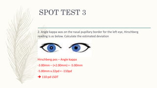
Angle kappa was on the nasal pupillary border for the left eye, indicating exotropia. The Hirschberg reading was -3.00mm.
To calculate the estimated deviation:
- Subtract the angle kappa position (+2.00mm nasal) from the Hirschberg reading (-3.00mm) giving -5.00mm
- Multiply the result (-5.00mm) by 22 (prism diopters per millimeter) giving -110 prism diopters
Therefore, the estimated deviation is 110 prism diopters of left exotropia.
Accommodative and vergence dysfunctions can cause symptoms like blurred vision, difficulty reading, and asthenopia. Key diagnostic tests include cover test, versions, near point of convergence, and fusional vergence amplitudes. Accommodative issues include insufficiency, fatigue, and infacility. Vergence issues include convergence insufficiency, divergence excess, and basic exophoria. Early treatment is important to prevent amblyopia or learning problems from vergence anomalies.
An orthoptic evaluation systematically evaluates the function of eye muscles during binocular eye movements to maintain fusion. It identifies accommodative, vergence, or fusional vergence anomalies to guide orthoptic exercises for treatment. The evaluation includes tests to check for single vision, diplopia, suppression, alignment, and accommodation. Based on symptoms like headaches or blurry vision, further tests are done to diagnose conditions like convergence insufficiency. The evaluation involves tests of phoria, near point of convergence, accommodation, fusional vergence, and accommodative function and facility.
The document provides an overview of eye anatomy and examination procedures. It describes the external structures of the eye including the eyelids, muscles, and lacrimal apparatus. Internally, it outlines the three layers of the eye - sclera, choroid, and retina. Examination steps are detailed including visual acuity tests, pupil examination, eye muscle function, ophthalmoscopy, and visual field testing. Common eye signs and conditions like strabismus, cataracts, and hemorrhages are also summarized.
Optic nerve (cn ii) eXAMINATION and etc.pptxafdalriza1
The document discusses examination of the pupils and visual fields. It describes:
1) The pupillary examination includes observing the pupil size, shape, and reaction to light via tests like light reflex and swinging flashlight. Near reflex is also tested.
2) Visual field examination includes screening tests like confrontation fields and Amsler grid to check for defects. Perimetry provides more detailed assessment of the visual field boundaries.
3) Mars contrast sensitivity testing evaluates a patient's ability to perceive low contrasts from high to low. It is a rapid test used to establish baselines and monitor diseases affecting low contrast vision.
Retinoscopy is an objective method to determine a person's refractive error by observing the movement of light reflected from the retina. A retinoscope projects light into the eye and the examiner observes the movement of the reflected light called the ret reflex. By introducing lenses and observing changes in the ret reflex, the examiner can determine the refractive error. There are different types of retinoscopy used for different purposes and patients. Careful technique and understanding ret reflex characteristics are important for accurate results.
This document discusses the treatment of suppression and arc. It defines suppression as a cortical phenomenon that eliminates visual confusion and diplopia in strabismus. There are various types and causes of suppression. The purpose of suppression is to avoid diplopia and confusion. Treatment aims to eliminate suppression and establish binocular vision through techniques like occlusion, prism adaptation, and use of instruments like the TV trainer and bar reader that break suppression by manipulating target parameters.
This document summarizes the key steps in examining a case of squint. It outlines obtaining the patient's presenting signs and symptoms, medical history, and previous treatments. Tests are described to assess visual acuity, fixation, refractive error, and the anterior segment. The motor status is examined through head posture, ocular movements, and fusional vergences. Ocular deviation is detected using cover tests and quantified. The sensory status is evaluated for binocularity, diplopia, retinal correspondence, suppression, amblyopia, and stereopsis. Both objective and subjective examination methods are outlined to thoroughly evaluate squint.
Estes são os slides do Exame Físico Neurológico que apresentei no evento da Liga de Neurologia e Neurocirurgia da Ulbra em 215. Mais slides no slideshare e no blog da nossa Neuroliga Ulbra: http://neuroligaulbra.blogspot.com.br/
A Liga de Neurologia e Neurocirurgia traz um evento inovador:
Uma monitoria de Neurologia Clínica diferente, focada nas dúvidas -e curiosidades- dos acadêmicos. Teremos 5 temas, em 5 quintas-feiras
às 12h pelos monitores da Neuro e membros da NeuroLiga e pelos nossos professores experts da Neurologia I e II presentes.
Quintas-feiras, 12h-13h
26/03 – Exame Físico na neuro 16/04 – Tumores Cranianos 07/05 – Cefaleia28/05 – AVC 18/06 – Infecções SNC e S. de Guillain Barré
http://neuroligaulbra.blogspot.com.br/2015/04/revisando-topicos-essenciais-em.html
1. Retinoscopy is an objective refraction technique used to determine a patient's refractive error without their subjective response. It involves examining the movement of the patient's retinal reflex seen through a retinoscope.
2. Several factors must be considered to perform an accurate retinoscopy, including working distance, lighting conditions, the fixation target, and patient and examiner positioning. The characteristics of the retinal reflex, including direction of movement, speed, width and brightness provide clues about the refractive error.
3. Spherical refractive errors are neutralized by increasing or decreasing lens power until reversal of movement is seen. For astigmatism, each principal meridian must be neutralized separately using the same technique. Estim
This document describes an orthoptic instrument called a haploscope. A haploscope is an optical device that presents different images to each eye, allowing examination of binocular vision. The haploscope described has adjustable components to measure fusion, stereopsis, deviations in different gazes, and more. It can be used for both diagnostic and therapeutic purposes in orthoptic treatment and management of conditions like strabismus and amblyopia.
Orthoptics involves the evaluation and non-surgical treatment of disorders of the visual system, with a focus on binocular vision and eye movements. Orthoptists are trained professionals who work with ophthalmologists to examine patients' vision, eye alignment, and eye muscle function and treat issues through specialized exercises. Common conditions treated include convergence insufficiency, accommodative insufficiency, and binocular instability. Exercises aim to improve fusion, eye alignment control and coordination, and may involve targets, lenses, or stereoscopes. Care must be taken to only use exercises appropriately for motivated patients without underlying issues requiring medical or surgical care.
The near point of convergence is defined as the point of intersection of the lines of sight of the eyes when maximum convergence is used. It is measured as the distance from this point of intersection to the midpoint between the eyes. Measuring the near point of convergence involves having a patient fixate on a target that is slowly moved toward them while maintaining fusion until their eyes can no longer converge or they see double. This distance is used to assess a patient's ability to converge their eyes for near vision tasks. A near point further than 10cm may indicate convergence insufficiency while one closer than 5cm suggests convergence excess.
The document discusses various techniques for examining the interior of the eye including ophthalmoscopy. It describes the indirect ophthalmoscopy technique where a convex lens is used to make the eye highly myopic, allowing examination of the retina. The key steps of the technique are outlined including using a dark room, convex lens, illumination source and obtaining an inverted, magnified view of the retina between the lens and examiner's eye. Advantages of indirect ophthalmoscopy are also provided.
Dr. Pushkar Dhir presented on retinoscopy. Retinoscopy is an objective refraction technique used to assess refractive error by observing the movement of the red reflex in the pupil when a light source is moved. It can be used for infants, children, and uncooperative patients. There are different types of retinoscopes. Retinoscopy involves illuminating the retina and observing the light reflex. Various factors are assessed like size, speed and brightness of the reflex to determine refractive error. Cycloplegic retinoscopy is used with young children and hyperopes to paralyze accommodation. Problems like small pupils or hazy media can make the reflex difficult to see. Subjective refraction then refines the
This document discusses various techniques for subjective refraction including determining the best vision sphere and using the duochrome test. It provides details on:
1. Using plus and minus lenses to find the maximum plus or minimum minus that can be tolerated without blurring vision, known as the best vision sphere.
2. The duochrome test which takes advantage of chromatic aberration, using a split red-green filter to refine the endpoint by comparing clarity on red and green backgrounds.
3. Additional methods like pinhole and fogging are described to control accommodation and improve accuracy.
This document provides guidance on performing a physical eye exam, including:
1. Testing visual acuity using a Snellen chart and recording results in standard notation. Near and distance vision are assessed.
2. Examining the pupils for size, shape, reaction to light, and swinging flashlight test.
3. Testing extraocular eye movements through the six cardinal positions of gaze.
4. Using a slit lamp biomicroscope to examine ocular tissues under magnification and illumination.
5. Performing fundoscopy to systematically examine the ocular media, optic disc, retinal vasculature, background, and macula using an ophthalmoscope.
The document provides an overview of optometry and eye exams. It discusses the history of optometry, what a standard eye exam involves, including case history, refractive tests, binocular and accommodation tests, and ocular health evaluation. It then describes how some optometrists are modernizing exams with new technologies like optical coherence tomography, fundus photography, and automated testing to provide more detailed analysis of the eye.
This document provides instructions for examining a patient's fundus using an ophthalmoscope. It describes how to position the patient and ophthalmoscope, how to focus on different parts of the fundus including the optic disc, blood vessels, periphery, and macula, and what features to assess for each area such as shape and clarity of the optic disc or signs of hypertensive retinopathy. The goal is to be able to properly examine the fundus and recognize common abnormalities.
The document provides instructions for performing a cranial nerve examination, including which equipment is needed and the steps to assess each of the 12 cranial nerves. It details how to test the olfactory, optic, oculomotor, trochlear, trigeminal, abducens, facial, vestibulocochlear, glossopharyngeal, vagus, accessory, and hypoglossal nerves through sensory and motor function tests like smell, vision, eye and facial muscle movement, hearing, taste, swallowing, and tongue movement. The examination assesses for any abnormalities, deficits, or asymmetries in cranial nerve function.
- Visual field examination tests the peripheral sensitivity of the retina and visual pathways. It is important for assessing topographic sensitivity and detecting visual field defects.
- Automated perimetry provides standardized, quantitative tests to measure threshold sensitivity across the visual field. It allows for reliable long-term monitoring to detect glaucomatous progression.
- Interpretation of visual field tests involves analyzing parameters like total deviation plots, pattern deviation plots, and global indices to identify patterns indicative of glaucoma according to established criteria. Clinical correlation with optic nerve examination is also important.
This document discusses visual field examination and interpretation of automated perimetry in glaucoma. It provides details on the physiology of the visual field and different types of visual field defects. It also describes various methods of visual field examination including kinetic and static perimetry as well as clinical techniques. Automated perimetry devices like Humphrey Field Analyzer and their advantages are discussed. Important aspects of visual field test interpretation including reliability indices, total and pattern deviation plots, and global indices are summarized.
DIRECT DOWNLOAD LINK ❤❤https://healthkura.com/retinoscopy/❤❤
Dear viewers Check Out my other piece of works at ❤❤❤ https://healthkura.com ❤❤❤
Retinoscopy and Objective Refraction and Subjective Refraction in spherical ametropia and astigmatism
Retinoscopy (Principle & Techniques of Retinoscopy) and objective refraction, Subjective Refracition
Best presentation about retinoscopy and objective refraction techniques, and basis of subjective refraction. If you want to master the technique of retinoscopy, this presentation can be your guidance and partner in your journey to retinoscopy, objective refraction and subjective refraction.
Presentation Layout:
Retinoscope, types of retinoscope and uses of retinoscope
-Introduction to retinoscopy and objective refraction
-Retinoscopy
- In spherical ametropia
- In astigmatism
- Others: strabismus, amblyopia, pediatric pt.,
cycloplegic refraction
-Static and Dynamic Retinoscopy
-Problems seeing reflex during retinoscopy
-Errors in retinoscopy
Objective of retinoscopy and objective refraction
-To locate the far point of the eye conjugate to the retina
- Myopia or hyperopia
-Bring far point to the infinity by using appropriate lenses
- Determines amount of ametropia by retinoscopy and objective refraction
References:
-Clinical Procedures in Optometry by Eskridge, Amos and Bartlett ,
-Primary Care Optometry by Grosvenor T.,
-Borish’s Clinical Refraction by Benjamin W. J.,
-Theory And Practice Of Optics And Refraction by AK Khurana
-Retinoscopy-Student Manual by ICEE Refractive Error Training Package (2009)
-Clinical Optics and Refraction By Andrew Keirl, Caroline Christie
-Clinical Refraction Guide - A Kumar Bhootra
-Clinical Procedures in Primary Eye Care by David B. Elliott
-Internet
Follow me to get in touch with optometric and ophthalmic updates.
The document discusses the field of vision, including its anatomy and testing methods. It notes that the field of vision is like an island surrounded by blindness, with the fovea being the summit of highest sensitivity and the blind spot being the trough of zero sensitivity. It describes kinetic and static perimetry testing methods and different types of visual field defects seen in conditions like glaucoma and neurological disorders. Global indices, reliability indices, and corrected pattern deviation maps are used to analyze perimetry results. Factors affecting testing and new techniques like FDT perimetry are also mentioned.
This document provides information about conducting an eye exam, including:
1) Taking a case history to understand a patient's vision needs, eye health history, and general health conditions.
2) Performing objective and subjective refraction tests to determine a patient's prescription for distance and near vision.
3) Evaluating binocular vision by testing motor and sensory functions like eye movements, stereopsis, and fusional reserves.
4) Prescribing corrective lenses or prisms as needed based on the refraction and binocular vision results.
Similar to 2 CASE HISTORY & PRELIMINARY TESTS w spot.pptx (20)
This document provides an outline for a module on personality theories. It will cover major approaches like psychodynamic, behavioral, trait, humanistic, and cognitive theories. Key topics include the relevance of personality psychology in South Africa, the influence of culture on personality, and commonalities across approaches. The first theory discussed in more depth is Freud's psychodynamic theory, covering its background, the structure of personality with id, ego and superego, and the psychosexual stages of development. Limitations of this theory are also evaluated.
The document discusses the properties and functions of dietary fats and lipids. It explains that fats provide energy density and are stored in adipose tissue. Dietary fats include saturated, monounsaturated, and polyunsaturated fatty acids. Lipids include triglycerides, phospholipids, and sterols like cholesterol. Lipids play structural and functional roles in cells and as carriers of fat-soluble vitamins and minerals. Omega-3 and omega-6 fatty acids are essential but must be obtained through diet.
The document discusses blood typing and genetics. It explains that the ABO blood type is determined by alleles inherited from each parent. The A and B alleles are dominant, while O is recessive. A person's genotype refers to their specific allele combination, while their phenotype is their observable blood type. Careful blood type matching between donors and recipients is important to avoid immune reactions from mismatched antigens on red blood cells.
Cardiovascular disease can manifest in the eyes through retinal vascular diseases like central retinal artery occlusion caused by emboli from the heart or carotid artery. Hypertension is a major risk factor for central retinal vein occlusion and can also cause non-arteritic anterior ischemic optic neuropathy. The document further discusses anatomical details of the heart and blood vessels and various ocular manifestations of hypertension in the retina like cotton wool spots, exudates, and papilledema.
The document describes the slit lamp biomicroscope, an instrument used to examine the anterior segment of the eye. It consists of an illumination system that produces a focused beam of light, an observation system using a microscope, and a mechanical system to support the patient and control the device. Various techniques can be used like diffuse illumination, retroillumination and vital staining to examine different ocular structures. Proper maintenance of the device is important to ensure optimal performance.
The digestive system is a series of tubes that transports food from the mouth to the anus. It includes organs like the mouth, esophagus, stomach, and intestines. Accessory organs like the liver, gallbladder and pancreas also play important roles in digestion. As food moves through the tubes, it is broken down mechanically and chemically. Enzymes and hormones help regulate digestion. The nervous and endocrine systems control movement and secretions to efficiently break down food into nutrients that can be absorbed and used by the body.
This document discusses various aspects of accommodation including:
1. It provides background on accommodation and defines terms like amplitude of accommodation.
2. It summarizes several theories of accommodation including Helmholtz's relaxation theory and Schachar's theory.
3. It describes the accommodation pathway from visual stimuli to contraction of the ciliary muscle.
4. It outlines different types of accommodation like tonic, reflex, and convergence accommodation.
5. It discusses tests to measure amplitude of accommodation and normal values.
6. It explains accommodation response and the lag of accommodation.
A workshop hosted by the South African Journal of Science aimed at postgraduate students and early career researchers with little or no experience in writing and publishing journal articles.
A review of the growth of the Israel Genealogy Research Association Database Collection for the last 12 months. Our collection is now passed the 3 million mark and still growing. See which archives have contributed the most. See the different types of records we have, and which years have had records added. You can also see what we have for the future.
বাংলাদেশের অর্থনৈতিক সমীক্ষা ২০২৪ [Bangladesh Economic Review 2024 Bangla.pdf] কম্পিউটার , ট্যাব ও স্মার্ট ফোন ভার্সন সহ সম্পূর্ণ বাংলা ই-বুক বা pdf বই " সুচিপত্র ...বুকমার্ক মেনু 🔖 ও হাইপার লিংক মেনু 📝👆 যুক্ত ..
আমাদের সবার জন্য খুব খুব গুরুত্বপূর্ণ একটি বই ..বিসিএস, ব্যাংক, ইউনিভার্সিটি ভর্তি ও যে কোন প্রতিযোগিতা মূলক পরীক্ষার জন্য এর খুব ইম্পরট্যান্ট একটি বিষয় ...তাছাড়া বাংলাদেশের সাম্প্রতিক যে কোন ডাটা বা তথ্য এই বইতে পাবেন ...
তাই একজন নাগরিক হিসাবে এই তথ্য গুলো আপনার জানা প্রয়োজন ...।
বিসিএস ও ব্যাংক এর লিখিত পরীক্ষা ...+এছাড়া মাধ্যমিক ও উচ্চমাধ্যমিকের স্টুডেন্টদের জন্য অনেক কাজে আসবে ...
How to Setup Warehouse & Location in Odoo 17 InventoryCeline George
In this slide, we'll explore how to set up warehouses and locations in Odoo 17 Inventory. This will help us manage our stock effectively, track inventory levels, and streamline warehouse operations.
Walmart Business+ and Spark Good for Nonprofits.pdfTechSoup
"Learn about all the ways Walmart supports nonprofit organizations.
You will hear from Liz Willett, the Head of Nonprofits, and hear about what Walmart is doing to help nonprofits, including Walmart Business and Spark Good. Walmart Business+ is a new offer for nonprofits that offers discounts and also streamlines nonprofits order and expense tracking, saving time and money.
The webinar may also give some examples on how nonprofits can best leverage Walmart Business+.
The event will cover the following::
Walmart Business + (https://business.walmart.com/plus) is a new shopping experience for nonprofits, schools, and local business customers that connects an exclusive online shopping experience to stores. Benefits include free delivery and shipping, a 'Spend Analytics” feature, special discounts, deals and tax-exempt shopping.
Special TechSoup offer for a free 180 days membership, and up to $150 in discounts on eligible orders.
Spark Good (walmart.com/sparkgood) is a charitable platform that enables nonprofits to receive donations directly from customers and associates.
Answers about how you can do more with Walmart!"
it describes the bony anatomy including the femoral head , acetabulum, labrum . also discusses the capsule , ligaments . muscle that act on the hip joint and the range of motion are outlined. factors affecting hip joint stability and weight transmission through the joint are summarized.
How to Make a Field Mandatory in Odoo 17Celine George
In Odoo, making a field required can be done through both Python code and XML views. When you set the required attribute to True in Python code, it makes the field required across all views where it's used. Conversely, when you set the required attribute in XML views, it makes the field required only in the context of that particular view.
हिंदी वर्णमाला पीपीटी, hindi alphabet PPT presentation, hindi varnamala PPT, Hindi Varnamala pdf, हिंदी स्वर, हिंदी व्यंजन, sikhiye hindi varnmala, dr. mulla adam ali, hindi language and literature, hindi alphabet with drawing, hindi alphabet pdf, hindi varnamala for childrens, hindi language, hindi varnamala practice for kids, https://www.drmullaadamali.com
Main Java[All of the Base Concepts}.docxadhitya5119
This is part 1 of my Java Learning Journey. This Contains Custom methods, classes, constructors, packages, multithreading , try- catch block, finally block and more.
2. WHAT IS A TENTATIVE
DIAGNOSIS?
– Provisional, Not fixed, Not certain
– Refers to one of several potential diagnoses of a patient’s illness
– Based on case history
– Developed as you postulate a number of different disease processes/possible causes
to your patient’s signs/symptoms that must be explored further
– It is included in a differential diagnosis
– There can be multiple tentative diagnoses and you need to perform all relevant tests
to confirm a diagnosis
– In essence a tentative diagnosis is a diagnosis that you suspect, that is yet to be
confirmed, however it must be supported by the history/background
3. HOW TO DEVELOP A
TENTATIVE DIAGNOSIS?
– Take into consideration the detailed objective and subjective case history
– Postulate likely causes for your patients complaint
– Require knowledge of presenting signs and symptoms of ocular
diseases/ conditions
4. EXAMPLES OF TENTATIVES
BASED ON COMMON SYMPTOMS
Visual Complaints e.g
– Blurry vx
– Dbl vx
– Photophobia
– Asthenopia
– H/A
Non Visual Complaints e.g
– Itching
– Pain
– Swelling
– Discharge
– Redness
5. CONVERGENCE
– Disjunctive movement of the eyes where the eyes synchronously adduct so that the
lines of sight intersect in front of eyes
– Assists in maintaining bifoveal single vision at any fixation distance
– It is a fusional movement that may be stimulated by volition, disparate stimulation
and accommodation
– Unlike Accommodation, the amplitude of convergence doesn’t deteriorate with age.
It may deteriorate due to a decrease in ACC Convergence
– Some convergence may reduce under certain abnormal circumstances
– Power or reserve of convergence can be increased by orthoptic exercises
– Symmetrical vs Asymmetrical convergence?
8. NPC
– Point of intersection of the lines of sight of the eyes when maximum
convergence is utilized
– NPC Distance: Is the distance from the near point of convergence to the
midpoint of the line connecting the centre of rotation of the eyes
– Pxs with receeded NPC distances may have visual and ocular discomfort when
performing near point vision tasks
10. CLINICAL PROCEDURE
Equipment
– RAF Ruler
– Accommodative target
– 30cm Ruler
– Red lens/ R&G lens
– Penlight
Procedure
– Conducted in free space
– Patient is seated comfortably with
habitual Rx
– Ruler is held against outer cathus
– The patient is encouraged to
maintain fixation on target and
report double
12. CLINICAL PROCEDURE
Accommodative NPC
– Px to focus on dot on target and
report when line is double
– Ensure to check that line is initially
single before proceeding to
measure
– Slowly move the target along
midline, 15 degrees below eye level
until px reports to seeing double
Non-Accommodative NPC
– Shine light at bridge of nose
– Px to focus on penlight with red lens infront
of one eye
– Ensure to check the light is the same color as
the lens in each eye, and becomes a mixture
when both eyes are open
– Slowly move the penlight along midline, 15
degrees below eye level until the light breaks
into two colors, or patient no longer reports
a mixture
13. CLINICAL PROCEDURE
– When the patient reports diplopia (subjective), ensure to instruct the patient to
blink! If still reports diplopia, measure. If no diplopia after blink, continue
towards patient
– The sustained double is the break value
*If the px doesn’t report diplopia, note the fixation distance if one eye loses
fixation on the target (objective)
(The eye that maintains fixation is the dominant eye)
– Measure the subjective or objective break value then slowly move the target
away and ask px to report fusion or you see the eye regain fixation Recovery
value
14. CLINICAL PROCEDURE
– This is recorded as Break/Recovery in cm
– E.g 6cm/9cm
– Repeat a minimum of two times and take an average of those results
– When NPC is repeated 5 or more times, convergence ability significantly
decreases with symptomatic patients and minimally with asymptomatic
patients
15. CLINICAL SIGNIFICANCE/
INTERPRETATION
– Pxs with a convergence problem may express symptoms such as diplopia,
frontal headaches, decreases reading comprehension, asthenopia, fatigue when
performing near tasks
– NPC has a normal range of 6 to 10cm
– Closer than 5cm is considered to be convergence excess
– A remote/receeded NPC is suspected to have Convergence Insufficiency
16. ANOMALIES OF
CONVERGENCE
Convergence Insufficiency (CI):
– Inability to obtain or maintain adequate
convergence over a certain period of
time without undue effort
– Commonest cause of asthenopia
Convergence Excess
Convergence Spasm
Convergence Paralysis
17. VISUAL FIELD
Contains:
– Centeral or foveal which is measured by visual acuity test, contrast sensitivity and
macular function tests and concerned with Resolution, forms
– Foveal (central 3 degrees)
– Parafoveal (within 5 degrees)
– Macular (within 10 degrees)
– Central (fixation and within 30 degree circle, includes physiological blind spot)
– Peripheral which is concerned with Peripheral sensitivity , motion and light
detection (Beyond 30 degrees to outer field of vision)
– Normal visual field is reversed inverted map of corresponding retinal points
18. VISUAL FIELD
Normal monocular visual field limitations:
– 1. 60° Superiorly
– 2. 60° Nasally
– 3. 75° Inferiorly
– 4. 100° Temporally.
19. PERIMETRY
– It is making of a visual field using stimulus. Stimulus can be:
1. Moving (kinetic) determine the visual threshold along edge of visual field
2. Static in which the static object has different level of brightness.
20. CONFRONTATION TEST
– Simple preliminary test which is done for gross visual field screening
– Falls under kinetic perimetry
– Used as screening for moderate to severe visual field defects
– Un reliable in identifying Mild visual field defects
– Results are quantitative and test can be viewed as a pass fail criteria
21. CLINICAL PROCEDURE
– Prior to testing of visual field insure central vision and visual acuity is intact
– Pt faces the examiner at a dx of 1m with his/her eyes same level as examiner’s.
– You each focus on the other’s opposite eye while covering the contralateral eye
with palm of hand
– Avoid confusion by using 1,2,5 fingers
– Correct position : 2 finger side by side in front of the eye field . Incorrect
position: One finger behind the other
23. CLINICAL PROCEDURE
Facial Amsler
– Checks for central scotoma
– Instruct patient to focus on Examiners nose
– Ask the pt, whilst they are focussed on your nose “is there anything that
appears to be missing on your face”
24. CLINICAL PROCEDURE
Finger Counting
– Checks for paracentral and peripheral scotoma in four quadrants
– Px instructed to focus on examiners open eye throughout this procedure
– Quickly project and retract (0,1,2 or 5) fingers in each quadrant and ask the
patient to report how many fingers they see
– Repeat twice in each quadrant, one projection paracentrally and the other more
peripherally
– Ensure px does not look at the fingers directly but maintains fixation on Ex open
eye
25. CLINICAL PROCEDURE
Simultaneous Finger Counting
– Checks for Extinction phenomenon :
(Harrington) ‘The presence of the stimulus
in the seeing field gives the impression of
“extinguishing” the test object in the non-
seeing field’
– Project (1,1 or 1,2 or 2,2) fingers in
opposition quadrants
– Instruct px to sum up the fingers projected
26. CLINICAL PROCEDURE
– Simultaneous hand comparison
– Hold up both hands with backs of palms facing the patient
– Px maintains fixation on your open eye
– Instruct px to report any difference in brightness or darkness of your hands
– Perform central, eccentric
– Hemianopsia, Altitudinal
27. KINETIC CONFRONTATION
– Monocularly, patient fixated on examiners open eye
– Target (preferably 1cm Red or Green) is moved in a flat plane midway between
the pt and examiner
– Begin from an unseen (outside of VF) to a seen position moving inwards
– Px instructed to report when they can see the target
– Process to be repeated in each of the quadrants for each eye separately
28. INTERPRETATION
– Compare patients field to examiners field, assumed the examiner has normal
field
– If pt cant see object and ex can, pt is interpreted to have visual field defect
– If defect is detected, re-examine the area and define further
– Discrepancies between the examiner’s and patient’s visual field after repetitions
should prompt further field examinations
29. CONFRONTATION
DRAWBACKS
– Early visual field defects can go unnoticed, particularly if one eye is affected
– Subjective test and misinterpretations of report may be presented if the patient
has poor compliance Paeds, geriatric pts
– Screening test, further examination required to diagnose
31. SPOT TEST 3
1. Angle Kappa was at the physiological position, Hirschberg reading is as below.
Calculate the estimated deviation
2. Angle kappa was on the nasal pupillary border for the left eye, Hirschberg
reading is as below. Calculate the estimated deviation
32. SPOT TEST 3
1. Angle Kappa was at the physiological position, Hirschberg reading is as below.
Calculate the estimated deviation
Hirschberg pos – Angle kappa
-4.00 mm-(+0.50mm)= -4.50mm
-4.50mm x 22pd= -99pd
99 pd LSOT
33. SPOT TEST 3
2. Angle kappa was on the nasal pupillary border for the left eye, Hirschberg
reading is as below. Calculate the estimated deviation
Hirschberg pos – Angle kappa
-3.00mm – (+2.00mm) = -5.00mm
-5.00mm x 22pd = -110pd
110 pd LSOT