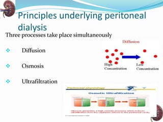
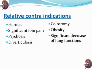
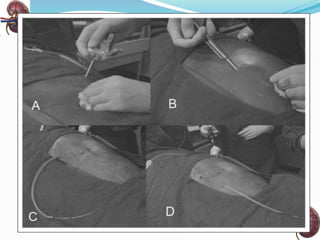
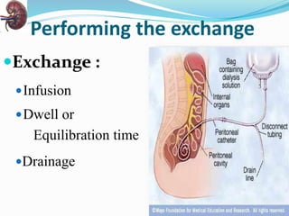
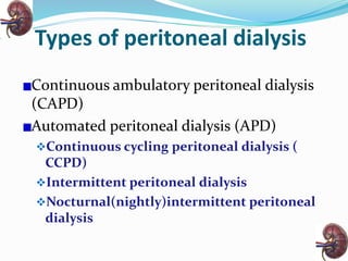
MODERATOR : PRESENTOR:
Madam Manju Singh Yogesh Kumar Tiwari
LECTURER M.Sc .Nursing 1st year
CON,AIIMS
Objectives
At the end of the class students will be able to :
 Define peritoneal dialysis
 Anatomy and physiology of peritoneal membrane
 The goals of peritoneal dialysis
 Indications and contra indications of PD
 Peritoneal dialysis access and insertion procedure
 The procedure of PD
 Different types of PD
 Complications of PD
 The advantages of PD over HD
 Nursing management of patients undergoing PD
Anatomy
Physiology
 The semipermeable peritoneal membrane allows
solutes and water to be transported from the vascular
system to the peritoneal cavity and vice versa.
Peritoneal dialysis:
Introduction
 Peritoneal dialysis(PD) is a treatment for
patients with severe chronic kidney disease.
 A dialysis technique that uses the patient's own body
tissue-peritoneal membrane inside the abdominal
cavity as a filter.
Goals of PD
• Remove toxic substances and metabolic wastes
• Reverse the symptoms of uremia
• Reestablish normal fluid and electrolyte balance
• Maintain a positive nitrogen balance
• Prolong life
• Have the maximum level of quality of life
Principles underlying peritoneal
dialysis
Three processes take place simultaneously
 Diffusion
 Osmosis
 Ultrafiltration
Treatment of choice for….
 Patients with RF unable or unwilling to undergo HD or renal
transplantation
• Diabetic patients
• Patients with cardiovascular diseases eg:heart failure
• Older patients
• Patients at risk of adverse effects of systemic heparin
• Patients with severe hypertension
Contra indications
Absolute contra indications
 Peritoneal fibrosis and adhesions following
intra abdominal operations
 Inflammatory gut diseases
Relative contra indications
Hernias
Significant loin pain
Psychosis
Diverticulosis
Colostomy
Obesity
Significant decrease
of lung functions
PD catheters
Catheters for long-term use (Tenckhoff, Swan, Cruz) are usually
made of silicone and are radioopaque to permit visualization
on x-ray. These catheters have three sections:
(1) An intraperitoneal section with numerous openings and
an open tip to let dialysate flow freely;
(2) A subcutaneous section that passes from the peritoneal
membrane and tunnels through muscle and subcutaneous
fat to the skin
(3) An external section for connection to the dialysate section
Catheters have two cuffs, which are made of Dacron polyester.
The cuffs:
 stabilize the catheter
 limit movement
 prevent leak
 provide a barrier against microorganism
Cuff placement :
 adjacent to the peritoneum
 subcutaneously.
The subcutaneous tunnel (5 to 10 cm long) further protects
against bacterial infection
Types of catheters
 The design of a peritoneal catheter need to be such
that
 It should give maximum inflow and output
 Discourage infection
 Four main types
o Straight Tenckhoff
o Curled Tenckhoff
o Swan-neck
o T- fluted
PD catheters
PD catheters
T fluted catheter Two –cuff tenckhoff
catheter
Insertion techniques
 Blind placement using Tenckhoff trocar
 Blind placement using guide wire
 Surgical placement by dissection
 Mini trocar placement using peritoneoscopy
Preinsertion preparation
of the patient
 Determine the catheter exit site
Site :
 Midline
 3 cm below umbilicus
 Lateral site
 At the lateral border of the rectus muscles
 On a line, half way between the umbilicus and anterior
superior iliac spine
 Left lateral side is preferred as it avoids caecum
Pre operative care of the
patient
 Take bath or have a shower in the morning
 Abdominal hair should be clipped
 Empty bowel and bladder before catheter insertion
 Enema can be given
 Staphylococcus aureus screening
 Administer antibiotics prophylactically
• A trocar is used to puncture the peritoneum as the patient
tightens the abdominal muscles by raising the head.
• The catheter is threaded through the trocar and
positioned.
• Previously prepared dialysate is infused into the peritoneal
cavity, pushing the omentum (peritoneal lining extending
from the abdominal organs) away from the catheter.
• The physician may then secure the catheter with a
purse-string suture and apply antibacterial ointment
and a sterile dressing over the site
Post operative care of the
patient
Goals:
 Minimise bacterial colonisation of exit site
during early healing period
 Prevent trauma to exit site and traction on
cuffs by immobilization of catheter
 Minimise intra abdominal pressure to prevent
leakage
 Do not disturb the exit site for 7-10 days
 Flush the catheter with 500-1500 ml of PD fluid to check
patency
Composition of PD solution
Components
Na 132 mmol/l
Ca 1.25mmol/l
Mg 0.5mmol/l
Cl 100mmol/l
Lactate 35mmol/l
Glucose 1.36-4.25g/dl
Osmolarity 347-486
pH 5.2
PROCEDURE
Patient preparation
Explain the procedure & obtain informed consent.
Baseline vital signs, weight, serum electrolyte
levels are recorded.
Assess patient’s anxiety about the procedure.
Broad spectrum antibiotics prophylactically.
PROCEDURE
Equipment preparation
 Assemble the equipments needed
 Check physician’s order for the concentration of dialysate
and medications to be added
 Heparin : to prevent clotting
 KCl: to prevent hypokalemia
 Antibiotics : peritonitis
 Insulin : for diabetic patients
PROCEDURE
 Warm the dialysate solution to body
temperature:
 To prevent patient discomfort and abdominal
pain
 To dilate the vessels of peritoneum
 Dry heating should be done
 Too cold solution causes pain, cramping, and
reduce clearance
Not recommended....
 Soaking the bag of solution in warm water
 Use of microwave to heat the fluid
 Avoid too cold solution
Performing the exchange
Exchange :
Infusion
Dwell or
Equilibration time
Drainage
PERITONEAL DIALYSIS
Performing the exchange
Infusion
 The dialysate is infused by gravity into the
peritoneal cavity.
 A period of about 5 to 10 minutes is usually
required to infuse 2 L of fluid.
•Dwell or equilibration and
drainage of dialysate
• The prescribed dwell, or equilibration, time allows diffusion
and osmosis to occur.
• Diffusion of small molecules, such as urea and creatinine,
peaks in the first 5 to 10 minutes of the dwell time.
• At the end of the dwell time, the drainage portion of
the exchange begins.
• The tube is unclamped and the solution drains from the
peritoneal cavity by gravity through a closed system.
• Drainage is usually completed in 10 to 30 minutes.
Performing the exchange
Drainage fluid
• Colorless or straw-colored
• Should not be cloudy
• Bloody drainage may be seen in the first few exchanges
Entire exchange time
 1 to 4 hours (depending on prescribed dwell time)
Performing the exchange
• No.of exchanges
According to patient’s physical status and acuity of
illness
• Dextrose solutions
Dextrose solutions of 1.5%, 2.5%, and 4.25% are
available in several volumes, from 500 mL to 3,000
mL, allowing the dialysate selection to fit the patient’s
tolerance, size, and physiologic need
Types of peritoneal dialysis
Continuous ambulatory peritoneal dialysis
(CAPD)
Automated peritoneal dialysis (APD)
Continuous cycling peritoneal dialysis (
CCPD)
Intermittent peritoneal dialysis
Nocturnal(nightly)intermittent peritoneal
dialysis
Continuous ambulatory peritoneal
dialysis (CAPD)
 Carried out during day time , manually by patients or
by caregivers
 Dialysis fluid is infused to the peritoneal cavity
 Dwell time for between 3 - 10 hrs
 Most suitable for patients whose membrane transport
solutes at a slow to average rate
Automated peritoneal dialysis
 -is performed through a cycler machine.
 -during the night when the patient is asleep.
Continuous cycling peritoneal
dialysis
 Patient carries PD solution in the abdominal cavity
through out the day but performs no exchanges.
 At bedtime ,patient hooks upto the cycler ,which
drains and refills the abdomen with solution three or
more times in the course of the night.
Intermittent peritoneal
dialysis(IPD)
• Intermittent peritoneal dialysis is offered to patients on a
temporary basis when their blood pressure is low or in
children with acute renal failure to tide over a crisis.
• It is performed for a short period of 12-24 hours,2-3 times
weekly.
• Common routine hourly exchange consists of 10 min
infusion, 30min dwell time and a 20 min drain time.
Nocturnal intermittent
peritoneal dialysis
 Patient drains out fully at the end of the cycling
period, so the abdomen is dry all day.
 Clearances are lower on NIPD.
Assessing peritoneal dialysis
adequacy
 Creatinine clearance
 A solute removal test based on the body surface area
 General well being
Dialysis related problems
 Protein loss
 Protein loss through the peritoneal membrane @ 6-
12g/day in a PD patient
 To compensate for this PD patients need to eat between
1 – 1.2 g/ kg body weight/ day
 The loss is increased during peritonitis
 Other substances lost in dialysate are amino acids,
water-soluble vitamins, hormones and some
medications
Dialysis related problems …
 Cardio vascular and lipid problems
 Increased glucose absorption from the PD fluid
 Raised intra abdominal pressure
 Can cause hernias & dialysate leakage around the
insertion site
 If leakage occurs , PD must be ceased for a short period
Dialysis related problems
 Drainage problems
Reasons can be
 Kinks in the tubing
 Constipation
 Fibrin formation
 Milking can be done
 Heparin administration
 Streptokinase or urokinase in 0.9% NaCl
 Malpositioned catheter
Dialysis related problems
 Blood stained effluent
 In menstruating females; due to endometriosis or
retrograde bleeding through fallopian tube
 Severe intraperitoneal bleeding ; due to straining while
lifting a heavy object or suffering trauma to abdomen
 Shoulder pain
 Following infusion of fresh dialysate
 Referred pain caused by intra abdominal pressure or air
under the diaphragm
 Resolve within 20 min; analgesics can be given
Infectious complications
Peritonitis
Most common and most serious complication
 Diagnosis
 Cloudy PD effluent
 Abdominal pain , tenderness , pyrexia
 Identification of micro organisms in PD effluent
in culture or positive gram staining
Infectious complications
 Treatment
 Initial one to three rapid exchanges with 1.5%
dextrose solution – to wash out mediators of
inflammation
 Drainage fluid – examined for cell count ,
Gram’s stain, culture
 Intraperitoneal or intravenous antibiotics
 Unresolved peritonitis after 4 days of
appropriate therapy necessitates catheter
removal
Infectious complications
Exit site infection
 The presence of purulent drainage with or without
erythema of the skin at catheter epidermal interface.
Tunnel infection
 Can present as an extension of the exit site infection
into the catheter tunnel. Swelling, pain and redness
over the subcutaneous tunnel may be observed.
 Management
 Culture of drainage
 Antibiotic therapy
Advantages of PD over HD
1. Easy to use without sophisticated equipments
2. Easy to manage in home and community health care
facilities
3. more independence and mobility
4. Dialysis treatment of choice for children
5. May allow better blood pressure and volume control
with cardiovascular benefits
6. May give better quality of life
7. Lower risk of Hepatitis C
8. Equal or better survival in early years
Nursing management
 Potential for developing infection related to the
catheter
 Assess the site for any signs of infection; any
redness, rebound tenderness, swelling, drainage from
the exit site or change in vital signs
 Maintain strict aseptic technique while carrying
out the procedure
Nursing management
 Potential for developing cardiac and respiratory
complications related to the uremic state and
presence of fluid in the peritoneum
 Frequent cardiac and respiratory assessment
 Watch for signs of fluid accumulation; heart
failure, and pulmonary edema
 Auscultate the base of lungs for crackles
 Assess for signs of pericarditis; substernal pain,
low grade fever, and pericardial friction rub.
Nursing management
 Acute pain and abdominal discomfort related to
the dialysate infusion
 Warm the dialysate to body temperature
 Altered nutrition less than body requirement
related to the protein loss
 High protein, fiber rich well balanced diet
 Limit carbohydrate intake
Nursing management
 Knowledge deficit related to care of catheter site
 Teach the patient the possible signs of infection
 Catheter care should be done daily
 Avoid tub bath and exit site should not be
submerged in water
Nursing management
 Altered body image related to the abdominal
catheter and bag and tubing
 Assess for any such problem
 Allow the patient to express his feelings and
concerns about body image disturbances.
 Assist in selecting of proper clothing
 Provide an opportunity to the patient to meet
similar patients who are well adjusted with the
condition
Nursing management
 Altered sexual patterns and sexual dysfunction
 Provide privacy to the patient so that he can
discuss his problem
 Nurse can start the discussion by asking about any
concerns related to sexuality
Conclusion
 Long term outcomes associated with peritoneal
dialyses are good.
 The treatment is usually effective for years.
 However scarring of the peritoneum and repeated
infections may require a change to hemodialysis.
14 peritoneal dialysis

14 peritoneal dialysis

  • 1.
    MODERATOR : PRESENTOR: MadamManju Singh Yogesh Kumar Tiwari LECTURER M.Sc .Nursing 1st year CON,AIIMS
  • 2.
    Objectives At the endof the class students will be able to :  Define peritoneal dialysis  Anatomy and physiology of peritoneal membrane  The goals of peritoneal dialysis  Indications and contra indications of PD  Peritoneal dialysis access and insertion procedure  The procedure of PD  Different types of PD  Complications of PD  The advantages of PD over HD  Nursing management of patients undergoing PD
  • 3.
  • 4.
    Physiology  The semipermeableperitoneal membrane allows solutes and water to be transported from the vascular system to the peritoneal cavity and vice versa.
  • 5.
    Peritoneal dialysis: Introduction  Peritonealdialysis(PD) is a treatment for patients with severe chronic kidney disease.  A dialysis technique that uses the patient's own body tissue-peritoneal membrane inside the abdominal cavity as a filter.
  • 6.
    Goals of PD •Remove toxic substances and metabolic wastes • Reverse the symptoms of uremia • Reestablish normal fluid and electrolyte balance • Maintain a positive nitrogen balance • Prolong life • Have the maximum level of quality of life
  • 7.
    Principles underlying peritoneal dialysis Threeprocesses take place simultaneously  Diffusion  Osmosis  Ultrafiltration
  • 8.
    Treatment of choicefor….  Patients with RF unable or unwilling to undergo HD or renal transplantation • Diabetic patients • Patients with cardiovascular diseases eg:heart failure • Older patients • Patients at risk of adverse effects of systemic heparin • Patients with severe hypertension
  • 9.
    Contra indications Absolute contraindications  Peritoneal fibrosis and adhesions following intra abdominal operations  Inflammatory gut diseases
  • 10.
    Relative contra indications Hernias Significantloin pain Psychosis Diverticulosis Colostomy Obesity Significant decrease of lung functions
  • 11.
    PD catheters Catheters forlong-term use (Tenckhoff, Swan, Cruz) are usually made of silicone and are radioopaque to permit visualization on x-ray. These catheters have three sections: (1) An intraperitoneal section with numerous openings and an open tip to let dialysate flow freely; (2) A subcutaneous section that passes from the peritoneal membrane and tunnels through muscle and subcutaneous fat to the skin (3) An external section for connection to the dialysate section
  • 12.
    Catheters have twocuffs, which are made of Dacron polyester. The cuffs:  stabilize the catheter  limit movement  prevent leak  provide a barrier against microorganism Cuff placement :  adjacent to the peritoneum  subcutaneously. The subcutaneous tunnel (5 to 10 cm long) further protects against bacterial infection
  • 13.
    Types of catheters The design of a peritoneal catheter need to be such that  It should give maximum inflow and output  Discourage infection  Four main types o Straight Tenckhoff o Curled Tenckhoff o Swan-neck o T- fluted
  • 14.
  • 15.
    PD catheters T flutedcatheter Two –cuff tenckhoff catheter
  • 16.
    Insertion techniques  Blindplacement using Tenckhoff trocar  Blind placement using guide wire  Surgical placement by dissection  Mini trocar placement using peritoneoscopy
  • 17.
    Preinsertion preparation of thepatient  Determine the catheter exit site Site :  Midline  3 cm below umbilicus  Lateral site  At the lateral border of the rectus muscles  On a line, half way between the umbilicus and anterior superior iliac spine  Left lateral side is preferred as it avoids caecum
  • 18.
    Pre operative careof the patient  Take bath or have a shower in the morning  Abdominal hair should be clipped  Empty bowel and bladder before catheter insertion  Enema can be given  Staphylococcus aureus screening  Administer antibiotics prophylactically
  • 19.
    • A trocaris used to puncture the peritoneum as the patient tightens the abdominal muscles by raising the head. • The catheter is threaded through the trocar and positioned. • Previously prepared dialysate is infused into the peritoneal cavity, pushing the omentum (peritoneal lining extending from the abdominal organs) away from the catheter. • The physician may then secure the catheter with a purse-string suture and apply antibacterial ointment and a sterile dressing over the site
  • 21.
    Post operative careof the patient Goals:  Minimise bacterial colonisation of exit site during early healing period  Prevent trauma to exit site and traction on cuffs by immobilization of catheter  Minimise intra abdominal pressure to prevent leakage  Do not disturb the exit site for 7-10 days  Flush the catheter with 500-1500 ml of PD fluid to check patency
  • 22.
    Composition of PDsolution Components Na 132 mmol/l Ca 1.25mmol/l Mg 0.5mmol/l Cl 100mmol/l Lactate 35mmol/l Glucose 1.36-4.25g/dl Osmolarity 347-486 pH 5.2
  • 23.
    PROCEDURE Patient preparation Explain theprocedure & obtain informed consent. Baseline vital signs, weight, serum electrolyte levels are recorded. Assess patient’s anxiety about the procedure. Broad spectrum antibiotics prophylactically.
  • 24.
    PROCEDURE Equipment preparation  Assemblethe equipments needed  Check physician’s order for the concentration of dialysate and medications to be added  Heparin : to prevent clotting  KCl: to prevent hypokalemia  Antibiotics : peritonitis  Insulin : for diabetic patients
  • 25.
    PROCEDURE  Warm thedialysate solution to body temperature:  To prevent patient discomfort and abdominal pain  To dilate the vessels of peritoneum  Dry heating should be done  Too cold solution causes pain, cramping, and reduce clearance
  • 26.
    Not recommended....  Soakingthe bag of solution in warm water  Use of microwave to heat the fluid  Avoid too cold solution
  • 27.
    Performing the exchange Exchange: Infusion Dwell or Equilibration time Drainage
  • 28.
  • 29.
    Performing the exchange Infusion The dialysate is infused by gravity into the peritoneal cavity.  A period of about 5 to 10 minutes is usually required to infuse 2 L of fluid.
  • 30.
    •Dwell or equilibrationand drainage of dialysate • The prescribed dwell, or equilibration, time allows diffusion and osmosis to occur. • Diffusion of small molecules, such as urea and creatinine, peaks in the first 5 to 10 minutes of the dwell time. • At the end of the dwell time, the drainage portion of the exchange begins. • The tube is unclamped and the solution drains from the peritoneal cavity by gravity through a closed system. • Drainage is usually completed in 10 to 30 minutes.
  • 31.
    Performing the exchange Drainagefluid • Colorless or straw-colored • Should not be cloudy • Bloody drainage may be seen in the first few exchanges Entire exchange time  1 to 4 hours (depending on prescribed dwell time)
  • 32.
    Performing the exchange •No.of exchanges According to patient’s physical status and acuity of illness • Dextrose solutions Dextrose solutions of 1.5%, 2.5%, and 4.25% are available in several volumes, from 500 mL to 3,000 mL, allowing the dialysate selection to fit the patient’s tolerance, size, and physiologic need
  • 33.
    Types of peritonealdialysis Continuous ambulatory peritoneal dialysis (CAPD) Automated peritoneal dialysis (APD) Continuous cycling peritoneal dialysis ( CCPD) Intermittent peritoneal dialysis Nocturnal(nightly)intermittent peritoneal dialysis
  • 34.
    Continuous ambulatory peritoneal dialysis(CAPD)  Carried out during day time , manually by patients or by caregivers  Dialysis fluid is infused to the peritoneal cavity  Dwell time for between 3 - 10 hrs  Most suitable for patients whose membrane transport solutes at a slow to average rate
  • 35.
    Automated peritoneal dialysis -is performed through a cycler machine.  -during the night when the patient is asleep.
  • 36.
    Continuous cycling peritoneal dialysis Patient carries PD solution in the abdominal cavity through out the day but performs no exchanges.  At bedtime ,patient hooks upto the cycler ,which drains and refills the abdomen with solution three or more times in the course of the night.
  • 37.
    Intermittent peritoneal dialysis(IPD) • Intermittentperitoneal dialysis is offered to patients on a temporary basis when their blood pressure is low or in children with acute renal failure to tide over a crisis. • It is performed for a short period of 12-24 hours,2-3 times weekly. • Common routine hourly exchange consists of 10 min infusion, 30min dwell time and a 20 min drain time.
  • 38.
    Nocturnal intermittent peritoneal dialysis Patient drains out fully at the end of the cycling period, so the abdomen is dry all day.  Clearances are lower on NIPD.
  • 39.
    Assessing peritoneal dialysis adequacy Creatinine clearance  A solute removal test based on the body surface area  General well being
  • 40.
    Dialysis related problems Protein loss  Protein loss through the peritoneal membrane @ 6- 12g/day in a PD patient  To compensate for this PD patients need to eat between 1 – 1.2 g/ kg body weight/ day  The loss is increased during peritonitis  Other substances lost in dialysate are amino acids, water-soluble vitamins, hormones and some medications
  • 41.
    Dialysis related problems…  Cardio vascular and lipid problems  Increased glucose absorption from the PD fluid  Raised intra abdominal pressure  Can cause hernias & dialysate leakage around the insertion site  If leakage occurs , PD must be ceased for a short period
  • 42.
    Dialysis related problems Drainage problems Reasons can be  Kinks in the tubing  Constipation  Fibrin formation  Milking can be done  Heparin administration  Streptokinase or urokinase in 0.9% NaCl  Malpositioned catheter
  • 43.
    Dialysis related problems Blood stained effluent  In menstruating females; due to endometriosis or retrograde bleeding through fallopian tube  Severe intraperitoneal bleeding ; due to straining while lifting a heavy object or suffering trauma to abdomen  Shoulder pain  Following infusion of fresh dialysate  Referred pain caused by intra abdominal pressure or air under the diaphragm  Resolve within 20 min; analgesics can be given
  • 44.
    Infectious complications Peritonitis Most commonand most serious complication  Diagnosis  Cloudy PD effluent  Abdominal pain , tenderness , pyrexia  Identification of micro organisms in PD effluent in culture or positive gram staining
  • 45.
    Infectious complications  Treatment Initial one to three rapid exchanges with 1.5% dextrose solution – to wash out mediators of inflammation  Drainage fluid – examined for cell count , Gram’s stain, culture  Intraperitoneal or intravenous antibiotics  Unresolved peritonitis after 4 days of appropriate therapy necessitates catheter removal
  • 46.
    Infectious complications Exit siteinfection  The presence of purulent drainage with or without erythema of the skin at catheter epidermal interface. Tunnel infection  Can present as an extension of the exit site infection into the catheter tunnel. Swelling, pain and redness over the subcutaneous tunnel may be observed.  Management  Culture of drainage  Antibiotic therapy
  • 47.
    Advantages of PDover HD 1. Easy to use without sophisticated equipments 2. Easy to manage in home and community health care facilities 3. more independence and mobility 4. Dialysis treatment of choice for children 5. May allow better blood pressure and volume control with cardiovascular benefits 6. May give better quality of life 7. Lower risk of Hepatitis C 8. Equal or better survival in early years
  • 48.
    Nursing management  Potentialfor developing infection related to the catheter  Assess the site for any signs of infection; any redness, rebound tenderness, swelling, drainage from the exit site or change in vital signs  Maintain strict aseptic technique while carrying out the procedure
  • 49.
    Nursing management  Potentialfor developing cardiac and respiratory complications related to the uremic state and presence of fluid in the peritoneum  Frequent cardiac and respiratory assessment  Watch for signs of fluid accumulation; heart failure, and pulmonary edema  Auscultate the base of lungs for crackles  Assess for signs of pericarditis; substernal pain, low grade fever, and pericardial friction rub.
  • 50.
    Nursing management  Acutepain and abdominal discomfort related to the dialysate infusion  Warm the dialysate to body temperature  Altered nutrition less than body requirement related to the protein loss  High protein, fiber rich well balanced diet  Limit carbohydrate intake
  • 51.
    Nursing management  Knowledgedeficit related to care of catheter site  Teach the patient the possible signs of infection  Catheter care should be done daily  Avoid tub bath and exit site should not be submerged in water
  • 52.
    Nursing management  Alteredbody image related to the abdominal catheter and bag and tubing  Assess for any such problem  Allow the patient to express his feelings and concerns about body image disturbances.  Assist in selecting of proper clothing  Provide an opportunity to the patient to meet similar patients who are well adjusted with the condition
  • 53.
    Nursing management  Alteredsexual patterns and sexual dysfunction  Provide privacy to the patient so that he can discuss his problem  Nurse can start the discussion by asking about any concerns related to sexuality
  • 54.
    Conclusion  Long termoutcomes associated with peritoneal dialyses are good.  The treatment is usually effective for years.  However scarring of the peritoneum and repeated infections may require a change to hemodialysis.

Editor's Notes

  • #4 Peritoneum covers abdominal cavity,,semi permeable membrane.,,.highly vascular,,,BSA1.5-2 m2.two layers parietal &visceral...pariet-attached to abdo wall,,,viscer is wrapped arund internal organs,,,space b/w these layers –peritoneal cavity,,large no. Of arteries &veins in cavity
  • #6 Very simple and effective technique...HD uses an artificial membrane...here natural membrane is used
  • #7 Uremia-urea in the blood,,,disorientation,confusion,drowsiness,,slurred speech,,coma//seizure,,hypertension,,hypocalcemia,hyponatremia,hyperphosphatemia,hyperkalemia
  • #9 During HD rapid fluid & electrolyte changes occur,,pts. Who r susceptible to these prob..experience fewer prob. With PD..bcz it occurs in a slower rate...
  • #10 1)Fibrotic encasement of the small intestine,,some toxins stimulate fibro blastic proliferation&reactive fibrosis 2)fibrous bands dt form b/w tissues &organs3)grp.of inflammatory condi.. Of small intes &colon 4)excess breakdown of specific substance or body tissue leads to wt.loss&wasting
  • #23 To ensure hyper osmolarity.
  • #26 To increase urea clearance.. Dry heat-heating cabinet,incubator,heating pad
  • #27 Introduce bacteria to the exterior of bag and increase chance of peritonitis....increase the danger of burning peritoneum...to reduce amnt of air entering the catheter and peritoneal cavity
  • #28 defined as the infusion, dwell and drainage of the dialysate....clearance-proces of removal of substances from blood
  • #40 Creat clara >70/L/wk/1.73 m2....urea clear >2.1/wk creat molecular wt.113Da urea mole wt, 60 Da