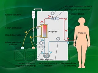
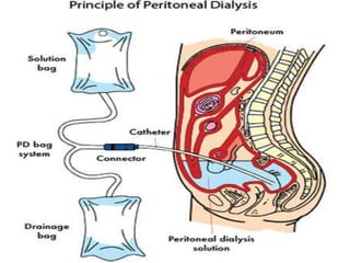
Hussein Alhawari, MD, FASN
Hemodialysis
Peritoneal Dialysis
Kidney Transplantation
Hemodialysis is an extracorporeal
therapy that is prescribed to reduce
the signs and symptoms of uremia and
to replace partially a number of the
key functions of the kidneys when
kidney function is no longer sufficient
to maintain the patient’s well-being or
life.
Hemodialysis used in the treatment of ESRD is
effective in:
(a) Reducing the concentration of uremic toxins,
particularly small and medium-sized molecules,
primarily by diffusion.
(b) Reducing excess fluid volume by convection.
(c) Correcting some of the metabolic
abnormalities, such as acidosis and
hyperkalemia, by use of dialysate solutions with
variable solute concentrations.
The apparatus used to conduct hemodialysis
consists of the following components:
Dialyzer
Dialysis solution (dialysate)
Tubing for transport of blood and dialysis
solution
Machine to power and mechanically monitor
the procedure
Dialyzers are composed of a polyurethane
capsule or shell within which hollow fibers or
parallel membrane plates are suspended in
dialysate. The fibers or plates function as a
semipermeable membrane across which blood
and dialysate flow. By crossing this membrane,
solutes and water move between a patient's
intravascular compartment and the dialysis fluid
contained within the dialyzer
Solute is cleared from the intravascular
compartment by either diffusive or convective
transport.
Such transport depends upon multiple factors
including the concentration gradient between
the blood and dialysate for a particular solute,
the type and amount of blood and dialysate
flow, the properties of the dialysis membrane,
and the size and physicochemical property of
the solute being removed.
Diffusive transport is the primary means of
metabolic waste removal in patients undergoing
hemodialysis. Diffusion of solutes proceeds
down a concentration gradient from blood to
dialysate or from dialysate to blood
(bidirectional).
Blood and dialysate flow in opposite directions
through the dialyzer at rates of 300 to 500
mL/min and 500 to 800 mL/min, respectively.
Such countercurrent flow is integral to
maximizing the diffusive clearance of metabolic
waste solutes.
Convective transport — With high rates of fluid
transport (ie, ultrafiltration) from blood to
dialysate, convective transport of solute also
occurs, thereby augmenting diffusive solute
transport.
In this mechanism, solutes are effectively
dragged along with the fluid as it moves across
the membrane, depending upon their size
relative to the size of the membrane pores.
Fluid removal occurs via a hydrostatic pressure
gradient across the membrane, which is
generated by the dialysis machine.
The TMP is set manually or, with newer
machines, automatically for each patient at
each dialysis treatment so that fluid can be
removed at a desired volume over a designated
time.
Dialysis machines mix different components
with water to produce the final solution of
dialysate.
The exposure of dialysis patients to certain
contaminants frequently found in water may
result in significant complications, including
central nervous system dysfunction, bone
disease, hemolysis, infection, and death.
Copper and chloramine — The presence of
either copper or chloramine in dialysate water
may result in significant hemolytic anemia. The
use of carbon tanks to prevent chloramine
exposure is essential.
Bacteria — Levels of bacteria are suppressed
within the dialysate via the use of disinfectants
and/or filters. Maximum standard level for
bacteria of 200 colony-forming units (CFU)/mL,
while the European Pharmacopoeia standard is
100 CFU/mL.
AV fistula is the best access! Standard is a distal
cephalic vein to radial artery end to side
anastomosis near the wrist. The best next is a
more proximal brachiocephalic or brachiobasilic
fistulas (upper arm fistulas).
Catheters… If you have to!
With peritoneal dialysis, the peritoneal
membrane is used as the dialyzing surface. The
visceral peritoneal membrane tightly covers the
intestine and mesentery, whereas the parietal
peritoneum lines the remaining surfaces of the
abdominal cavity. The peritoneal membrane
consists of a single layer of mesothelial cells
overlying an interstitium in which the blood and
lymphatic vessels lie.
The mesothelial cells are covered by microvilli
that markedly increase the nominal surface area
(2 m2) of the peritoneum, but the effective
peritoneal surface area available for dialysis is
estimated to be about one third of this.
Peritoneal dialysis primarily represents solute
and fluid exchange across the peritoneal
membrane between the peritoneal capillary
blood and the dialysis solution that is instilled
into the peritoneal cavity. There is also a small
amount of fluid and solute resorption via the
lymphatics.
Solute movement occurs as a result of both
diffusion and convective transport, whereas
fluid shifts relate largely to osmosis created by
the addition of osmotic agents to the dialysis
solutions.
During peritoneal dialysis, solutes such as urea,
creatinine, and potassium move from the
peritoneal capillaries across the peritoneal
membrane to the peritoneal cavity, whereas
other solutes, such as lactate and bicarbonate,
usually move in the opposite direction.
Continuous ambulatory peritoneal dialysis
(CAPD) involves multiple exchanges during the
day (usually three), followed by an overnight
dwell.
Automated peritoneal dialysis (APD) refers to
techniques that use an automated device to do
multiple exchanges overnight, with or without a
daytime dwell, such as continuous cycler
peritoneal dialysis (CCPD), nightly intermittent
peritoneal dialysis (NIPD), and tidal peritoneal
dialysis (TPD). Of these, CCPD, the most
commonly used, has a long daytime dwell and
several cycles overnight.
Peritoneal dialysis solutions primarily consist of
water, osmotic agents, electrolytes, and
minerals.
Osmotic agents allow net water removal by
altering the osmotic pressure gradient between
the peritoneal dialysis solution and plasma
water. Dextrose is the most commonly used
osmotic agent. Available dextrose
concentrations include 1.5, 2.5, and 4.25
percent solutions. Amino acids may be used as
an alternative to dextrose to improve nutritional
status in peritoneal dialysis patients.
Icodextrin-containing solutions can cause falsely
elevated glucose levels. In patients using
icodextrin, blood glucose monitoring must be
done with glucose-specific methods to prevent
falsely elevated levels and subsequent
inappropriate treatment of presumed
hyperglycemia.
Peritonitis
PD catheter exit site infection and tunnel
infection.
Catheter malfunction.
Hernias.
Fluid leaks.
Peritoneal membrane changes… Encapsulating
peritoneal sclerosis and UF failure.
Peritonitis remains a major complication of PD
despite advances in equipment and aseptic
technique.
Peritonitis accounts for 15% to 35% of hospital
admissions for these patients and is the major
cause of catheter loss and technique failure
resulting in transfer to hemodialysis.
Entry of bacteria into the catheter during an
exchange procedure (touch contamination) is
the most common source, but organisms can
also track along the external surface of the
catheter or migrate into the peritoneum from
another abdominal viscus.
Diagnosis of peritonitis requires the presence of
two of the following criteria in any combination:
1. Organisms identified on Gram staining or
subsequent culture
2. Cloudy fluid (white cell count >100/mm3; >50%
neutrophils)
3. Symptoms and signs of peritoneal
inflammation.
Cloudy dialysate effluent is almost invariably
present, and abdominal pain is present in about
80% to 95% of cases. Gastrointestinal
symptoms, chills, and fever are present in as
many as 25% of the cases, and abdominal
tenderness in 75%. Bacteremia is rare. Gram
staining of the effluent is seldom helpful, except
with fungal peritonitis, but cultures are usually
positive.
The majority of peritonitis cases are caused by
bacteria; a small percentage of cases are caused
by fungi, mostly Candida species.
Historically, coagulase-negative staphylococci
were the most common cause of peritonitis in
continuous ambulatory peritoneal dialysis
(CAPD), presumably due to touch
contamination or infection via the pericatheter
route. The use of Y systems (or flush before fill)
has reduced the incidence of coagulase-
negative staphylococcal peritonitis, with a
relative increase in the incidence of peritonitis
due to Staphylococcus aureus.
Initial empiric antibiotic coverage for peritoneal
dialysis-associated peritonitis consists of
coverage for gram-positive organisms (by
vancomycin or a first-generation cephalosporin)
and gram-negative organisms (by a third-
generation cephalosporin or an
aminoglycoside). Subsequently, the regimen
should be adjusted based on culture and
sensitivity data.
Indications for catheter removal include
relapsing or refractory peritonitis, refractory
catheter infection, fungal or mycobacterial
peritonitis, and peritonitis associated with intra-
abdominal pathology.
Peritonitis due to multiple enteric organisms or
mixed gram-negative/gram-positive organisms
should raise concern for a concurrent intra-
abdominal condition such as ischemic bowel or
diverticular disease.
Culture-negative peritonitis should be treated
with empiric antibiotic therapy; a repeat cell
count and culture should be obtained after
three to five days of empiric therapy. If there is
improvement in cell count and clinical status,
empiric antibiotic therapy should be continued
for two weeks. If the repeat cell count suggests
persistent infection, the catheter should be
removed, evaluation for intra-abdominal
disease should be pursued, and culture for fungi
and mycobacteria should be performed.
Most episodes of peritoneal dialysis-associated
peritonitis resolve with outpatient antibiotic
treatment; overall cure rates are approximately
75 percent.
Most DM patients will require insulin while they
are on PD, even if they did not require it before
PD. This partly the result of glucose absorption
from the dialysate and associated weight gain.
Insulin can be given via IP route or SC.
Kidney transplantation is the treatment of
choice for most patients with end-stage renal
disease (ESRD) because it prolongs survival,
improves quality of life, and is less costly than
the alternative therapy of dialysis. However,
only a small percentage of ESRD patients
actually receive a transplant; many patients are
not suitable candidates, and for those patients
who are eligible, there are simply not enough
organs available.
Active infections.
Active malignancy.
Active substance abuse.
Reversible renal failure.
Uncontrolled psychiatric disease.
Documented active and ongoing treatment
nonadherence.
A significantly shortened life expectancy.
In the United States, the United Network for
Organ Sharing (UNOS) allows listing for
transplantation when a patient’s estimated
glomerular filtration rate (eGFR) falls below 20
mL/min, whereas organizations in other
countries have established stage 5 CKD (eGFR
below 15 mL/min) as the upper limit for listing.
Thus patients
In Jordan… Only Living Related
Donors.
ABO and Tissue matching…
Questions??

Renal Replacement Therapies.pptx

  • 1.
  • 2.
  • 3.
    Hemodialysis is anextracorporeal therapy that is prescribed to reduce the signs and symptoms of uremia and to replace partially a number of the key functions of the kidneys when kidney function is no longer sufficient to maintain the patient’s well-being or life.
  • 5.
    Hemodialysis used inthe treatment of ESRD is effective in: (a) Reducing the concentration of uremic toxins, particularly small and medium-sized molecules, primarily by diffusion. (b) Reducing excess fluid volume by convection. (c) Correcting some of the metabolic abnormalities, such as acidosis and hyperkalemia, by use of dialysate solutions with variable solute concentrations.
  • 6.
    The apparatus usedto conduct hemodialysis consists of the following components: Dialyzer Dialysis solution (dialysate) Tubing for transport of blood and dialysis solution Machine to power and mechanically monitor the procedure
  • 7.
    Dialyzers are composedof a polyurethane capsule or shell within which hollow fibers or parallel membrane plates are suspended in dialysate. The fibers or plates function as a semipermeable membrane across which blood and dialysate flow. By crossing this membrane, solutes and water move between a patient's intravascular compartment and the dialysis fluid contained within the dialyzer
  • 9.
    Solute is clearedfrom the intravascular compartment by either diffusive or convective transport. Such transport depends upon multiple factors including the concentration gradient between the blood and dialysate for a particular solute, the type and amount of blood and dialysate flow, the properties of the dialysis membrane, and the size and physicochemical property of the solute being removed.
  • 10.
    Diffusive transport isthe primary means of metabolic waste removal in patients undergoing hemodialysis. Diffusion of solutes proceeds down a concentration gradient from blood to dialysate or from dialysate to blood (bidirectional).
  • 11.
    Blood and dialysateflow in opposite directions through the dialyzer at rates of 300 to 500 mL/min and 500 to 800 mL/min, respectively. Such countercurrent flow is integral to maximizing the diffusive clearance of metabolic waste solutes.
  • 12.
    Convective transport —With high rates of fluid transport (ie, ultrafiltration) from blood to dialysate, convective transport of solute also occurs, thereby augmenting diffusive solute transport. In this mechanism, solutes are effectively dragged along with the fluid as it moves across the membrane, depending upon their size relative to the size of the membrane pores.
  • 13.
    Fluid removal occursvia a hydrostatic pressure gradient across the membrane, which is generated by the dialysis machine. The TMP is set manually or, with newer machines, automatically for each patient at each dialysis treatment so that fluid can be removed at a desired volume over a designated time.
  • 14.
    Dialysis machines mixdifferent components with water to produce the final solution of dialysate.
  • 15.
    The exposure ofdialysis patients to certain contaminants frequently found in water may result in significant complications, including central nervous system dysfunction, bone disease, hemolysis, infection, and death.
  • 16.
    Copper and chloramine— The presence of either copper or chloramine in dialysate water may result in significant hemolytic anemia. The use of carbon tanks to prevent chloramine exposure is essential. Bacteria — Levels of bacteria are suppressed within the dialysate via the use of disinfectants and/or filters. Maximum standard level for bacteria of 200 colony-forming units (CFU)/mL, while the European Pharmacopoeia standard is 100 CFU/mL.
  • 18.
    AV fistula isthe best access! Standard is a distal cephalic vein to radial artery end to side anastomosis near the wrist. The best next is a more proximal brachiocephalic or brachiobasilic fistulas (upper arm fistulas). Catheters… If you have to!
  • 19.
    With peritoneal dialysis,the peritoneal membrane is used as the dialyzing surface. The visceral peritoneal membrane tightly covers the intestine and mesentery, whereas the parietal peritoneum lines the remaining surfaces of the abdominal cavity. The peritoneal membrane consists of a single layer of mesothelial cells overlying an interstitium in which the blood and lymphatic vessels lie.
  • 20.
    The mesothelial cellsare covered by microvilli that markedly increase the nominal surface area (2 m2) of the peritoneum, but the effective peritoneal surface area available for dialysis is estimated to be about one third of this. Peritoneal dialysis primarily represents solute and fluid exchange across the peritoneal membrane between the peritoneal capillary blood and the dialysis solution that is instilled into the peritoneal cavity. There is also a small amount of fluid and solute resorption via the lymphatics.
  • 22.
    Solute movement occursas a result of both diffusion and convective transport, whereas fluid shifts relate largely to osmosis created by the addition of osmotic agents to the dialysis solutions. During peritoneal dialysis, solutes such as urea, creatinine, and potassium move from the peritoneal capillaries across the peritoneal membrane to the peritoneal cavity, whereas other solutes, such as lactate and bicarbonate, usually move in the opposite direction.
  • 23.
    Continuous ambulatory peritonealdialysis (CAPD) involves multiple exchanges during the day (usually three), followed by an overnight dwell. Automated peritoneal dialysis (APD) refers to techniques that use an automated device to do multiple exchanges overnight, with or without a daytime dwell, such as continuous cycler peritoneal dialysis (CCPD), nightly intermittent peritoneal dialysis (NIPD), and tidal peritoneal dialysis (TPD). Of these, CCPD, the most commonly used, has a long daytime dwell and several cycles overnight.
  • 24.
    Peritoneal dialysis solutionsprimarily consist of water, osmotic agents, electrolytes, and minerals.
  • 25.
    Osmotic agents allownet water removal by altering the osmotic pressure gradient between the peritoneal dialysis solution and plasma water. Dextrose is the most commonly used osmotic agent. Available dextrose concentrations include 1.5, 2.5, and 4.25 percent solutions. Amino acids may be used as an alternative to dextrose to improve nutritional status in peritoneal dialysis patients.
  • 28.
    Icodextrin-containing solutions cancause falsely elevated glucose levels. In patients using icodextrin, blood glucose monitoring must be done with glucose-specific methods to prevent falsely elevated levels and subsequent inappropriate treatment of presumed hyperglycemia.
  • 29.
    Peritonitis PD catheter exitsite infection and tunnel infection. Catheter malfunction. Hernias. Fluid leaks. Peritoneal membrane changes… Encapsulating peritoneal sclerosis and UF failure.
  • 30.
    Peritonitis remains amajor complication of PD despite advances in equipment and aseptic technique. Peritonitis accounts for 15% to 35% of hospital admissions for these patients and is the major cause of catheter loss and technique failure resulting in transfer to hemodialysis.
  • 31.
    Entry of bacteriainto the catheter during an exchange procedure (touch contamination) is the most common source, but organisms can also track along the external surface of the catheter or migrate into the peritoneum from another abdominal viscus.
  • 32.
    Diagnosis of peritonitisrequires the presence of two of the following criteria in any combination: 1. Organisms identified on Gram staining or subsequent culture 2. Cloudy fluid (white cell count >100/mm3; >50% neutrophils) 3. Symptoms and signs of peritoneal inflammation.
  • 33.
    Cloudy dialysate effluentis almost invariably present, and abdominal pain is present in about 80% to 95% of cases. Gastrointestinal symptoms, chills, and fever are present in as many as 25% of the cases, and abdominal tenderness in 75%. Bacteremia is rare. Gram staining of the effluent is seldom helpful, except with fungal peritonitis, but cultures are usually positive.
  • 34.
    The majority ofperitonitis cases are caused by bacteria; a small percentage of cases are caused by fungi, mostly Candida species.
  • 35.
    Historically, coagulase-negative staphylococci werethe most common cause of peritonitis in continuous ambulatory peritoneal dialysis (CAPD), presumably due to touch contamination or infection via the pericatheter route. The use of Y systems (or flush before fill) has reduced the incidence of coagulase- negative staphylococcal peritonitis, with a relative increase in the incidence of peritonitis due to Staphylococcus aureus.
  • 36.
    Initial empiric antibioticcoverage for peritoneal dialysis-associated peritonitis consists of coverage for gram-positive organisms (by vancomycin or a first-generation cephalosporin) and gram-negative organisms (by a third- generation cephalosporin or an aminoglycoside). Subsequently, the regimen should be adjusted based on culture and sensitivity data.
  • 37.
    Indications for catheterremoval include relapsing or refractory peritonitis, refractory catheter infection, fungal or mycobacterial peritonitis, and peritonitis associated with intra- abdominal pathology. Peritonitis due to multiple enteric organisms or mixed gram-negative/gram-positive organisms should raise concern for a concurrent intra- abdominal condition such as ischemic bowel or diverticular disease.
  • 38.
    Culture-negative peritonitis shouldbe treated with empiric antibiotic therapy; a repeat cell count and culture should be obtained after three to five days of empiric therapy. If there is improvement in cell count and clinical status, empiric antibiotic therapy should be continued for two weeks. If the repeat cell count suggests persistent infection, the catheter should be removed, evaluation for intra-abdominal disease should be pursued, and culture for fungi and mycobacteria should be performed.
  • 39.
    Most episodes ofperitoneal dialysis-associated peritonitis resolve with outpatient antibiotic treatment; overall cure rates are approximately 75 percent.
  • 40.
    Most DM patientswill require insulin while they are on PD, even if they did not require it before PD. This partly the result of glucose absorption from the dialysate and associated weight gain. Insulin can be given via IP route or SC.
  • 41.
    Kidney transplantation isthe treatment of choice for most patients with end-stage renal disease (ESRD) because it prolongs survival, improves quality of life, and is less costly than the alternative therapy of dialysis. However, only a small percentage of ESRD patients actually receive a transplant; many patients are not suitable candidates, and for those patients who are eligible, there are simply not enough organs available.
  • 42.
    Active infections. Active malignancy. Activesubstance abuse. Reversible renal failure. Uncontrolled psychiatric disease. Documented active and ongoing treatment nonadherence. A significantly shortened life expectancy.
  • 43.
    In the UnitedStates, the United Network for Organ Sharing (UNOS) allows listing for transplantation when a patient’s estimated glomerular filtration rate (eGFR) falls below 20 mL/min, whereas organizations in other countries have established stage 5 CKD (eGFR below 15 mL/min) as the upper limit for listing. Thus patients
  • 44.
    In Jordan… OnlyLiving Related Donors. ABO and Tissue matching…
  • 45.