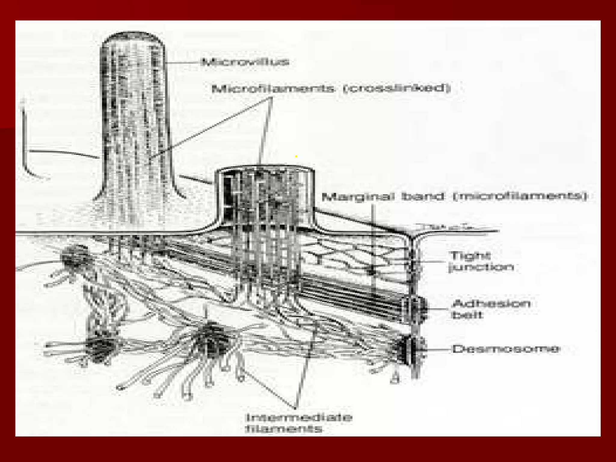
Microvilli, cilia, and flagella are cell surface projections that serve important functions.
Microvilli increase cell surface area and aid in absorption. They cover the intestinal surface and form the brush border. Cilia are shorter projections that beat in a coordinated way to move substances over cell surfaces, such as mucus in the respiratory tract.
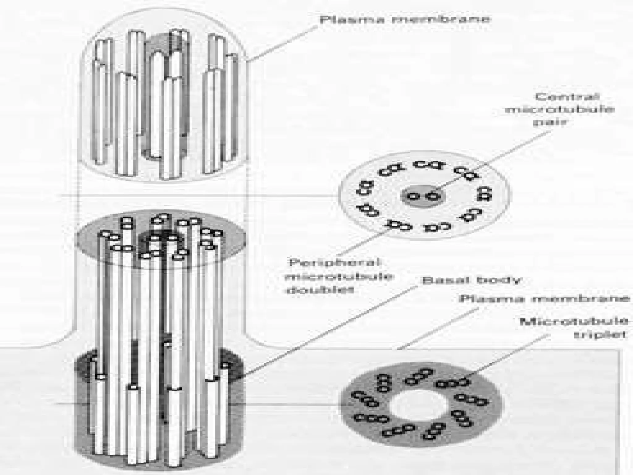
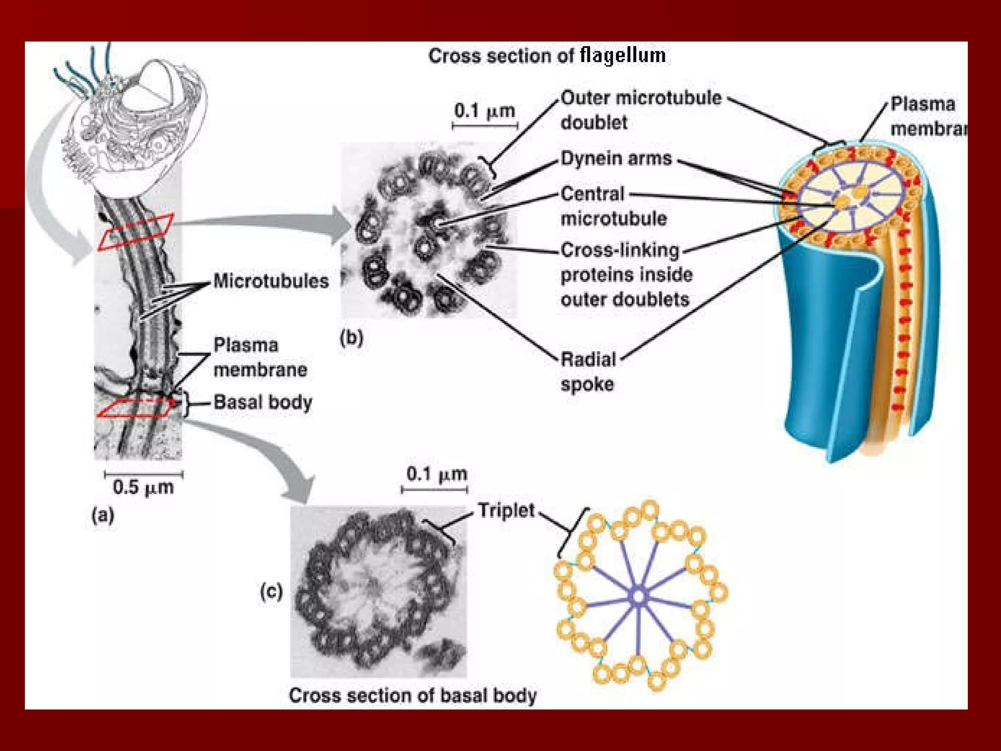
Flagella are much longer than cilia and power sperm cell motility through whip-like beating. Both cilia and flagella contain microtubules in their core that extend from basal bodies anchored in the cell.