
Recommended
More Related Content
Similar to Epithelium.pptx
Similar to Epithelium.pptx (20)
More from VishwajitDeshmukh4
Recently uploaded
Recently uploaded (20)
Epithelium.pptx
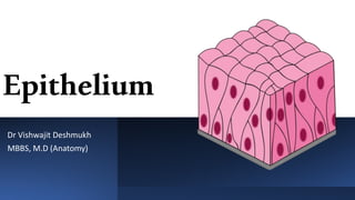
- 1. Epithelium Dr Vishwajit Deshmukh MBBS, M.D (Anatomy)
- 2. • Epithelium is an avascular tissue composed of cells that cover the exterior body surfaces and line internal closed cavities (including the vascular system) and body tubes that communicate with the exterior (the alimentary, respiratory, and genitourinary tracts). • Epithelium also forms the secretory portion (parenchyma) of glands and their ducts. In addition, specialized epithelial cells function as receptors for the special senses (smell, taste, hearing, and vision) Introduction
- 3. Diverse epithelial functions can be found in different organs of the body. • Secretion, as in the columnar epithelium of the stomach and the gastric glands; • Absorption, as in the columnar epithelium of the intestines and proximal convoluted tubules in the kidney; • Transportation, as in the transport of materials or cells along the surface of an epithelium by motile cilia or in the transport of materials across an epithelium to and from the connective tissue; • Protection, as in the stratified squamous epithelium of the skin (epidermis) and the transitional epithelium of the urinary bladder; and • Receptor function to receive and transduce external stimuli, as in the taste buds of the tongue, olfactory epithelium of the nasal mucosa, and the retina of the eye.
- 4. • Epithelia involved in secretion or absorption are typically simple or, in a few cases, pseudostratified. • The height of the cells often reflects the level of secretory or absorptive activity. Simple squamous epithelia are compatible with a high rate of transepithelial transport. • Stratification of the epithelium usually correlates with transepithelial impermeability. • Finally, in some pseudostratified epithelia, basal cells are the stem cells that give rise to the mature functional cells of the epithelium, thus balancing cell turnover.
- 5. 3 Characteristic features • They are closely apposed and adhere to one another by means of specific cell-to-cell adhesion molecules that form specialized cell junctions • They exhibit functional and morphologic polarity. In other words, different functions are associated with three distinct morphologic surface domains: a free surface or apical domain, a lateral domain, and a basal domain. • Their basal surface is attached to an underlying basement membrane, a noncellular, protein–polysaccharide-rich layer demonstrable at the light microscopic level by histochemical methods
- 7. • Epithelioid organization is typical of most endocrine glands; examples of such tissue include the interstitial cells of Leydig in the testis, the lutein cells of the ovary, the islets of Langerhans in the pancreas, the parenchyma of the adrenal gland, and the anterior lobe of the pituitary gland • In some locations, cells are closely apposed to one another but lack a free surface. Although the close apposition of these cells and the presence of a basement membrane would classify them as epithelium, the absence of a free sur- face more appropriately classifies such cell aggregates as epithelioid tissues. Epithelioid cells
- 8. Endothelium and mesothelium are the simple squamous epithelia lining the vascular system and body cavities 1.Specific names are given to epithelium in certain locations • Endothelium • Endocardium • Mesothelium
- 9. CLASSIFICATION OF EPITHELIUM The traditional classification of epithelium is descriptive and based on two factors: the number of cell layers and the shape of the surface cells. The terminology, therefore, reflects only structure, not function. • Simple and stratified • Cuboidal, columnar and squamous • Pseudostratified • Transitional epithelium
- 11. Pseudostratified epithelium and transitional epithelium are special classifications of epithelium •Pseudostratified epithelium appears stratified, although some of the cells do not reach the free surface; all rest on the basement membrane. Thus, it is actually a simple epithelium. •Transitional epithelium (urothelium) is a term applied to the epithelium lining the lower urinary tract, extending from the minor calyces of the kidney down to the proximal part of the urethra. Urothelium is a stratified epithelium with specific morphologic characteristics that allow it to distend
- 13. • Both endothelium and endocardium, as well as mesothelium, are almost always simple squamous epithelia. • An exception is found in postcapillary venules of certain lymphatic tissues in which the endothelium is cuboidal. • These venules are called high endothelial venules (HEV). Another exception is found in the spleen in which endothelial cells of the venous sinuses are rod-shaped and arranged like the staves of a barrel
- 14. Cell Polarity Epithelial cells exhibit distinct polarity. They have an apical domain, a lateral domain, and a basal domain • Microvilli, cytoplasmic processes containing a core of actin filaments; • Stereocilia (stereovilli), microvilli of unusual length and • Cilia, cytoplasmic processes containing bundle of micro-tubules.
- 15. Microvilli Microvilli are finger-like cytoplasmic projections on the apical surface of most epithelial cells • In intestinal absorptive cells, this surface structure was originally called the striated border; • in the kidney tubule cells, it is called the brush border
- 16. • In other cell types, they are tall, closely packed, uniform projections that greatly increase the free cell surface area. • the number and shape of the microvilli of a given cell type correlate with the cell’s absorptive capacity. • In intestinal absorptive cells, this surface structure was originally called the striated border; in the kidney tubule cells, it is called the brush border. • The internal structure of microvilli contains a core of actin filaments that are cross-linked by several actin- bundling proteins.
- 17. Stereocilia
- 18. • Stereocilia are not widely distributed among epithelia. They are, in fact, limited to the epididymis, the proximal part of the ductus deferens of the male reproductive system, and the sensory (hair) cells of the inner ear. • Stereocilia of the genital ducts are extremely long processes that extend from the apical surface of the cell and facilitate absorption. • Stereocilia develop from microvilli by the lateral addition of actin filaments to the actin bundle as well as elongation of the actin filaments. • A striking difference between microvilli and stereocilia, other than size and the presence of ezrin, is the absence of villin from the tip of the stereocilium. • Stereocilia of the sensory epithelium of the ear also derive from microvilli. They are exquisitely sensitive to mechanical vibration and serve as sensory mechanoreceptors rather than absorptive structures.
- 19. Cilia
- 20. Motile cilia have been historically the most studied of all cilia. They are found in large numbers on the apical domain of many epithelial cells. Motile cilia and their counterparts, flagella, possess a typical 9 +2 axonemal organization with microtubule-associated motor proteins that are necessary for the generation of forces needed to induce motility. Primary cilia (monocilia) are solitary projections found on almost all eukaryotic cells. The term monocilia implies that only a single cilium per cell is usually present. Primary cilia are immotile. They function as chemosensors, and mechanosensors, and they mediate light sensation, odorant, and sound perception in multiple organs in the body. Nodal cilia are found in the embryo on the bilaminar embryonic disc at the time of gastrulation. They are concentrated in the area that surrounds the primitive node, hence their name nodal cilia. They have a similar ax- onemal internal architecture as primary cilia; however, they are distinct in their ability to perform rotational movement. They play an important role in early embryonic development.
- 21. Basal Body
- 25. There are three types of junctional complexes •Occluding junctions •Anchoring •Communicating
- 27. Occluding Junctions • Occluding junctions are impermeable and allow epithelial cells to function as a barrier. Also called tight junctions, occluding junctions form the primary intercellular diffusion barrier between adjacent cells. • By limiting the movement of water and other molecules through the intercellular space, they maintain physicochemical separation of tissue compartments. • Examination of the zonula occludens or tight junction with the transmission electron microscope (TEM) reveals a narrow region in which the plasma membranes of adjoining cells come in close contact to seal off the intercellular space. • At high resolution, the zonula occludens appears not as a continuous seal but as a series of focal fusions between the cells. • Several proteins are involved in the formation of zonula occludens strands • Occludin • Claudin • Junctional adhesion molecules
- 29. • Anchoring junctions provide mechanical stability to epithelial cells by linking the cytoskeleton of one cell to the cytoskeleton of an adjacent cell. • These junctions are important in creating and maintaining the structural unity of the epithelium. • Anchoring junctions interact with both actin and intermediate filaments and can be found not only on the lateral cell surface but also on the basal domain of the epithelial cell. • Through their signal transduction capability, anchoring junctions also play important roles in cell-to-cell recognition, morphogenesis, and differentiation. Anchoring Junctions • Zonula Adherens which inter- acts with the network of actin filaments inside the cell • Macula Adherens or desmosome, which interacts with intermediate filaments In addition, two other types of anchoring junctions can be found where epithelial cells rest on the connective tissue matrix. These focal adhesions (focal contacts) and hemidesmosomes
- 33. Communicating Junctions/ Gap junctions or Nexuses • The only known cellular structures that permit the direct passage of signaling molecules from one cell to another. • Gap junctions are important in tissues in which activity of adjacent cells must be coordinated, such as epithelia engaged in fluid and electrolyte transport, vascular and intestinal smooth muscle, and heart muscle. • Various procedures have been used to study gap junctions, including the injection of dyes and fluorescent or radiolabeled compounds. • High-resolution imaging techniques such as cryo-electron microscopy have been used to examine the structure of gap junctions. These studies reveal groups of tightly packed channels, each formed by two half- channels called connexons embedded in the facing mem- branes. • Each connexon contains six symmetrical subunits of an integral membrane protein called connexin (Cx) that is paired with a similar structure from the adjacent membrane.
- 35. Atomic force microscopic image of the gap junctions
- 36. Basal Domain • The basement membrane is a specialized structure located next to the basal domain of epithelial cells and the underlying connective tissue stroma. • Cell-to-extracellular matrix junctions anchor the cell to the extracellular matrix; they are represented by focal adhesions and hemidesmosomes. • Basal cell membrane infoldings increase the cell surface area and facilitate morphologic interactions between adjacent cells and extracellular matrix proteins.
- 37. This section was stained by the PAS method
- 39. Electron micrograph of epithelial cells
- 40. Thanks VDsnaps