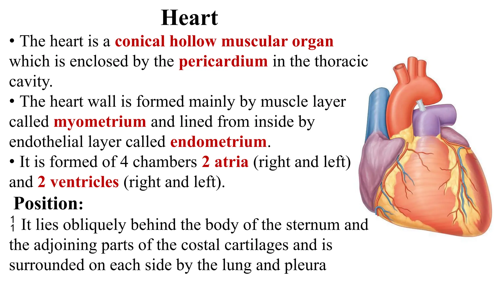
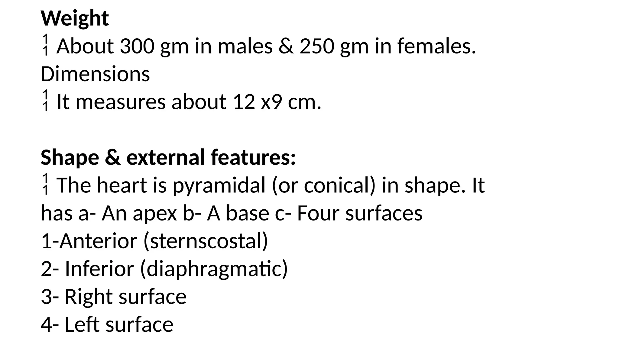
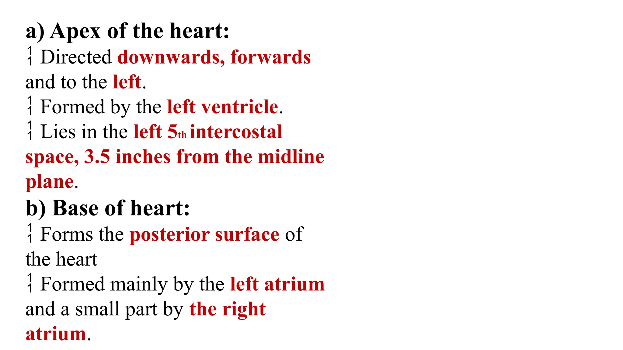
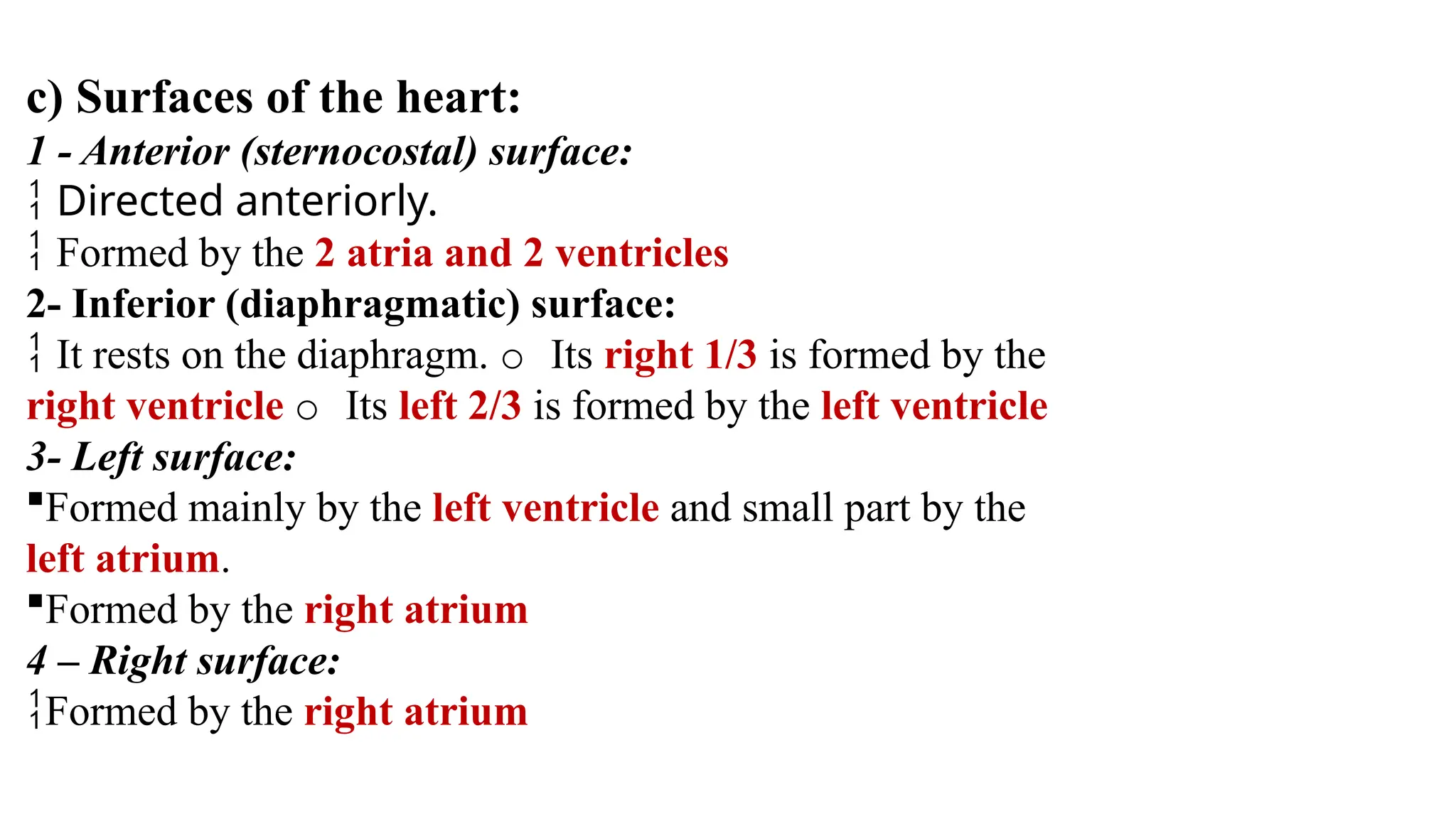
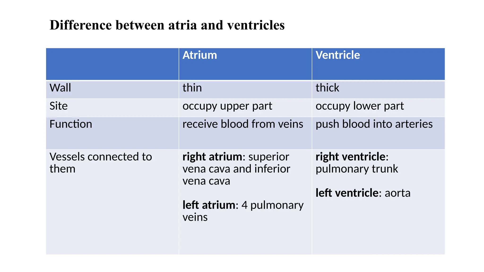
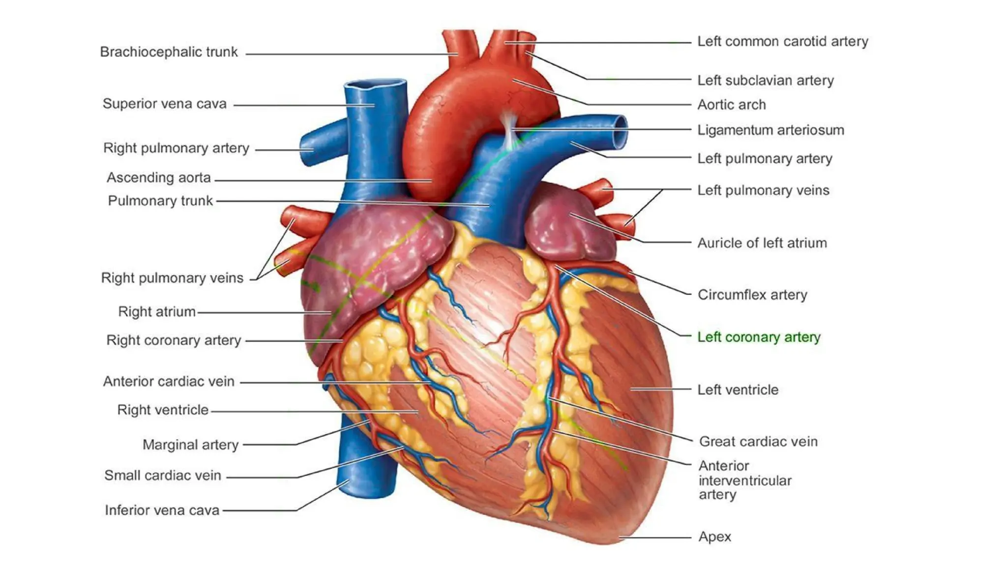
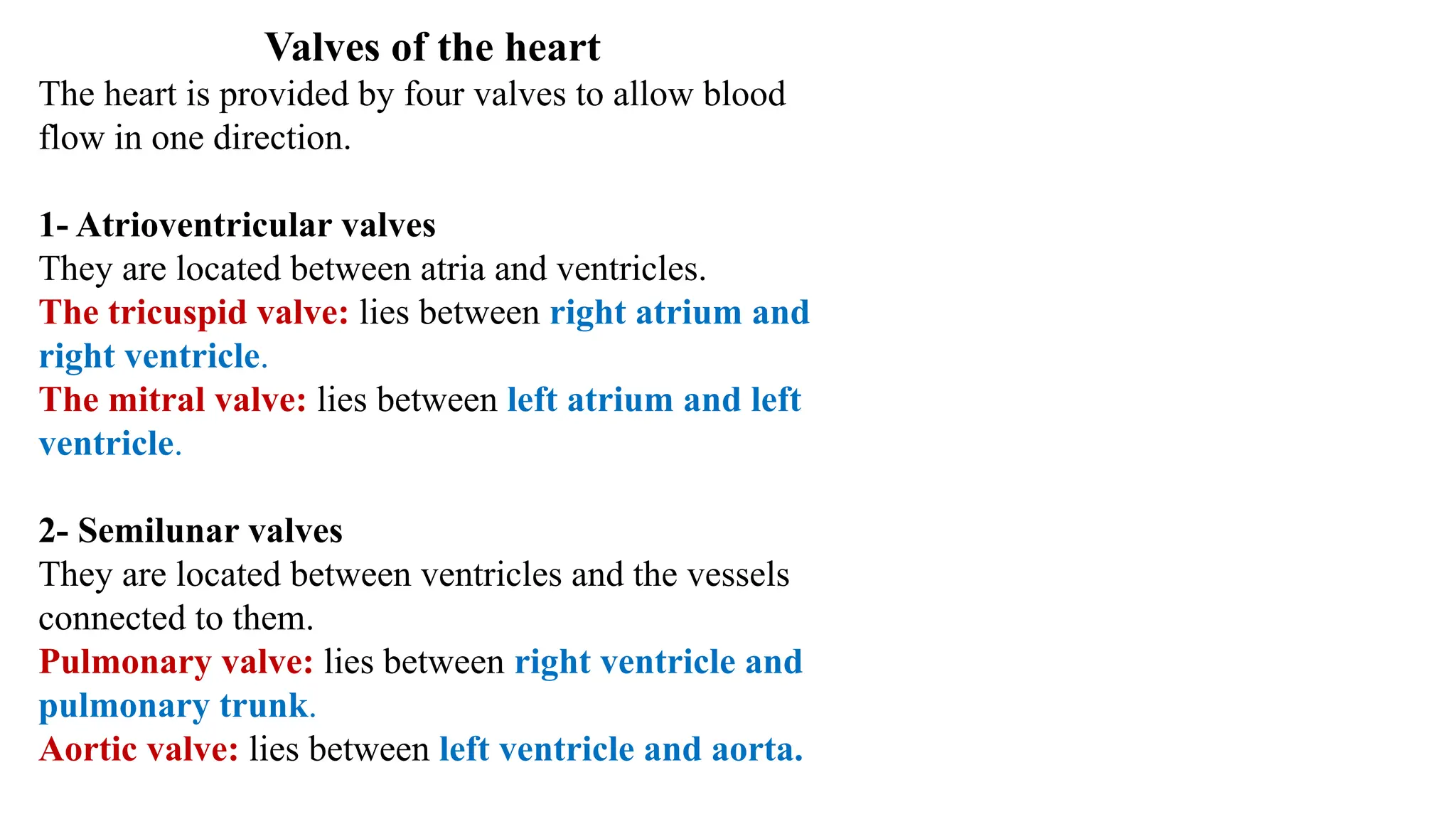
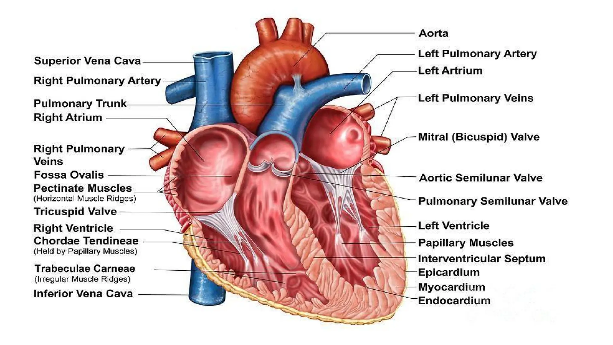
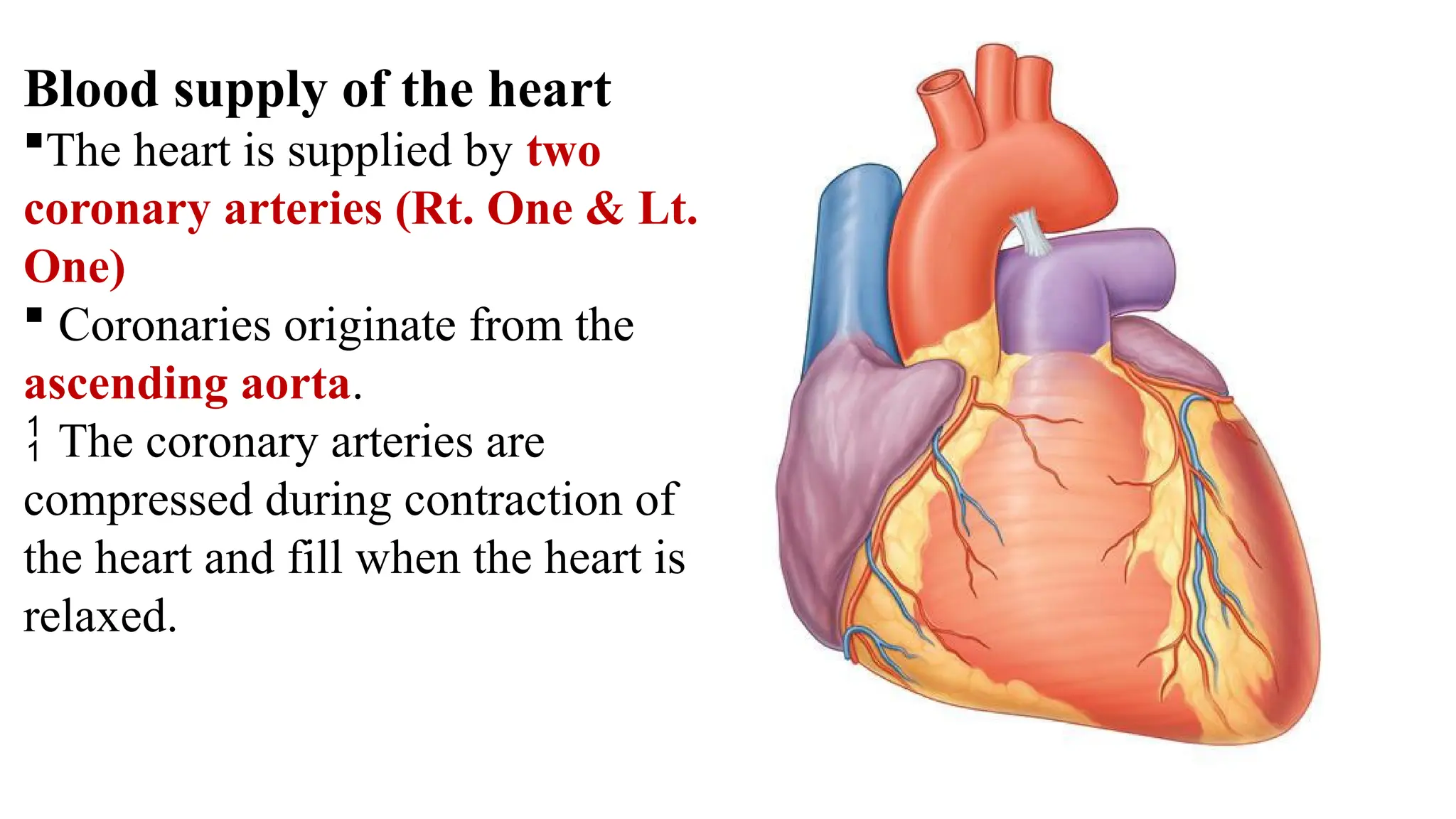
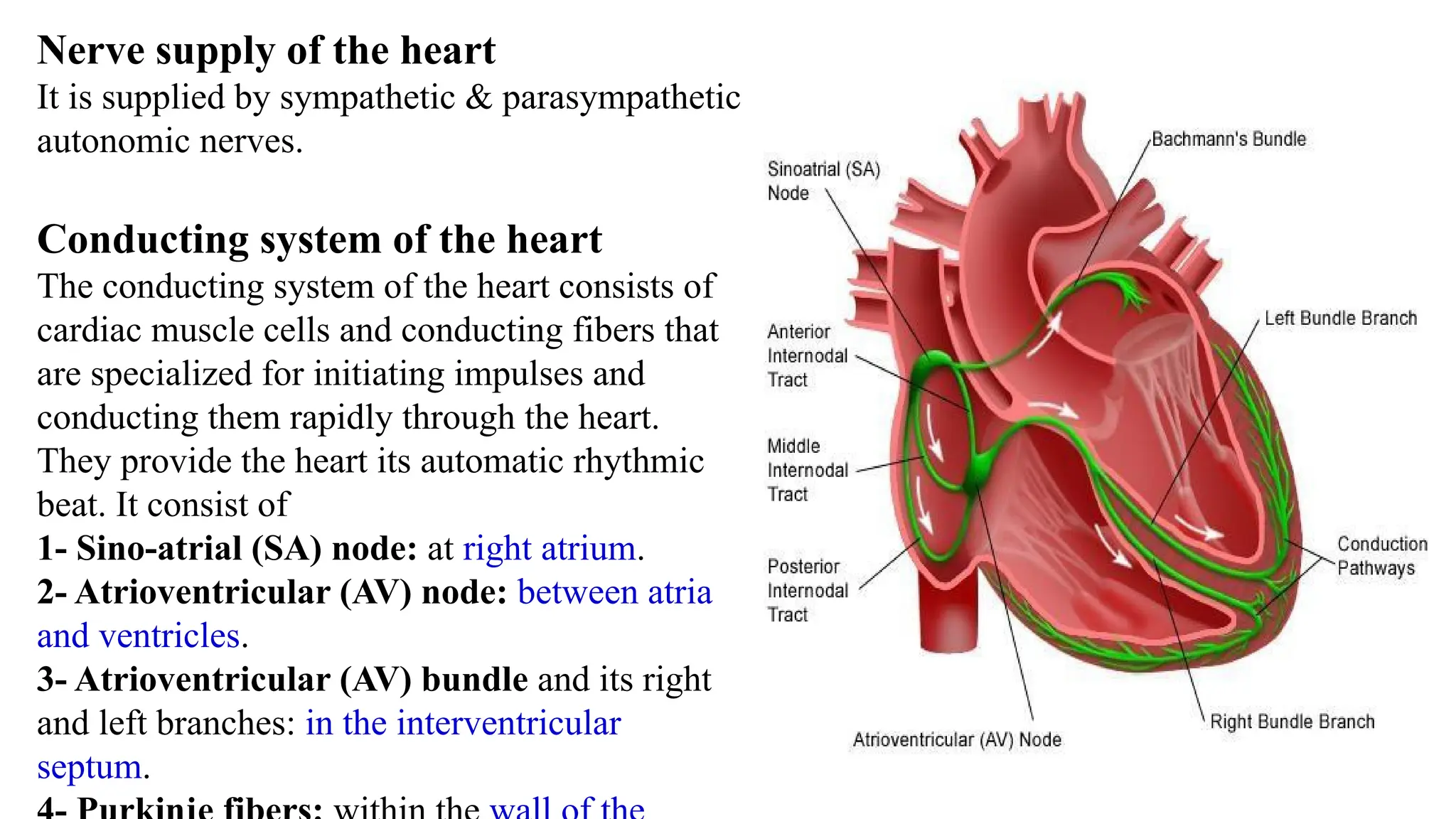
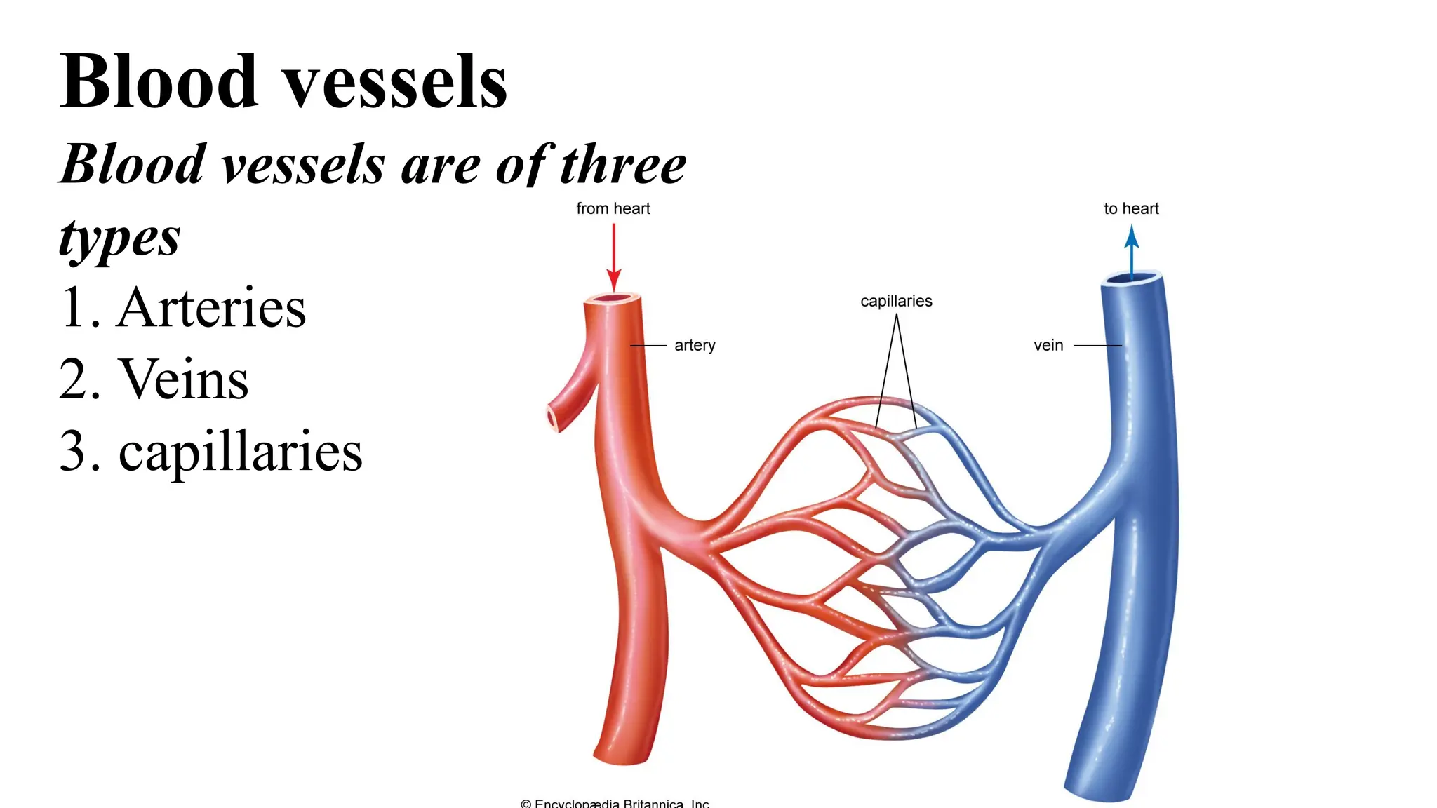
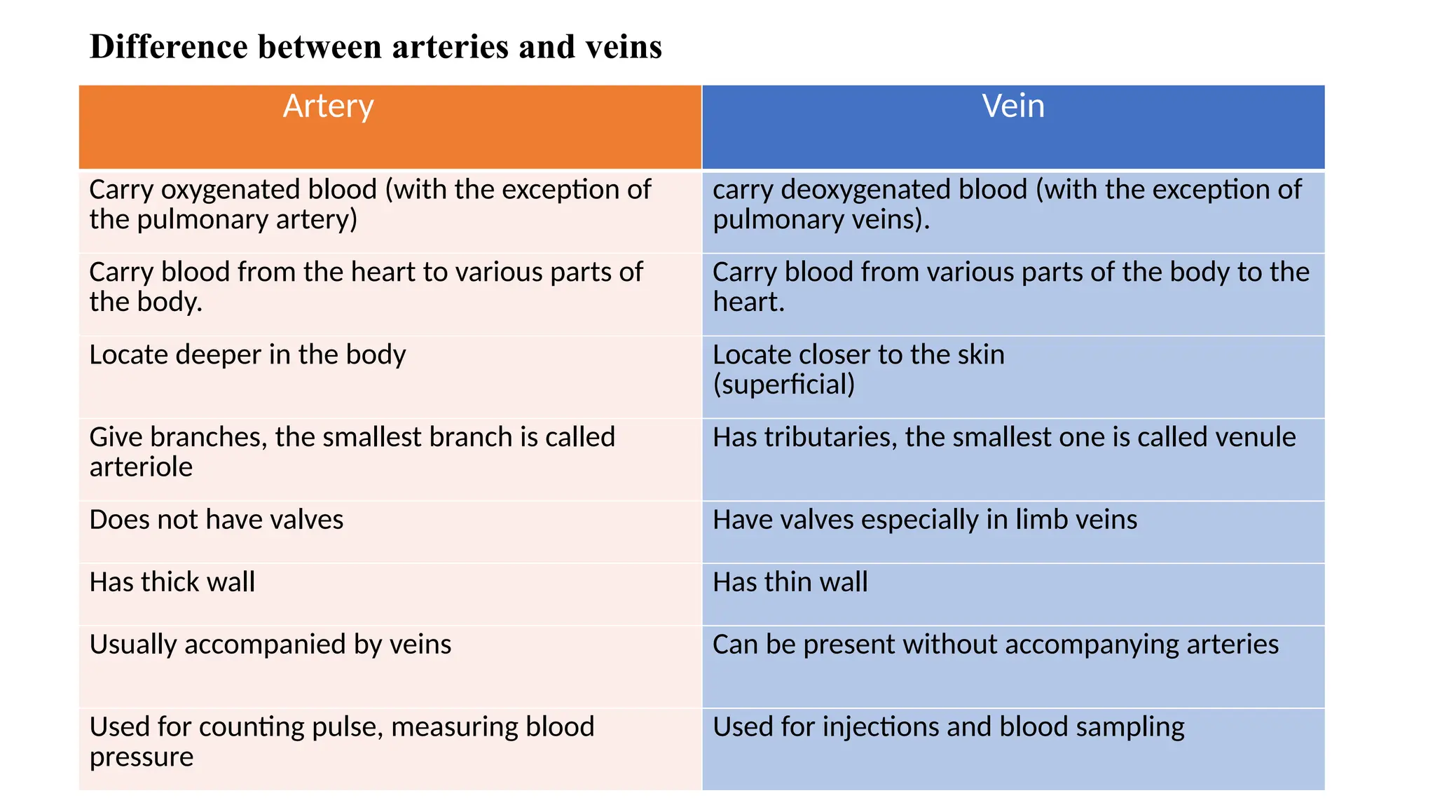
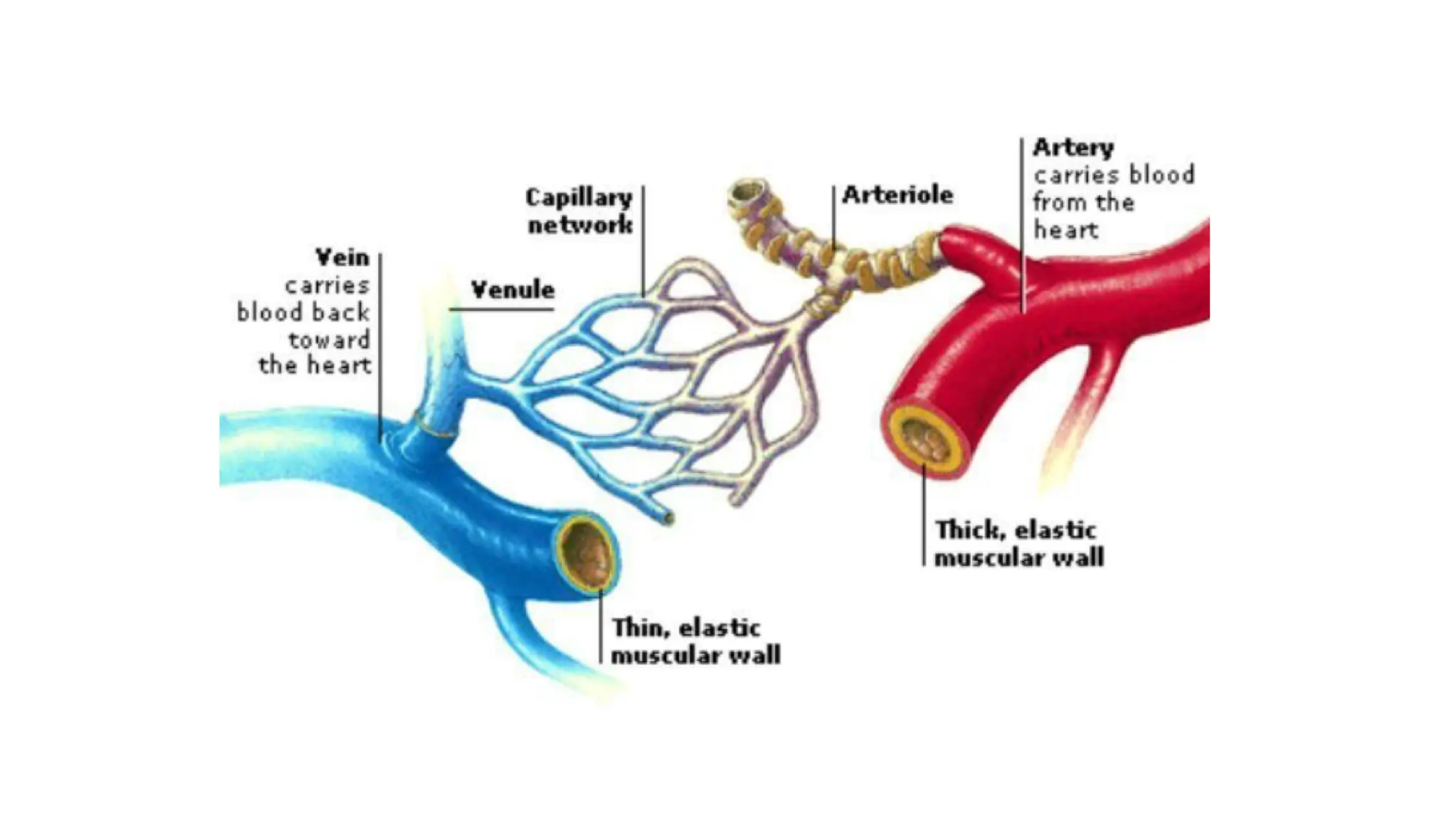
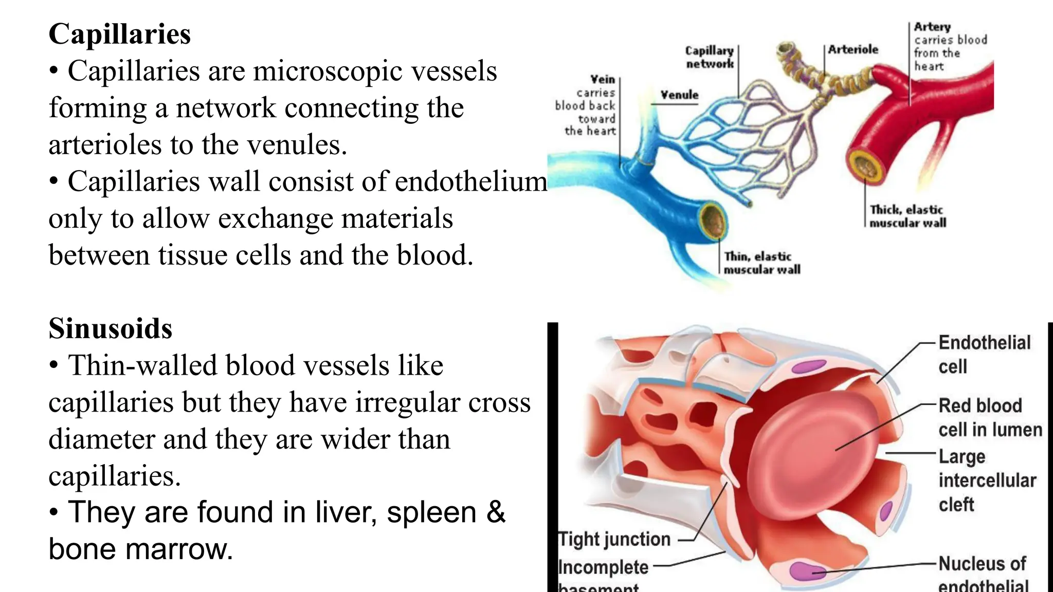
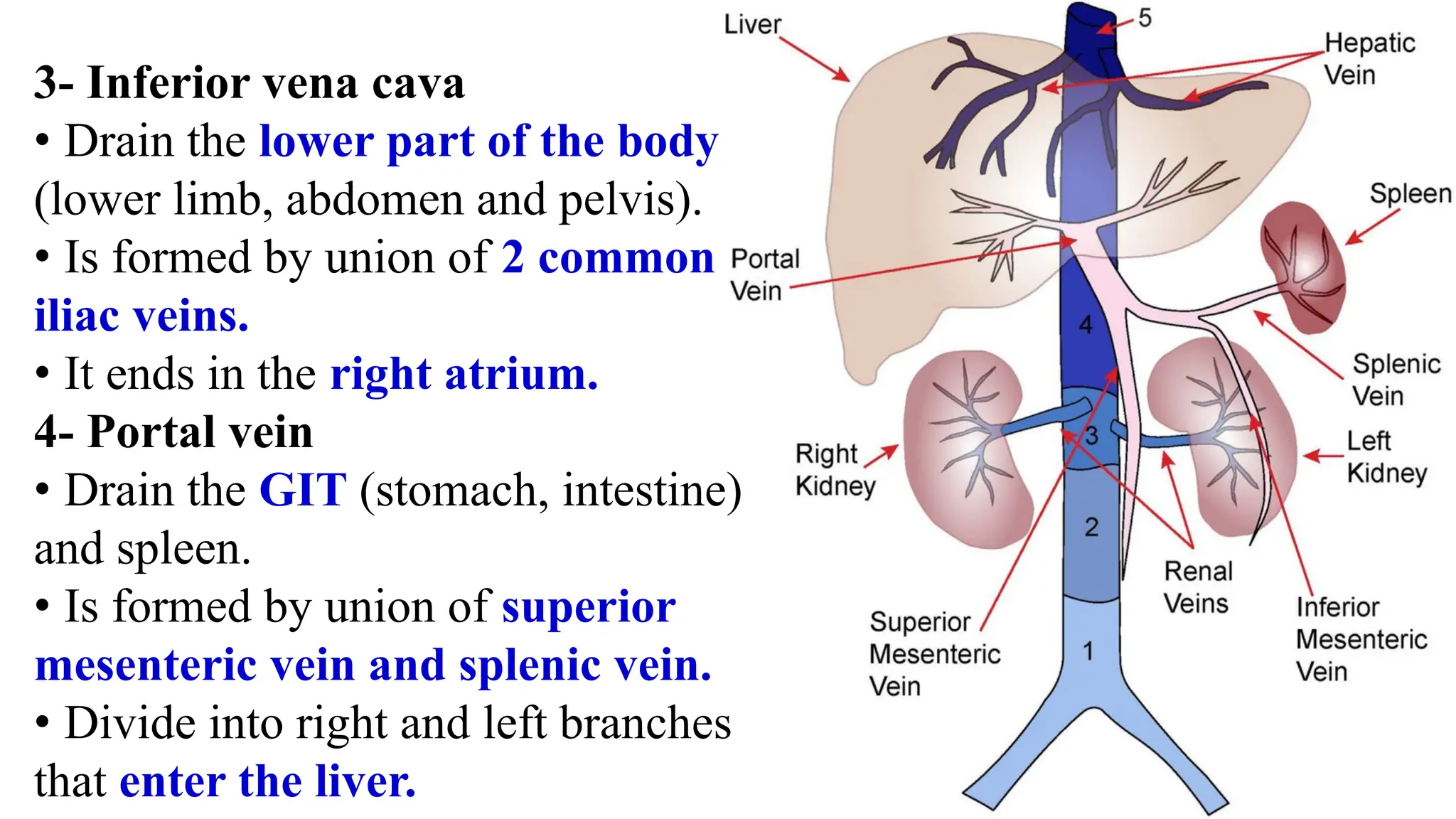
CARDIOVASCULAR
SYSTEM
CARDIOVASCULAR SYSTEM
The cardiovascular system transports blood to and
from capillary beds in the tissues for the exchange
of respiratory gases, nutrients and metabolites.
The cardiovascular system consists of the heart &
blood vessels.
Heart
• The heart is a conical hollow muscular organ
which is enclosed by the pericardium in the thoracic
cavity.
• The heart wall is formed mainly by muscle layer
called myometrium and lined from inside by
endothelial layer called endometrium.
• It is formed of 4 chambers 2 atria (right and left)
and 2 ventricles (right and left).
Position:
 It lies obliquely behind the body of the sternum and
the adjoining parts of the costal cartilages and is
surrounded on each side by the lung and pleura
Weight
 About 300 gm in males & 250 gm in females.
Dimensions
 It measures about 12 x9 cm.
Shape & external features:
 The heart is pyramidal (or conical) in shape. It
has a- An apex b- A base c- Four surfaces
1-Anterior (sternscostal)
2- Inferior (diaphragmatic)
3- Right surface
4- Left surface
a) Apex of the heart:
 Directed downwards, forwards
and to the left.
 Formed by the left ventricle.
 Lies in the left 5th intercostal
space, 3.5 inches from the midline
plane.
b) Base of heart:
 Forms the posterior surface of
the heart
 Formed mainly by the left atrium
and a small part by the right
atrium.
c) Surfaces of the heart:
1 - Anterior (sternocostal) surface:
 Directed anteriorly.
 Formed by the 2 atria and 2 ventricles
2- Inferior (diaphragmatic) surface:
 It rests on the diaphragm. o Its right 1/3 is formed by the
right ventricle o Its left 2/3 is formed by the left ventricle
3- Left surface:
Formed mainly by the left ventricle and small part by the
left atrium.
Formed by the right atrium
4 – Right surface:
Formed by the right atrium
Difference between atria and ventricles
Atrium Ventricle
Wall thin thick
Site occupy upper part occupy lower part
Function receive blood from veins push blood into arteries
Vessels connected to
them
right atrium: superior
vena cava and inferior
vena cava
left atrium: 4 pulmonary
veins
right ventricle:
pulmonary trunk
left ventricle: aorta
Valves of the heart
The heart is provided by four valves to allow blood
flow in one direction.
1- Atrioventricular valves
They are located between atria and ventricles.
The tricuspid valve: lies between right atrium and
right ventricle.
The mitral valve: lies between left atrium and left
ventricle.
2- Semilunar valves
They are located between ventricles and the vessels
connected to them.
Pulmonary valve: lies between right ventricle and
pulmonary trunk.
Aortic valve: lies between left ventricle and aorta.
Blood supply of the heart
The heart is supplied by two
coronary arteries (Rt. One & Lt.
One)
 Coronaries originate from the
ascending aorta.
 The coronary arteries are
compressed during contraction of
the heart and fill when the heart is
relaxed.
Nerve supply of the heart
It is supplied by sympathetic & parasympathetic
autonomic nerves.
Conducting system of the heart
The conducting system of the heart consists of
cardiac muscle cells and conducting fibers that
are specialized for initiating impulses and
conducting them rapidly through the heart.
They provide the heart its automatic rhythmic
beat. It consist of
1- Sino-atrial (SA) node: at right atrium.
2- Atrioventricular (AV) node: between atria
and ventricles.
3- Atrioventricular (AV) bundle and its right
and left branches: in the interventricular
septum.
Blood vessels
Blood vessels
Blood vessels are of three
types
1. Arteries
2. Veins
3. capillaries
Difference between arteries and veins
Artery Vein
Carry oxygenated blood (with the exception of
the pulmonary artery)
carry deoxygenated blood (with the exception of
pulmonary veins).
Carry blood from the heart to various parts of
the body.
Carry blood from various parts of the body to the
heart.
Locate deeper in the body Locate closer to the skin
(superficial)
Give branches, the smallest branch is called
arteriole
Has tributaries, the smallest one is called venule
Does not have valves Have valves especially in limb veins
Has thick wall Has thin wall
Usually accompanied by veins Can be present without accompanying arteries
Used for counting pulse, measuring blood
pressure
Used for injections and blood sampling
Capillaries
• Capillaries are microscopic vessels
forming a network connecting the
arterioles to the venules.
• Capillaries wall consist of endothelium
only to allow exchange materials
between tissue cells and the blood.
Sinusoids
• Thin-walled blood vessels like
capillaries but they have irregular cross
diameter and they are wider than
capillaries.
• They are found in liver, spleen &
bone marrow.
Portal systems
• A portal system is a system of
vessels present between two
capillary beds.
• Example: Veins draining the
capillary beds of the gastro-
intestinal tract do not go directly
to the heart but collect in the
portal vein which enters the liver
and divides again forming a second
set of capillaries (sinusoids).
Important arteries in the body
1- Aorta
• The aorta is the largest artery
in the body.
• It arises from the left ventricles
• It has three parts
a- The ascending aorta: gives rise
to the right and left coronary
arteries, which supply the heart.
b- The aortic arch: gives rise to 3 branches
 The brachiocephalic artery that divides
into right common carotid artery and
right subclavian artery
 The left common carotid artery
 The left subclavian artery
(The subclavian arteries supply the upper
limb, while the common carotid arteries
supply the head and neck and the brain)
c- Descending aorta: which has 2 parts
Descending thoracic aorta: gives branches to
diaphragm, lung, thoracic wall.
Descending abdominal aorta: gives branches
supplying GIT, kidney, suprarenal gland and gonads.
The branches of the descending abdominal aorta that
supply GIT are
1- Cealiac trunk: supply stomach, liver, spleen.
2- Superior mesenteric artery: supply small intestine
and part of large intestine.
3- Inferior mesenteric artery: supply large intestine.
The abdominal aorta divides into 2 terminal branches
called common iliac arteries that divide into
External iliac artery: supply the lower limb
Internal iliac artery: supply the pelvic organs (urinary
bladder, rectum, sex organs)
2- Pulmonary trunk
= It originates from the right
ventricle.
= It divides into right and left
pulmonary arteries that enter the
lung.
= They carry nonoxygenated
blood to the lungs.
Important veins in the body
1- Superior vena cava
• Drain the upper part of the body
( head and neck, upper limb and chest).
• Is formed by union of right and left
brachiocephalic veins.
• It ends the right atrium.
2- Brachiocephalic veins
• Are formed by the union of internal
jugular vein (drain the head and neck
and brain) and subclavian vein (drain
the upper limb).
3- Inferior vena cava
• Drain the lower part of the body
(lower limb, abdomen and pelvis).
• Is formed by union of 2 common
iliac veins.
• It ends in the right atrium.
4- Portal vein
• Drain the GIT (stomach, intestine)
and spleen.
• Is formed by union of superior
mesenteric vein and splenic vein.
• Divide into right and left branches
that enter the liver.

Introduction to cardiovascular system.pptx

  • 1.
  • 2.
    CARDIOVASCULAR SYSTEM The cardiovascularsystem transports blood to and from capillary beds in the tissues for the exchange of respiratory gases, nutrients and metabolites. The cardiovascular system consists of the heart & blood vessels.
  • 3.
    Heart • The heartis a conical hollow muscular organ which is enclosed by the pericardium in the thoracic cavity. • The heart wall is formed mainly by muscle layer called myometrium and lined from inside by endothelial layer called endometrium. • It is formed of 4 chambers 2 atria (right and left) and 2 ventricles (right and left). Position:  It lies obliquely behind the body of the sternum and the adjoining parts of the costal cartilages and is surrounded on each side by the lung and pleura
  • 4.
    Weight  About 300gm in males & 250 gm in females. Dimensions  It measures about 12 x9 cm. Shape & external features:  The heart is pyramidal (or conical) in shape. It has a- An apex b- A base c- Four surfaces 1-Anterior (sternscostal) 2- Inferior (diaphragmatic) 3- Right surface 4- Left surface
  • 5.
    a) Apex ofthe heart:  Directed downwards, forwards and to the left.  Formed by the left ventricle.  Lies in the left 5th intercostal space, 3.5 inches from the midline plane. b) Base of heart:  Forms the posterior surface of the heart  Formed mainly by the left atrium and a small part by the right atrium.
  • 6.
    c) Surfaces ofthe heart: 1 - Anterior (sternocostal) surface:  Directed anteriorly.  Formed by the 2 atria and 2 ventricles 2- Inferior (diaphragmatic) surface:  It rests on the diaphragm. o Its right 1/3 is formed by the right ventricle o Its left 2/3 is formed by the left ventricle 3- Left surface: Formed mainly by the left ventricle and small part by the left atrium. Formed by the right atrium 4 – Right surface: Formed by the right atrium
  • 7.
    Difference between atriaand ventricles Atrium Ventricle Wall thin thick Site occupy upper part occupy lower part Function receive blood from veins push blood into arteries Vessels connected to them right atrium: superior vena cava and inferior vena cava left atrium: 4 pulmonary veins right ventricle: pulmonary trunk left ventricle: aorta
  • 9.
    Valves of theheart The heart is provided by four valves to allow blood flow in one direction. 1- Atrioventricular valves They are located between atria and ventricles. The tricuspid valve: lies between right atrium and right ventricle. The mitral valve: lies between left atrium and left ventricle. 2- Semilunar valves They are located between ventricles and the vessels connected to them. Pulmonary valve: lies between right ventricle and pulmonary trunk. Aortic valve: lies between left ventricle and aorta.
  • 11.
    Blood supply ofthe heart The heart is supplied by two coronary arteries (Rt. One & Lt. One)  Coronaries originate from the ascending aorta.  The coronary arteries are compressed during contraction of the heart and fill when the heart is relaxed.
  • 12.
    Nerve supply ofthe heart It is supplied by sympathetic & parasympathetic autonomic nerves. Conducting system of the heart The conducting system of the heart consists of cardiac muscle cells and conducting fibers that are specialized for initiating impulses and conducting them rapidly through the heart. They provide the heart its automatic rhythmic beat. It consist of 1- Sino-atrial (SA) node: at right atrium. 2- Atrioventricular (AV) node: between atria and ventricles. 3- Atrioventricular (AV) bundle and its right and left branches: in the interventricular septum.
  • 13.
  • 14.
    Blood vessels Blood vesselsare of three types 1. Arteries 2. Veins 3. capillaries
  • 15.
    Difference between arteriesand veins Artery Vein Carry oxygenated blood (with the exception of the pulmonary artery) carry deoxygenated blood (with the exception of pulmonary veins). Carry blood from the heart to various parts of the body. Carry blood from various parts of the body to the heart. Locate deeper in the body Locate closer to the skin (superficial) Give branches, the smallest branch is called arteriole Has tributaries, the smallest one is called venule Does not have valves Have valves especially in limb veins Has thick wall Has thin wall Usually accompanied by veins Can be present without accompanying arteries Used for counting pulse, measuring blood pressure Used for injections and blood sampling
  • 17.
    Capillaries • Capillaries aremicroscopic vessels forming a network connecting the arterioles to the venules. • Capillaries wall consist of endothelium only to allow exchange materials between tissue cells and the blood. Sinusoids • Thin-walled blood vessels like capillaries but they have irregular cross diameter and they are wider than capillaries. • They are found in liver, spleen & bone marrow.
  • 18.
    Portal systems • Aportal system is a system of vessels present between two capillary beds. • Example: Veins draining the capillary beds of the gastro- intestinal tract do not go directly to the heart but collect in the portal vein which enters the liver and divides again forming a second set of capillaries (sinusoids).
  • 19.
  • 20.
    1- Aorta • Theaorta is the largest artery in the body. • It arises from the left ventricles • It has three parts a- The ascending aorta: gives rise to the right and left coronary arteries, which supply the heart.
  • 21.
    b- The aorticarch: gives rise to 3 branches  The brachiocephalic artery that divides into right common carotid artery and right subclavian artery  The left common carotid artery  The left subclavian artery (The subclavian arteries supply the upper limb, while the common carotid arteries supply the head and neck and the brain)
  • 22.
    c- Descending aorta:which has 2 parts Descending thoracic aorta: gives branches to diaphragm, lung, thoracic wall. Descending abdominal aorta: gives branches supplying GIT, kidney, suprarenal gland and gonads. The branches of the descending abdominal aorta that supply GIT are 1- Cealiac trunk: supply stomach, liver, spleen. 2- Superior mesenteric artery: supply small intestine and part of large intestine. 3- Inferior mesenteric artery: supply large intestine. The abdominal aorta divides into 2 terminal branches called common iliac arteries that divide into External iliac artery: supply the lower limb Internal iliac artery: supply the pelvic organs (urinary bladder, rectum, sex organs)
  • 23.
    2- Pulmonary trunk =It originates from the right ventricle. = It divides into right and left pulmonary arteries that enter the lung. = They carry nonoxygenated blood to the lungs.
  • 24.
  • 25.
    1- Superior venacava • Drain the upper part of the body ( head and neck, upper limb and chest). • Is formed by union of right and left brachiocephalic veins. • It ends the right atrium. 2- Brachiocephalic veins • Are formed by the union of internal jugular vein (drain the head and neck and brain) and subclavian vein (drain the upper limb).
  • 26.
    3- Inferior venacava • Drain the lower part of the body (lower limb, abdomen and pelvis). • Is formed by union of 2 common iliac veins. • It ends in the right atrium. 4- Portal vein • Drain the GIT (stomach, intestine) and spleen. • Is formed by union of superior mesenteric vein and splenic vein. • Divide into right and left branches that enter the liver.