Pharynx Anatomy
•Download as PPTX, PDF•
1 like•360 views

Here anatomy of Nasopharynx, Oropharynx and Laryngopharynx has been discussed.

Recommended
More Related Content
What's hot
What's hot (20)
Similar to Pharynx Anatomy
Similar to Pharynx Anatomy (20)
More from Shraddha Joshi
More from Shraddha Joshi (20)
Recently uploaded
Recently uploaded (20)
Pharynx Anatomy
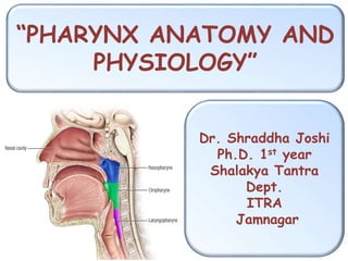
- 1. “PHARYNX ANATOMY AND PHYSIOLOGY” Dr. Shraddha Joshi Ph.D. 1st year Shalakya Tantra Dept. ITRA Jamnagar
- 2. Muscular tube. Situated behind Nose, Mouth and Larynx. Nasopharynx- Situated behind nose and above lower border of soft palate. Oropharynx- Middle part of pharynx. Hypopharynx- behind larynx from upper part of the epiglottis to lower border of cricoid cartilage. Pharynx
- 5. Naso- Pharynx Situated behind the nose and above the soft palate. Bony landmark is Hard palate- at C1 level. Rigid wall- Pharyngobasilar fascia and posterior median pharyngeal ligament. Anteriorly- Communicates with conchae. Inferiorly- Communicates with oropharynx at pharyngeal isthmus. Nasopharyngeal Isthmus- Passavant’s muscle. (Lower border of Soft palate & posterior wall of pharynx.
- 6. Lateral wall: • Pharyngeal opening of the eustachian tube. • Tubal elevation bounds the tubal opening. • Salpingopharyngeal fold. • Leveator palatani. • Fossa of Rosenmuller. Roof and Posterior wall: • Continuous slope, opposite the posterior part of body of sphenoid, basiocciput and anterior arch of atlas. • Adenoids- In the roof and the posterior wall of lymphoid tissue.
- 9. Oro-Pharynx Situated behind oral cavity Extend from the level of the soft palate to the level of the hyoid bone. Superiorly- communicates to the nasopharynx through the pharyngeal isthmus. Anteriorly- It communicates with the oral cavity Posteriorly- opposite to C2 and C3. Lateral wall- Palatine tonsils and anterior & posterior pillars. Inferior border- Hyoid bone.
- 11. Hypo- Pharynx Also known as Laryngopharynx. Situated behind the larynx. Extends from upper border of epiglottis/ level of hyoid bone to the lower border of cricoid cartilage. Anterior wall- • Inlet of larynx. • Posterior surface of cricoid and arytenoid cartilages. Posterior wall- • 4th and 5th cervical vertebrae.
- 12. Lateral wall- • Pyriform fossa, one on each side of the inlet of the larynx.
- 13. Pharyngeal wall 1. Mucosa- Lined by squamous epithelium except the nasopharyx (Nasopharynx- ciliated columnar epithelium) 2. Submucosa 3. Pharyngobasilar fascia- • Fibrous sheet deep to the pharyngeal muscle. • Fills the gap between upper border of superior constrictor and base of skull.
- 14. 4. Muscular coat- • Consist of outer circular layer made of 3 constrictors. • Inner longitudinal layer made up of Stylopharyngeus, Salpingopharyngeus, Palatopharyngeus muscles. 5. Buccopharyngeal fascia- • Covers the outer surface of the constrictors. • Below the buccopharyngeal fascia, on the muscular coat there lie the pharyngeal plexus of veins and nerves.
- 17. External Carotid Artery- Ascending pharyngeal branch. Facial artery- Ascending palatine & tonsillar branches. Lingual Artery- Dorsal lingual branches. Maxillary Artery- Greater Palatine, Pharyngeal , Pterygoid branches . Blood Supply
- 19. Venous Drainage
- 20. Lymphatic Drainage Retropharyngeal Lymphnodes. Deep Cervical Lymphnodes.
- 21. Nerve Supply Supplied by pharyngeal plexus of nerves chiefly on the middle constrictor. Plexus is formed by- • Pharyngeal branch of vagus (cranial accessory). • Pharyngeal branch of glossopharyngeal. • Pharyngeal branches of superior cervical sympathetic ganglion.