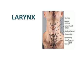
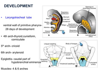
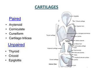
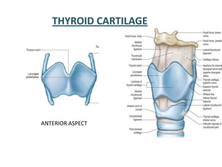
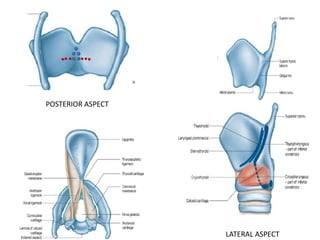
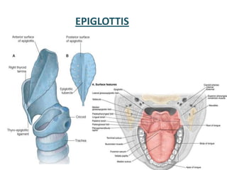
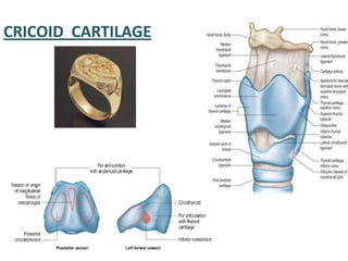
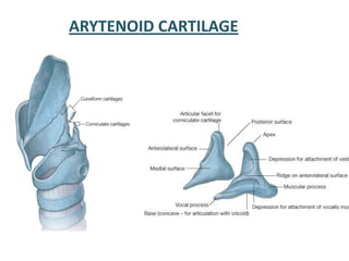
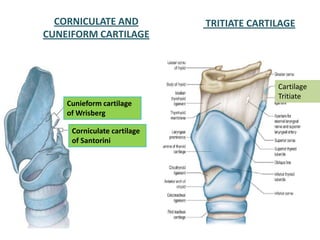
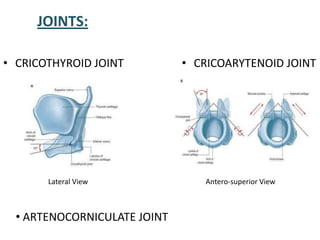
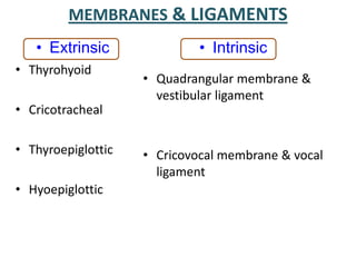
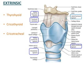
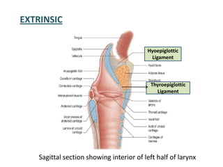
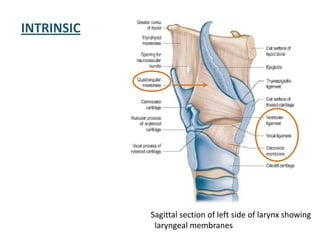
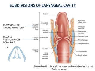
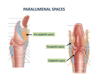
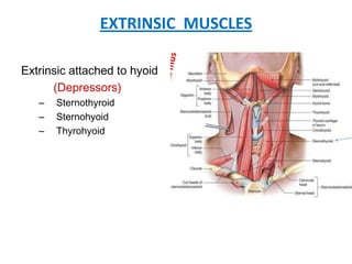
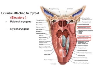
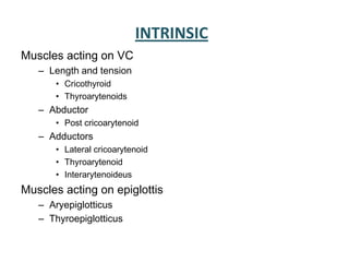
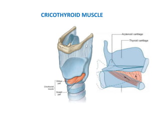
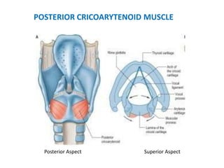
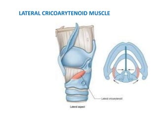
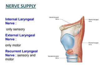
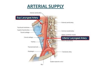
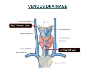
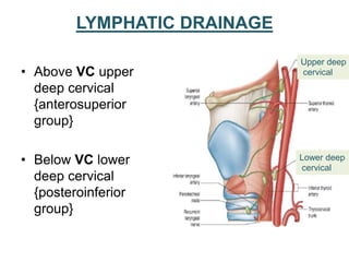
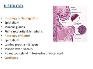
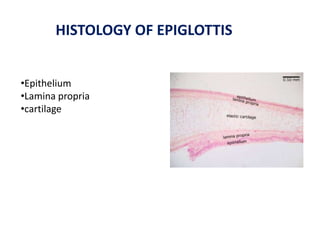
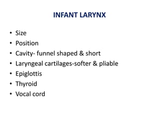
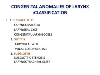
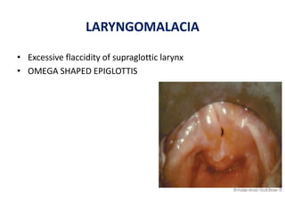
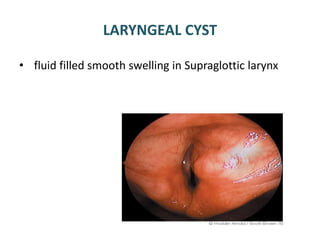
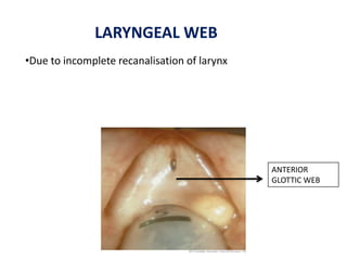
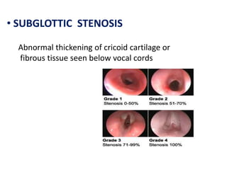
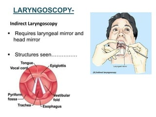
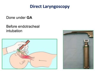
LARYNX
• INTRODUCTION
• DEVELOPMENT OF LARYNX
• SKELETAL FRAMEWORK
• SUBDIVISON OF LARYNX
• MUSCLES
• HISTOLOGY
• BLOOD SUPPLY
• NERVE SUPPLY
• LYMPHATIC DRAINAGE
• APPLIED ANATOMY
SITUATION AND EXTENT
MEASURMENTS-
Male larynx V=44mm,T=43 mm,AP=36mm
Female larynx V=36mm,T=41 mm,AP=26mm
DEVELOPMENT
• Laryngotracheal tube
ventral wall of primitive pharynx-
28 days of development
• 4th arch-thyroid,cunieform,
corniculate
5th arch- cricoid
6th arch- arytenoid
Epiglottis- caudal part of
hypobranchial eminence
Muscles- 4 & 6 arches
CARTILAGES
Paired
• Arytenoid
• Corniculate
• Cuneiform
• Cartilago triticea
Unpaired
• Thyroid
• Cricoid
• Epiglottis
5
THYROID CARTILAGE
ANTERIOR ASPECT
POSTERIOR ASPECT
LATERAL ASPECT
EPIGLOTTIS
CRICOID CARTILAGE
ARYTENOID CARTILAGE
CORNICULATE AND
CUNEIFORM CARTILAGE
Corniculate cartilage
of Santorini
Cunieform cartilage
of Wrisberg
Cartilage
Tritiate
TRITIATE CARTILAGE
JOINTS:
Lateral View Antero-superior View
• CRICOTHYROID JOINT • CRICOARYTENOID JOINT
• ARTENOCORNICULATE JOINT
MEMBRANES & LIGAMENTS
• Extrinsic
• Thyrohyoid
• Cricotracheal
• Thyroepiglottic
• Hyoepiglottic
• Intrinsic
• Quadrangular membrane &
vestibular ligament
• Cricovocal membrane & vocal
ligament
EXTRINSIC
• Thyrohyoid
• Cricothyroid
• Cricotracheal
Sagittal section showing interior of left half of larynx
EXTRINSIC
Hyoepiglottic
Ligament
Thyroepiglottic
Ligament
INTRINSIC
Sagittal section of left side of larynx showing
laryngeal membranes
Coronal section through the larynx and cranial end of trachea
Posterior aspect
SUBDIVISIONS OF LARYNGEAL CAVITY
LARYNGEAL INLET
ARYEPIGLOTTIC FOLD
SACCULE
VESTIBULAR FOLD
VOCAL FOLD
PARALUMENAL SPACES
Pre-epiglottic space
Paraglottic space
Subglottic space
EXTRINSIC MUSCLES
Extrinsic attached to hyoid
(Depressors)
– Sternothyroid
– Sternohyoid
– Thyrohyoid
Extrinsic attached to thyroid
(Elevators )
– Palatopharyngeus
– stylopharyngeus
INTRINSIC
Muscles acting on VC
– Length and tension
• Cricothyroid
• Thyroarytenoids
– Abductor
• Post cricoarytenoid
– Adductors
• Lateral cricoarytenoid
• Thyroarytenoid
• Interarytenoideus
Muscles acting on epiglottis
– Aryepiglotticus
– Thyroepiglotticus
CRICOTHYROID MUSCLE
Posterior Aspect Superior Aspect
POSTERIOR CRICOARYTENOID MUSCLE
LATERAL CRICOARYTENOID MUSCLE
OBLIQUE ARYTENOID MUSCLE and ARYEPIGLOTTICUS
TRANSVERSE ARYTENOID MUSCLE
THYROARYTENOID and VOCALIS MUSCLE
SHAPE OF RIMA GLOTTIDIS DURING DIFFERENT MOVEMENTS
OF VOCAL CORDS
DURING PHONATION DURING WHISPERING
NERVE SUPPLY
Internal Laryngeal
Nerve :
only sensory
External Laryngeal
Nerve :
only motor
Recurrent Laryngeal
Nerve : sensory and
motor
ARTERIAL SUPPLY
Sup Laryngeal Artery
Inferior Laryngeal Artery
VENOUS DRAINAGE
Sup Thyroid Vein
Inf Thyroid Vein
LYMPHATIC DRAINAGE
• Above VC upper
deep cervical
{anterosuperior
group}
• Below VC lower
deep cervical
{posteroinferior
group}
Upper deep
cervical
Lower deep
cervical
HISTOLOGY
• Histology of Supraglottis
• Epithelium
• Mucous glands
• Rich vascularity & lymphatic
• Histology of Glottis
• Epithelium
• Lamina propria – 3 layers
• Muscle layer- vocalis
• No mucous gland in free edge of vocal cord
• Cartilages
HISTOLOGY OF EPIGLOTTIS
•Epithelium
•Lamina propria
•cartilage
INFANT LARYNX
• Size
• Position
• Cavity- funnel shaped & short
• Laryngeal cartilages-softer & pliable
• Epiglottis
• Thyroid
• Vocal cord
APPLIED ANATOMY
CONGENITAL ANOMALIES OF LARYNX
:CLASSIFICATION
• 1. SUPRAGLOTTIS
LARYNGOMALACIA
LARYNGEAL CYST
CONGENITAL LARYNGOCELE
2. GLOTTIS
LARYNGEAL WEB
VOCAL CORD PARALYSIS
3. SUBGLOTTIS:
SUBGLOTTIC STENOSIS
LARYNGOTRACHEAL CLEFT
LARYNGOMALACIA
• Excessive flaccidity of supraglottic larynx
• OMEGA SHAPED EPIGLOTTIS
LARYNGEAL CYST
• fluid filled smooth swelling in Supraglottic larynx
LARYNGOCELE
Air filled dialatation of saccule
ANTERIOR
GLOTTIC WEB
•Due to incomplete recanalisation of larynx
LARYNGEAL WEB
• SUBGLOTTIC STENOSIS
Abnormal thickening of cricoid cartilage or
fibrous tissue seen below vocal cords
 Requires laryngeal mirror and
head mirror
 Structures seen……………
LARYNGOSCOPY-
Indirect Laryngoscopy
Done under GA
Before endotracheal
intubation
Direct Laryngoscopy
SINGERS /SCREAMERS/CLERGYMENS
NODULES
REINKE’S EDEMA
CLASSIFICATION OF LARYNGEAL PARALYSIS
• May be unilateral or
bilateral and may involve
1. Recurrent laryngeal nerve
2. Superior laryngeal nerve
a. External LN
b. Internal LN
LARYNGEAL OBSTRUCTION
LARYNGOTOMY
INFLAMMATION OF LARYNX
MALIGNANCY OF LARYNX
• Supraglottic
• Glottic
• Subglottic
Larynx

Larynx