2. PHARYNX
• The Nasal part-
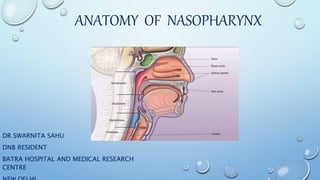
NASOPHARYNX/EPIPHARYNX:exte
nds from the base of the skull to
the soft palate.
• The Oral part-OROPHARYNX-
extends from the hard palate to
the hyoid bone.
• The laryngeal part-
LARYNGOPHARYNX/HYPOPHARYN
X-extends from the upper border
of epiglottis to lower border of
3. NASOPHARYNX
• UPPERMOST PART OF THE PHARYNX.
• CUBOIDAL CHAMBER.
• TRANSVERSE DIMENSION IS SLIGHTLY
BROADER THAN THE ANTERIOR-
POSTERIOR DIMENSION.
• LIES BEHIND THE NASAL CAVITY.
• HAS A PURE RESPIRATORY FUNCTION.
• LINED BY RESPIRATORY EPITHELIUM
(PSEUDOSTARTIFIED CILIATED
COLUMNAR EITHELIUM).
• SOFT PALATE AND UVULA CLOSES
NASOPHARYNX DURING
SWALLOWING AND PREVENTS
REGURGITATION OF FOOD.
4. PASSAVANT’S RIDGE
• It is the mucosal ridge raised by
fibres of palatopharyngeus.
• It encircles posterior and lateral
walls of nasopharyngeal isthmus.
• Soft palate during its contraction
makes firm contact with this ridge
to cut off nasopharynx from
oropharynx during deglutition or
speech.
9. SINUS OF MORGAGNI
• THE SUPERIOR CONSTRICTOR MUSCLE EXTENDS
SUPERIORLY TO THE SKULL BASE BUT ONLY IN THE
MIDLINE.
• LATERALLY THE PHARYNGOBASILAR FASCIA SERVES
TO ATTACH THE CONSTRICTOR MUSCLE TO THE
BASE OF SKULL AT THE BASIOCCIPUT AND PETROUS
PORTION OF THE TEMPORAL BONE.
• THIS LATERAL AREA OF MUSCULAR DEFICIENCY IS
THE SINUS OF MORGAGNI.
10. IMPORTANT STRUCTURES BETWEEN THE CONSTRICTORS
SPACE ORGAN MUSCLE ARTERY NERVE
SINUS OF
MORGAGNI
EUSTACHIAN
TUBE
LEVATOR PALITINI
TENSOR PALITINI
ASCENDING
PALATINE ARTERY
SUPERIOR AND
MIDDLE
STYLOPHARYNGE
US
GLOSSOPHARYNG
EAL
MIDDLE AND
INFERIOR
SUPERIOR
LARYNGEAL
INTERNAL
LARYNGEAL
BELOW INFERIOR INFERIOR
LARYNGEAL
RECURRENT
LARYNGEAL
11. • THE PHARYNGOBASILAR FASCIA IS CONTINOUS WITH THE FORAMEN LACERUM
AND IS IN CLOSE PROXIMITY TO THE-
• FORAMEN OVALE
• FORAMEN SPINOSUM
• JUGULAR FORAMEN
• HYPOGLOSSAL CANAL
• CAROTID SPACE
RESPONSIBLE FOR THE INTRACRANIAL
EXTENTION OF THE TUMOR OF
NASOPHARYNX.
12. FORAMINS-
FORAMEN/FIS
SURES
CN OTHER
NERVES
ARTERY VEIN
CRIBRIFORM
PLATE OF
ETHMOID
I ANTERIOR
ETHMOIDAL
OPTIC
FORAMEN
II OPTHALMIC
ARTERY
SUPERIOR
ORBITAL
FISSURE
III, IV, V1, VI SYMPATHETIC
PLEXUS
FILAMENTS
OF CAROTID
PLEXUS
• MIDDLE
MENINGEA
L ARTERY
• LACRIMAL
ARTERY
OPTHALMIC
VEIN
FORAMEN
ROTUNDUM
V2
FORAMEN
OVALE
V3 LESSER
SUPRFICIAL
PETROSAL
NERVE
ACCESSORY
MENINGEAL
ARTERY
FORAMEN
LACERUM
• INTERNAL
CAROTID
ART
• MENINGEA
L BR OF
ASC
13. FORAMINS/FI
SSURES
CRANIAL
NERVE
OTHER
NERVES
ARTERY VEIN
FORAMEN
SPINOSUM
RECURRENT
BR OF V3
MIDDLE
MENINGEAL
ART
MIDDLE
MENINGEAL
VEIN
STYLOMASTO
ID FORAMEN
VII
INTERNAL
ACOUSTIC
MEATUS
VIII INTERNAL
AUDITORY
ARTERY
JUGULAR
FORAMEN
IX X XI • MENINGEA
L BRNS
FROM
OCCIPITAL
AND ASC
PHARYNGE
AL
ARTERIES
• INFERIOR
PETROSAL
SINUS
• TEMPORAL
SINUS
HYPOGLOSSA
L CANAL
XII SPINAL CORD
SPINAL ACC
NERVE
• MENINGEA
L BR OF
ASC
PHARYNGE
AL ART
• VERTEBRA
L VESSELS
• ANT AND
14. NERVE SUPPLY
• SENSORY-
• ANTERIOR TO AUDITORY TUBE ORIFICE : V2 CN.
• POSTERIOR TO AUDITORY TUBE ORIFICE : GLOSSOPHARYNGEAL NERVE.
• MOTOR-
• IX CN
• X CN
• SYMPATHETIC FIBRES FROM THE SUPERIOR CERVICAL GANGLION
15. BLOOD SUPPLY
• ARTERIAL:
• ASCENDING PHARYNGEAL ARTERY
• SPHENOPALATINE ARTERY
• ARTERY TO THE PTERYGOID CANAL
• VENOUS:
• PHARYNGEAL PLEXUS TO INTERNAL JUGULAR VEINS (DIRECTLY OR VIA PTRYGOID
PLEXUS)
16. LYMPHATIC DRAINAGE
• 2 major lymphatics-
• Along posterior wall of pharynx along lateral wall of
pharynx
First node of the retropharyngeal lateral
pharyngeal node
Group jugulodigastric
node
3rd 4th 5th node
of
retropharyngeal grp
18. ANTERIORLY:
• NASAL CAVITY.
• PTERYGOID PLATES OF SPHENOID
(THROUGH LATERAL WALL OF
NASAL CAVITY).
• PTERYGOPALTINE FOSSA.
• PNS ( MAINLY POSTERIOR
ETHMOID AND MAXILLARY SINUS.)
• ORBITAL APEX (THRO INFERIOR
ORBITAL FISSURE).
19. • CONE-SHAPED DEPRESSION DEEP TO THE INFRATEMPORAL FOSSA
IT IS THE INDENTED AREA MEDIAL TO THE PTERYGOMAXILLARY FISSURE
20. POSTERIORLY
• PREVERTEBRAL MUSCLES.
• RETROPHARYNGEAL SPACE-
POSTERIOR TO PHARYNX AND
OESOPHAGUS
ANTERIOR TO ALAR FASCIA
SKULL BASE TO T1-T2
TWO CHAINS OF LYMPH NODES
ON EITHER SIDE OF THE MIDLINE
22. SUPERIORLY
• BASE OF SKULL
• SPHENOID SINUS
• CLIVUS
• CAVERNOUS SINUS
• MIDDLE CRANIAL FOSSA
• PETROUS PORTION OF TEMPORAL
BONE
23. The base of the skull (or skull base)
forms the floor of the cranial cavity
and separates the brain from the
structures of the neck and face.
The middle cranial fossa, deeper than
the anterior cranial fossa, is narrow
medially and widens laterally to the
sides of the skull. It is separated from
the posterior fossa by the clivus and
the petrous crest. It is bounded in
front by the posterior margins of the
lesser wings of the sphenoid bone
24.
25. CAVERNOUS SINUS
• A large channel of
venous blood creating a
"sinus" cavity bordered
by the sphenoid bone
and the temporal bone
of the skull.
• The cavernous sinus is
an important structure
because of its location
and its contents which
include the third cranial
(oculomotor) nerve, the
fourth cranial (trochlear)
nerve, parts 1 (the
ophthalmic nerve) and 2
(the maxillary nerve) of
the fifth cranial
(trigeminal) nerve, and
the sixth cranial
(abducens) nerve.