SlideShare a Scribd company logo
1 of 28
Temporal & Infratemporal Regions -I
Dr. Prabhakar Yadav
Associate Professor
Department of Human Anatomy
B.P. Koirala Institute of Health Sciences
Teporal Fossa:
Boundaries:
Anterior: Zygomatic & Frontal bones
Posterior: Inferior temporal line &
supramastoid crest
Superior: Superior temporal line
Inferior: Zygomatic arch
Floor: Parts of frontal, Parietal, Temporal &
greater wing of Sphenoid
Pterion: lies in anterior part of floor where
frontal, parietal, squamous temporal and
greater wing of sphenoid meet at an H-shaped
suture.
Zygomatic arch: formed
Anteriorly by-- Temporal process of zygomatic bone &
Posteriorly by-- Zygomatic process or zygoma of
temporal bone.
Contents:
• Temporal fascia
•Temporalis muscle
( attached to floor & inferior
temporal line)
•Middle temporal artery ( branch of
superficial temporal art.)
•Zygomaticotemopral nerve &
artery
•Deep temporal nerve
•Deep temporal artery
•Deep temporal nerve
Zygomaticotemopral nerve
Middle temporal artery
Infratemporal fossa: Lies below the skull, behind the
body of maxilla & lateral to lateral pterygoid plate.
Boundaries:
•Anterior: Posterior surface of body of maxilla &
medial surface of Zygomatic bone.
•Roof: Infra temporal surface of greater wing of
sphenoid.
•Medial: Lateral pterygoid plate & pyramidal process
of palatine bone
•Lateral: Ramus of mandible
• Floor & posterior wall: Open
Temporal fossa communicates with infratemporal
fossa through a gap deep to zygomatic arch.
Contents:
1. Lateral pterygoid muscle; Medial pterygoid muscle
2. Mandibular nerve & its branches
3. Maxillary nerve with post. Superior alveolar nerve
4. Chorda tympani nerve
5. 1st & 2nd part of maxillary artery with their branches
6. Posterior superior alveolar artery( br. Of 3rd part of maxillary artery)
7. Accompanying veins.
MUSCLES OF MASTICATION:
Masseter, Temporalis, Lateral & Medial pterygoid
develop from first branchial arch,-supplied
by mandibular nerve
Temporalis:
Origin: (a)Temporal fossa
(b) Temporal Fascia
Fibres pass through gap deep to zygomatic arch
Insertion:
(a) Margins and deep surface of coronoid process
(b) Anterior border of ramus of mandible
Nerve supply: Two deep temporal nerve- branches
from anterior division of mandibular nerve
Actions: (a) Elevates mandible (b) Posterior fibres
retract the protruded mandible (c) Helps in side
to side grinding movement
Masseter:
Origin:
(a) Superficial ayer- Anterior 2/3 of lower border of
zygomatic arch & adjoining zygomatic process of
maxilla
(b) Middle layer: posterior 1/3rd of lower border of
zygomatic arch
(c) Deep layer: from deep surface of zygomatic arch
Insertion:
(a) superficial layer: lower part of lateral surface of
ramus of mandible
(b) Middle layer: central part of ramus of mandible
(c )Deep layer: upper part of the ramus of mandible &
coronoid process
Nerve supply: Masseteric Nerve
Action:
•Elevates mandible to close mouth
• superficial fibres - protrusion
Lateral Pterygoid:
Origin:
upper (small) head: infratemporal surface & crest of
greater wing of sphenoid
Lower (large) head: lateral surface of lateral pterygoid
plate
Insertion:
Pterygoid fovea, Articular disc & capsule of
temporomandibular joint.
Nerve supply: nerve to latral pterygoid- branch of
anterior division of mandibular nerve.
Actions
1. Lateral pterygoids of two sides depress the mandible
2. Lateral and medial pterygoid muscles of two sides acting together protrude the mandible.
3. Lateral and medial pterygoid muscles of the two sides contract alternately to produce side-to-side
movements of the lower jaw as in chewing.
Medial Pterygoid:
Origin:
Superficial (small) head: Maxillary tuberosity & lateral
surface of pyramidal process of palatine bone
Deep (large) head: Medial surface of lateral pterygoid
plate & pyramidal process of palatine bone.
Insertion: Roughened area on posteroinferior part of
medial surface & angle of ramus of mandible.
Nerve supply: nerve to medial pterygoid
Actions
1.Medial pterygoids of two sides elevates mandible
2.Acting with lateral pterygoids protrude mandible
3. Lateral and medial pterygoids of two sides contract
alternately to produce side-to-side movements
Temporal fascia:
Thick aponeurotic sheet- covers temporalis muscle.
Superiorly: single layered - attached to superior
temporal line.
Inferiorly: Two layers- attached to inner and outer
lips of upper border of the zygomatic arch .
Gap contains fat, Middle temporal artery- branch
from superficial temporal artery & zygomatico-
temporal nerve.
• superficial surface receives an expansion from
epicranial aponeurosis.
•Superficial surface gives origin to auricularis
anterior and superior.
•Deep surface gives origin to some fibres of
temporalis muscle.
Relations of lateral pterygoid:
key muscle of infratemporal region
Superficial relation :
1. Masseter.
2. Ramus of the mandible.
3. Tendon of temporalis.
4. Superficial head of medial pterygoid.
5. Maxillary artery and its temporal &
masseteric branches.
Deep:
1. Mandibular nerve.
2. Middle meningeal artery.
3. Sphenomandibular ligament.
4. Deep head of medial pterygoid muscle.
Pterygoid plexus of veins surrounds the
lateral pterygoid.
Structures emerging at upper border:
1. Deep temporal nerves (two in number).
2. Masseteric nerve
Structures emerging at lower border:
1. Inferior alveolar nerve and artery.
2. Lingual nerve.
3. Middle meningeal artery (it passes up deep to the
lower border).
Structures passing through gap between two heads:
1. Maxillary artery- enter to reach pterygopalatine
fossa through pterygomaxillary fissure.
2. Buccal branch of mandibular nerve- leave to
provide sensory innervation to skin & mucus
membrane of the cheek.
Relations of Medial pterygoid:
Superficial :
1. Lingual nerve.
2. Inferior alveolar nerve.
3. Inferior alveolar vessels.
4. Lateral pterygoid plate
Deep:
1. Tensor veli palatini & Levator veli palatini
2. Superior constrictor of pharynx.
3. Styloglossus and stylopharyngeus
muscles.
MAXILLARY ARTERY:
arises behind neck of mandible.
Supplies: (a) Dura mater (b) External & Middle ears &
auditory tube; (c) upper and lower jaws; (d)Muscles
Of temporal and infratemporal regions; (e) Nose &
paranasal air sinuses; (f) Palate & (g) Root of pharynx
Course and Relations:
•Divided into three parts by lateral pterygoid.
First (mandibular) part: runs horizontally, first between
neck of mandible & sphenoniandibular ligament, and
then along lower border of lateral pterygoid.
second (pterygoid) part: runs upwards & forwards
superficial to lower head of lateral pterygoid
Third (pterygopalatine) part: passes between two
heads of lateral pterygoid & through pterygomaxillary
fissure, to enter pterygopalatine fossa.
Branches from the First Part:
1. Deep auricular artery- supplies:
(a) skin of external acoustic meatus, and
(b) outer surfaces of tympanic membrane.
(c) TMJ
2. Anterior tympanic artery- supplies
inner surface of the tympanic membrane & middle
ear
3. Middle meningeal artery-
Supplies meninges as well as the skull bone
-Artery ascends upwards deep to lateral pterygoid
- Enter cranial cavity through foramen spinosum.
-on the greater wing of sphenoid, it divides into frontal
and parietal branches.
-Middle meningeal artery and its branches lie outside
dura and deep to inner surface of skull. Both are
supplied by artery.
4. Accessory middle meningeal artery: supplies
meninges & structures in infratemporal fossa.
5. Inferior alveolar/dental artery- supplies
Molar & premolar teeth and adjoining gum.
Before entering the mandibular foramen, inferior
alveolar artery gives off two branches
(a) Lingual branch: accompanies lingual nerve -
supply mucous membrane of the cheek.
(b) Mylohyoid branch: supplies mylohyoid muscle.
It also gives off mental and incisive branches.
Incisive branch: supplies canine and incisor
Mental branch: supply skin of chin
Branches from Second Part
Deep temporal arteries (usually two in number):
supply- temporalis muscle
Pterygoid branches
Supply- medial & lateral pterygoid muscles.
Masseteric artery—supplies the masseter muscle.
Buccal artery —supplies buccinator muscle
Branches from Third Part
Posterior superior alveolar artery:
Supply- Molar & premolar teeth & mucus membrane
of maxillary air sinus.
Infraorbital artery: gives the following branches:
In the orbit:
(a) Some branches to orbital contents.
(b) Middle superior alveolar artery to premolar teeth.
(c) Anterior superior alveolar artery - supplies-
maxillary air sinus ,canine & incisor of upper jaw.
In the face:
Branches to supply lacrimal sac, medial angle of eye,
side of nose & upper lip.
Greater palatine artery:
•supplies roof of the mouth & adjoining gum,
•In greater palatine canal artery gives off lesser palatine arteries that emerge through lesser palatine
foramina and supply soft palate & tonsil
 Pharyngeal artery:
Supplies- mucus membrane of nasopharynx, auditory
tube, sphenoidal air sinus.
 Artery of pterygoid canal:
supplies - pharynx, auditory tube & tympanic cavity.
Sphenopalatine artery: continuation of maxillary art.
Enters nasal cavity in posterior part of superior meatus
through sphenopalatine foramen.
Here it divides into:
(a) posterior lateral nasal branch
Supply- lateral wall of nose;
Sphenoidal & ethmoidal air sinuses
(b) posterior septal/medial branches
Supply- nasal septum
Sphenopalatine artery- artery of epistaxis
Pterygoid Plexus of Veins:
Lies around and within lateral pterygoid muscle
Tributaries of plexus correspond to branches of
maxillary artery
Plexus is drained by maxillary vein(formed at
lower border of lateral pterygoid muscle) which
unites with superficial temporal vein to form
retromandibular vein.
Maxillary vein accompanies only first part of maxillary
artery.
 pterygoid venous plexus communicates with:
(a) Inferior ophthalmic vein via inferior orbital fissure,
(b)cavernous sinus by emissary veins via foramen
Ovale.
(c) Facial vein through deep facial vein
PTERYGOPALATINE FOSSA:
Pyramidal Space between body of maxilla & root of
pterygoid process, lateral to perpendicular plate of
palatine bone.
BOUNDARIES:
Anterior: superomedial part of posterior surface of
maxilla .
Posterior: Root of Pterygoid process and adjoining
part of anterior surface of the greater wing of
sphenoid.
Medial: Upper part of perpendicular plate of
palatine. orbital and sphenoidal process of palatine.
Lateral: Fossa opens into infratemporal fossa
through pterygomaxillary fissure.
Superior: Under surface of body of sphenoid
Inferior: Pyramidal process of the palatine bone (in
the angle between maxilla and pterygoid process)
COMMUNICATIONS:
Anteriorly:
With orbit through medial end of inferior orbital
fissure.
Posteriorly:
1. With middle cranial fossa via foramen rotundum.
2. With foramen lacerum via pterygoid canal.
3. With pharynx via palatovaginal(pharyngeal)
canal.
Medially:With nose through sphenopalatine
foramen.
Laterally: With infratemporal fossa via
Pterygomaxillary fissure.
Inferiorly: With oral cavity via greater palatine
canals
CONTENTS:
1. Maxillary nerve.
2. Pterygopalatine ganglion.
3. Third part of the maxillary artery.
THANK YOU

More Related Content

What's hot

anatomy - Scalp + face
anatomy - Scalp + faceanatomy - Scalp + face
anatomy - Scalp + face
MBBS IMS MSU
 
Pterygopalatine ganglion 1
Pterygopalatine ganglion 1Pterygopalatine ganglion 1
Pterygopalatine ganglion 1
Omar Eraky
 
Blood supply of face
Blood supply of faceBlood supply of face
Blood supply of face
1423262214
 

What's hot (20)

Posterior triangle of the neck
Posterior triangle of the neckPosterior triangle of the neck
Posterior triangle of the neck
 
Facial artery dr gosai
Facial artery dr gosaiFacial artery dr gosai
Facial artery dr gosai
 
Parasympathetic ganglia of Head and Neck
 Parasympathetic ganglia of Head and Neck Parasympathetic ganglia of Head and Neck
Parasympathetic ganglia of Head and Neck
 
Mandibular nerve and otic ganglion
Mandibular nerve and otic ganglionMandibular nerve and otic ganglion
Mandibular nerve and otic ganglion
 
Carotid sheath
Carotid sheathCarotid sheath
Carotid sheath
 
Pharynx Dr.N.Mugunthan.M.S.,
Pharynx  Dr.N.Mugunthan.M.S.,Pharynx  Dr.N.Mugunthan.M.S.,
Pharynx Dr.N.Mugunthan.M.S.,
 
DEEP CERVICAL FASCIA.pptx
DEEP CERVICAL FASCIA.pptxDEEP CERVICAL FASCIA.pptx
DEEP CERVICAL FASCIA.pptx
 
The hyoglossal muscle
The hyoglossal muscleThe hyoglossal muscle
The hyoglossal muscle
 
anatomy - Scalp + face
anatomy - Scalp + faceanatomy - Scalp + face
anatomy - Scalp + face
 
Pterygopalatine ganglion 1
Pterygopalatine ganglion 1Pterygopalatine ganglion 1
Pterygopalatine ganglion 1
 
Suboccipital triangle
Suboccipital triangleSuboccipital triangle
Suboccipital triangle
 
Blood supply of face
Blood supply of faceBlood supply of face
Blood supply of face
 
Suboccipital triangle
Suboccipital triangleSuboccipital triangle
Suboccipital triangle
 
Soft Palate
Soft PalateSoft Palate
Soft Palate
 
FACIAL ARTERY.pptx
FACIAL ARTERY.pptxFACIAL ARTERY.pptx
FACIAL ARTERY.pptx
 
The digastric triangle
The digastric triangleThe digastric triangle
The digastric triangle
 
Blood supply and nerve supply to head and neck
Blood supply and nerve supply to head and neckBlood supply and nerve supply to head and neck
Blood supply and nerve supply to head and neck
 
Scalp
Scalp Scalp
Scalp
 
Cervical fascia
Cervical fasciaCervical fascia
Cervical fascia
 
Submandibular salivary gland
Submandibular salivary glandSubmandibular salivary gland
Submandibular salivary gland
 

Similar to Temporal & Infratemporal Region -I

INFRATEMPORAL FOSSA.pptx
INFRATEMPORAL FOSSA.pptxINFRATEMPORAL FOSSA.pptx
INFRATEMPORAL FOSSA.pptx
praveen4uly
 
ANATOMY OF NOSE & PARANASAL SINUSES.ppt
ANATOMY OF NOSE &  PARANASAL SINUSES.pptANATOMY OF NOSE &  PARANASAL SINUSES.ppt
ANATOMY OF NOSE & PARANASAL SINUSES.ppt
srinivaspennam88
 
anatomy of Tmj
anatomy of Tmjanatomy of Tmj
anatomy of Tmj
ddert
 

Similar to Temporal & Infratemporal Region -I (20)

Temporal & infratemporal regions II
Temporal & infratemporal regions IITemporal & infratemporal regions II
Temporal & infratemporal regions II
 
Anatomy of the Temporal region & Temporomandibular joint
Anatomy of the Temporal region & Temporomandibular jointAnatomy of the Temporal region & Temporomandibular joint
Anatomy of the Temporal region & Temporomandibular joint
 
Infratemporal fossa
Infratemporal fossaInfratemporal fossa
Infratemporal fossa
 
BLOOD SUPPLY OF HEAD & NECK.pptx
BLOOD SUPPLY OF HEAD & NECK.pptxBLOOD SUPPLY OF HEAD & NECK.pptx
BLOOD SUPPLY OF HEAD & NECK.pptx
 
Salivary Glands
Salivary Glands   Salivary Glands
Salivary Glands
 
Anatomy of Internal Nose
Anatomy of Internal Nose  Anatomy of Internal Nose
Anatomy of Internal Nose
 
Temporal & infratemporal region
Temporal & infratemporal regionTemporal & infratemporal region
Temporal & infratemporal region
 
Nasal cavity and Paranasal Air Sinuses
Nasal cavity and Paranasal Air Sinuses Nasal cavity and Paranasal Air Sinuses
Nasal cavity and Paranasal Air Sinuses
 
INFRATEMPORAL FOSSA.pptx
INFRATEMPORAL FOSSA.pptxINFRATEMPORAL FOSSA.pptx
INFRATEMPORAL FOSSA.pptx
 
Middle ear anatomy
Middle ear anatomyMiddle ear anatomy
Middle ear anatomy
 
ANATOMY OF NOSE & PARANASAL SINUSES.ppt
ANATOMY OF NOSE &  PARANASAL SINUSES.pptANATOMY OF NOSE &  PARANASAL SINUSES.ppt
ANATOMY OF NOSE & PARANASAL SINUSES.ppt
 
Anatomy(2) The skull and face The temporal region Infratemporal and pterygopa...
Anatomy(2) The skull and face The temporal region Infratemporal and pterygopa...Anatomy(2) The skull and face The temporal region Infratemporal and pterygopa...
Anatomy(2) The skull and face The temporal region Infratemporal and pterygopa...
 
lateralis and frontalis-lecture 2.pptx
lateralis and frontalis-lecture 2.pptxlateralis and frontalis-lecture 2.pptx
lateralis and frontalis-lecture 2.pptx
 
Pterygopalatine fossa.ppt
Pterygopalatine fossa.pptPterygopalatine fossa.ppt
Pterygopalatine fossa.ppt
 
Surgical anatomy of Infratemporal fossa. by Dr. Aditya Tiwari
Surgical anatomy of Infratemporal fossa. by Dr. Aditya TiwariSurgical anatomy of Infratemporal fossa. by Dr. Aditya Tiwari
Surgical anatomy of Infratemporal fossa. by Dr. Aditya Tiwari
 
INFRATEMPORAL FOSSA AND PTERYGOPALATINE FOSSA NEW.pptx
INFRATEMPORAL FOSSA AND PTERYGOPALATINE FOSSA NEW.pptxINFRATEMPORAL FOSSA AND PTERYGOPALATINE FOSSA NEW.pptx
INFRATEMPORAL FOSSA AND PTERYGOPALATINE FOSSA NEW.pptx
 
Dr Hoda _Hassan_Anatomy of EAR 20-21.pptx
Dr Hoda _Hassan_Anatomy of EAR 20-21.pptxDr Hoda _Hassan_Anatomy of EAR 20-21.pptx
Dr Hoda _Hassan_Anatomy of EAR 20-21.pptx
 
anatomy of Tmj
anatomy of Tmjanatomy of Tmj
anatomy of Tmj
 
Pharynx & Palate
Pharynx & PalatePharynx & Palate
Pharynx & Palate
 
Anatomy of Facial Nerve
Anatomy of Facial NerveAnatomy of Facial Nerve
Anatomy of Facial Nerve
 

More from Prabhakar Yadav

More from Prabhakar Yadav (20)

Larynx
Larynx  Larynx
Larynx
 
Diaphragm
Diaphragm   Diaphragm
Diaphragm
 
Lungs,Pleura & Tracheo-bronchial Tree
Lungs,Pleura & Tracheo-bronchial TreeLungs,Pleura & Tracheo-bronchial Tree
Lungs,Pleura & Tracheo-bronchial Tree
 
Medulla oblongata
Medulla oblongata Medulla oblongata
Medulla oblongata
 
Tongue
TongueTongue
Tongue
 
Lacrimal apparatus & nose
Lacrimal apparatus & noseLacrimal apparatus & nose
Lacrimal apparatus & nose
 
Functional cortical area & white matter of cerebrum
Functional cortical area & white matter of cerebrumFunctional cortical area & white matter of cerebrum
Functional cortical area & white matter of cerebrum
 
Cerebrum external features
Cerebrum external featuresCerebrum external features
Cerebrum external features
 
Knee joint
Knee joint   Knee joint
Knee joint
 
Popliteal fossa & back of thigh
Popliteal fossa & back of thigh  Popliteal fossa & back of thigh
Popliteal fossa & back of thigh
 
Bilaminar & trilaminar embryonic germ disc
Bilaminar & trilaminar embryonic germ discBilaminar & trilaminar embryonic germ disc
Bilaminar & trilaminar embryonic germ disc
 
Perineum & external genitalia
Perineum & external genitaliaPerineum & external genitalia
Perineum & external genitalia
 
Boundaries & contents of Axilla
Boundaries & contents of Axilla   Boundaries & contents of Axilla
Boundaries & contents of Axilla
 
Temperomandibular joint
Temperomandibular  jointTemperomandibular  joint
Temperomandibular joint
 
Pharyngeal arches & pouches
Pharyngeal arches & pouches  Pharyngeal arches & pouches
Pharyngeal arches & pouches
 
Duodenum & pancreas
Duodenum & pancreas   Duodenum & pancreas
Duodenum & pancreas
 
Development of Hepatobiliary System
Development of Hepatobiliary System Development of Hepatobiliary System
Development of Hepatobiliary System
 
Development of ear
Development of ear  Development of ear
Development of ear
 
Development of eye
Development of eye  Development of eye
Development of eye
 
Spinal cord
Spinal cord Spinal cord
Spinal cord
 

Recently uploaded

Spellings Wk 4 and Wk 5 for Grade 4 at CAPS
Spellings Wk 4 and Wk 5 for Grade 4 at CAPSSpellings Wk 4 and Wk 5 for Grade 4 at CAPS
Spellings Wk 4 and Wk 5 for Grade 4 at CAPS
AnaAcapella
 

Recently uploaded (20)

Interdisciplinary_Insights_Data_Collection_Methods.pptx
Interdisciplinary_Insights_Data_Collection_Methods.pptxInterdisciplinary_Insights_Data_Collection_Methods.pptx
Interdisciplinary_Insights_Data_Collection_Methods.pptx
 
Exploring_the_Narrative_Style_of_Amitav_Ghoshs_Gun_Island.pptx
Exploring_the_Narrative_Style_of_Amitav_Ghoshs_Gun_Island.pptxExploring_the_Narrative_Style_of_Amitav_Ghoshs_Gun_Island.pptx
Exploring_the_Narrative_Style_of_Amitav_Ghoshs_Gun_Island.pptx
 
This PowerPoint helps students to consider the concept of infinity.
This PowerPoint helps students to consider the concept of infinity.This PowerPoint helps students to consider the concept of infinity.
This PowerPoint helps students to consider the concept of infinity.
 
SOC 101 Demonstration of Learning Presentation
SOC 101 Demonstration of Learning PresentationSOC 101 Demonstration of Learning Presentation
SOC 101 Demonstration of Learning Presentation
 
NO1 Top Black Magic Specialist In Lahore Black magic In Pakistan Kala Ilam Ex...
NO1 Top Black Magic Specialist In Lahore Black magic In Pakistan Kala Ilam Ex...NO1 Top Black Magic Specialist In Lahore Black magic In Pakistan Kala Ilam Ex...
NO1 Top Black Magic Specialist In Lahore Black magic In Pakistan Kala Ilam Ex...
 
Basic Intentional Injuries Health Education
Basic Intentional Injuries Health EducationBasic Intentional Injuries Health Education
Basic Intentional Injuries Health Education
 
Jamworks pilot and AI at Jisc (20/03/2024)
Jamworks pilot and AI at Jisc (20/03/2024)Jamworks pilot and AI at Jisc (20/03/2024)
Jamworks pilot and AI at Jisc (20/03/2024)
 
On National Teacher Day, meet the 2024-25 Kenan Fellows
On National Teacher Day, meet the 2024-25 Kenan FellowsOn National Teacher Day, meet the 2024-25 Kenan Fellows
On National Teacher Day, meet the 2024-25 Kenan Fellows
 
Mehran University Newsletter Vol-X, Issue-I, 2024
Mehran University Newsletter Vol-X, Issue-I, 2024Mehran University Newsletter Vol-X, Issue-I, 2024
Mehran University Newsletter Vol-X, Issue-I, 2024
 
Python Notes for mca i year students osmania university.docx
Python Notes for mca i year students osmania university.docxPython Notes for mca i year students osmania university.docx
Python Notes for mca i year students osmania university.docx
 
HMCS Max Bernays Pre-Deployment Brief (May 2024).pptx
HMCS Max Bernays Pre-Deployment Brief (May 2024).pptxHMCS Max Bernays Pre-Deployment Brief (May 2024).pptx
HMCS Max Bernays Pre-Deployment Brief (May 2024).pptx
 
Wellbeing inclusion and digital dystopias.pptx
Wellbeing inclusion and digital dystopias.pptxWellbeing inclusion and digital dystopias.pptx
Wellbeing inclusion and digital dystopias.pptx
 
21st_Century_Skills_Framework_Final_Presentation_2.pptx
21st_Century_Skills_Framework_Final_Presentation_2.pptx21st_Century_Skills_Framework_Final_Presentation_2.pptx
21st_Century_Skills_Framework_Final_Presentation_2.pptx
 
Tatlong Kwento ni Lola basyang-1.pdf arts
Tatlong Kwento ni Lola basyang-1.pdf artsTatlong Kwento ni Lola basyang-1.pdf arts
Tatlong Kwento ni Lola basyang-1.pdf arts
 
OSCM Unit 2_Operations Processes & Systems
OSCM Unit 2_Operations Processes & SystemsOSCM Unit 2_Operations Processes & Systems
OSCM Unit 2_Operations Processes & Systems
 
Simple, Complex, and Compound Sentences Exercises.pdf
Simple, Complex, and Compound Sentences Exercises.pdfSimple, Complex, and Compound Sentences Exercises.pdf
Simple, Complex, and Compound Sentences Exercises.pdf
 
Spellings Wk 4 and Wk 5 for Grade 4 at CAPS
Spellings Wk 4 and Wk 5 for Grade 4 at CAPSSpellings Wk 4 and Wk 5 for Grade 4 at CAPS
Spellings Wk 4 and Wk 5 for Grade 4 at CAPS
 
FSB Advising Checklist - Orientation 2024
FSB Advising Checklist - Orientation 2024FSB Advising Checklist - Orientation 2024
FSB Advising Checklist - Orientation 2024
 
How to Create and Manage Wizard in Odoo 17
How to Create and Manage Wizard in Odoo 17How to Create and Manage Wizard in Odoo 17
How to Create and Manage Wizard in Odoo 17
 
Towards a code of practice for AI in AT.pptx
Towards a code of practice for AI in AT.pptxTowards a code of practice for AI in AT.pptx
Towards a code of practice for AI in AT.pptx
 

Temporal & Infratemporal Region -I

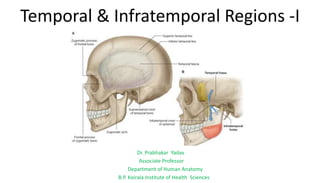
  • 1. Temporal & Infratemporal Regions -I Dr. Prabhakar Yadav Associate Professor Department of Human Anatomy B.P. Koirala Institute of Health Sciences
  • 2. Teporal Fossa: Boundaries: Anterior: Zygomatic & Frontal bones Posterior: Inferior temporal line & supramastoid crest Superior: Superior temporal line Inferior: Zygomatic arch Floor: Parts of frontal, Parietal, Temporal & greater wing of Sphenoid Pterion: lies in anterior part of floor where frontal, parietal, squamous temporal and greater wing of sphenoid meet at an H-shaped suture. Zygomatic arch: formed Anteriorly by-- Temporal process of zygomatic bone & Posteriorly by-- Zygomatic process or zygoma of temporal bone.
  • 3. Contents: • Temporal fascia •Temporalis muscle ( attached to floor & inferior temporal line) •Middle temporal artery ( branch of superficial temporal art.) •Zygomaticotemopral nerve & artery •Deep temporal nerve •Deep temporal artery •Deep temporal artery •Deep temporal nerve Zygomaticotemopral nerve Middle temporal artery
  • 4. Infratemporal fossa: Lies below the skull, behind the body of maxilla & lateral to lateral pterygoid plate. Boundaries: •Anterior: Posterior surface of body of maxilla & medial surface of Zygomatic bone. •Roof: Infra temporal surface of greater wing of sphenoid. •Medial: Lateral pterygoid plate & pyramidal process of palatine bone •Lateral: Ramus of mandible • Floor & posterior wall: Open Temporal fossa communicates with infratemporal fossa through a gap deep to zygomatic arch.
  • 5. Contents: 1. Lateral pterygoid muscle; Medial pterygoid muscle 2. Mandibular nerve & its branches 3. Maxillary nerve with post. Superior alveolar nerve 4. Chorda tympani nerve 5. 1st & 2nd part of maxillary artery with their branches 6. Posterior superior alveolar artery( br. Of 3rd part of maxillary artery) 7. Accompanying veins.
  • 6. MUSCLES OF MASTICATION: Masseter, Temporalis, Lateral & Medial pterygoid develop from first branchial arch,-supplied by mandibular nerve Temporalis: Origin: (a)Temporal fossa (b) Temporal Fascia Fibres pass through gap deep to zygomatic arch Insertion: (a) Margins and deep surface of coronoid process (b) Anterior border of ramus of mandible Nerve supply: Two deep temporal nerve- branches from anterior division of mandibular nerve Actions: (a) Elevates mandible (b) Posterior fibres retract the protruded mandible (c) Helps in side to side grinding movement
  • 7. Masseter: Origin: (a) Superficial ayer- Anterior 2/3 of lower border of zygomatic arch & adjoining zygomatic process of maxilla (b) Middle layer: posterior 1/3rd of lower border of zygomatic arch (c) Deep layer: from deep surface of zygomatic arch Insertion: (a) superficial layer: lower part of lateral surface of ramus of mandible (b) Middle layer: central part of ramus of mandible (c )Deep layer: upper part of the ramus of mandible & coronoid process Nerve supply: Masseteric Nerve Action: •Elevates mandible to close mouth • superficial fibres - protrusion
  • 8. Lateral Pterygoid: Origin: upper (small) head: infratemporal surface & crest of greater wing of sphenoid Lower (large) head: lateral surface of lateral pterygoid plate Insertion: Pterygoid fovea, Articular disc & capsule of temporomandibular joint. Nerve supply: nerve to latral pterygoid- branch of anterior division of mandibular nerve. Actions 1. Lateral pterygoids of two sides depress the mandible 2. Lateral and medial pterygoid muscles of two sides acting together protrude the mandible. 3. Lateral and medial pterygoid muscles of the two sides contract alternately to produce side-to-side movements of the lower jaw as in chewing.
  • 9. Medial Pterygoid: Origin: Superficial (small) head: Maxillary tuberosity & lateral surface of pyramidal process of palatine bone Deep (large) head: Medial surface of lateral pterygoid plate & pyramidal process of palatine bone. Insertion: Roughened area on posteroinferior part of medial surface & angle of ramus of mandible. Nerve supply: nerve to medial pterygoid Actions 1.Medial pterygoids of two sides elevates mandible 2.Acting with lateral pterygoids protrude mandible 3. Lateral and medial pterygoids of two sides contract alternately to produce side-to-side movements
  • 10. Temporal fascia: Thick aponeurotic sheet- covers temporalis muscle. Superiorly: single layered - attached to superior temporal line. Inferiorly: Two layers- attached to inner and outer lips of upper border of the zygomatic arch . Gap contains fat, Middle temporal artery- branch from superficial temporal artery & zygomatico- temporal nerve. • superficial surface receives an expansion from epicranial aponeurosis. •Superficial surface gives origin to auricularis anterior and superior. •Deep surface gives origin to some fibres of temporalis muscle.
  • 11. Relations of lateral pterygoid: key muscle of infratemporal region Superficial relation : 1. Masseter. 2. Ramus of the mandible. 3. Tendon of temporalis. 4. Superficial head of medial pterygoid. 5. Maxillary artery and its temporal & masseteric branches.
  • 12. Deep: 1. Mandibular nerve. 2. Middle meningeal artery. 3. Sphenomandibular ligament. 4. Deep head of medial pterygoid muscle. Pterygoid plexus of veins surrounds the lateral pterygoid.
  • 13. Structures emerging at upper border: 1. Deep temporal nerves (two in number). 2. Masseteric nerve Structures emerging at lower border: 1. Inferior alveolar nerve and artery. 2. Lingual nerve. 3. Middle meningeal artery (it passes up deep to the lower border). Structures passing through gap between two heads: 1. Maxillary artery- enter to reach pterygopalatine fossa through pterygomaxillary fissure. 2. Buccal branch of mandibular nerve- leave to provide sensory innervation to skin & mucus membrane of the cheek.
  • 14. Relations of Medial pterygoid: Superficial : 1. Lingual nerve. 2. Inferior alveolar nerve. 3. Inferior alveolar vessels. 4. Lateral pterygoid plate Deep: 1. Tensor veli palatini & Levator veli palatini 2. Superior constrictor of pharynx. 3. Styloglossus and stylopharyngeus muscles.
  • 15. MAXILLARY ARTERY: arises behind neck of mandible. Supplies: (a) Dura mater (b) External & Middle ears & auditory tube; (c) upper and lower jaws; (d)Muscles Of temporal and infratemporal regions; (e) Nose & paranasal air sinuses; (f) Palate & (g) Root of pharynx Course and Relations: •Divided into three parts by lateral pterygoid. First (mandibular) part: runs horizontally, first between neck of mandible & sphenoniandibular ligament, and then along lower border of lateral pterygoid. second (pterygoid) part: runs upwards & forwards superficial to lower head of lateral pterygoid Third (pterygopalatine) part: passes between two heads of lateral pterygoid & through pterygomaxillary fissure, to enter pterygopalatine fossa.
  • 16.
  • 17. Branches from the First Part: 1. Deep auricular artery- supplies: (a) skin of external acoustic meatus, and (b) outer surfaces of tympanic membrane. (c) TMJ 2. Anterior tympanic artery- supplies inner surface of the tympanic membrane & middle ear 3. Middle meningeal artery- Supplies meninges as well as the skull bone -Artery ascends upwards deep to lateral pterygoid - Enter cranial cavity through foramen spinosum. -on the greater wing of sphenoid, it divides into frontal and parietal branches. -Middle meningeal artery and its branches lie outside dura and deep to inner surface of skull. Both are supplied by artery.
  • 18. 4. Accessory middle meningeal artery: supplies meninges & structures in infratemporal fossa. 5. Inferior alveolar/dental artery- supplies Molar & premolar teeth and adjoining gum. Before entering the mandibular foramen, inferior alveolar artery gives off two branches (a) Lingual branch: accompanies lingual nerve - supply mucous membrane of the cheek. (b) Mylohyoid branch: supplies mylohyoid muscle. It also gives off mental and incisive branches. Incisive branch: supplies canine and incisor Mental branch: supply skin of chin
  • 19. Branches from Second Part Deep temporal arteries (usually two in number): supply- temporalis muscle Pterygoid branches Supply- medial & lateral pterygoid muscles. Masseteric artery—supplies the masseter muscle. Buccal artery —supplies buccinator muscle
  • 20. Branches from Third Part Posterior superior alveolar artery: Supply- Molar & premolar teeth & mucus membrane of maxillary air sinus. Infraorbital artery: gives the following branches: In the orbit: (a) Some branches to orbital contents. (b) Middle superior alveolar artery to premolar teeth. (c) Anterior superior alveolar artery - supplies- maxillary air sinus ,canine & incisor of upper jaw. In the face: Branches to supply lacrimal sac, medial angle of eye, side of nose & upper lip. Greater palatine artery: •supplies roof of the mouth & adjoining gum, •In greater palatine canal artery gives off lesser palatine arteries that emerge through lesser palatine foramina and supply soft palate & tonsil
  • 21.  Pharyngeal artery: Supplies- mucus membrane of nasopharynx, auditory tube, sphenoidal air sinus.  Artery of pterygoid canal: supplies - pharynx, auditory tube & tympanic cavity. Sphenopalatine artery: continuation of maxillary art. Enters nasal cavity in posterior part of superior meatus through sphenopalatine foramen. Here it divides into: (a) posterior lateral nasal branch Supply- lateral wall of nose; Sphenoidal & ethmoidal air sinuses (b) posterior septal/medial branches Supply- nasal septum Sphenopalatine artery- artery of epistaxis
  • 22. Pterygoid Plexus of Veins: Lies around and within lateral pterygoid muscle Tributaries of plexus correspond to branches of maxillary artery Plexus is drained by maxillary vein(formed at lower border of lateral pterygoid muscle) which unites with superficial temporal vein to form retromandibular vein. Maxillary vein accompanies only first part of maxillary artery.  pterygoid venous plexus communicates with: (a) Inferior ophthalmic vein via inferior orbital fissure, (b)cavernous sinus by emissary veins via foramen Ovale. (c) Facial vein through deep facial vein
  • 23.
  • 24.
  • 25. PTERYGOPALATINE FOSSA: Pyramidal Space between body of maxilla & root of pterygoid process, lateral to perpendicular plate of palatine bone. BOUNDARIES: Anterior: superomedial part of posterior surface of maxilla . Posterior: Root of Pterygoid process and adjoining part of anterior surface of the greater wing of sphenoid. Medial: Upper part of perpendicular plate of palatine. orbital and sphenoidal process of palatine. Lateral: Fossa opens into infratemporal fossa through pterygomaxillary fissure. Superior: Under surface of body of sphenoid Inferior: Pyramidal process of the palatine bone (in the angle between maxilla and pterygoid process)
  • 26. COMMUNICATIONS: Anteriorly: With orbit through medial end of inferior orbital fissure. Posteriorly: 1. With middle cranial fossa via foramen rotundum. 2. With foramen lacerum via pterygoid canal. 3. With pharynx via palatovaginal(pharyngeal) canal. Medially:With nose through sphenopalatine foramen. Laterally: With infratemporal fossa via Pterygomaxillary fissure. Inferiorly: With oral cavity via greater palatine canals
  • 27. CONTENTS: 1. Maxillary nerve. 2. Pterygopalatine ganglion. 3. Third part of the maxillary artery.