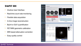
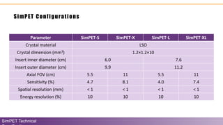
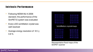
Download to read offline


![Oncologic PET/MRI with SUV (standard uptake value)
quantification
SimPET in Action
• [ 18F]FDG PET and T2
FSE MRI scans
conducted to
investigate the effects
of tumor associated
macrophages on
tumor hypoxia and
aerobic glycolysis
(Cancer Research,
2019)](https://image.slidesharecdn.com/brightonix-simpetv2-200722160542/85/SimPET-a-Preclinical-PET-Insert-for-Simultaneous-PET-MR-Imaging-38-320.jpg)
![High spatial-resolution PET/MR imaging in the mouse arthritis
model
SimPET in Action
• Simultaneously
acquired and matched
[18F]FDG PET (300
µCi, 60-min uptake)
and 3D GRE MR
images in arthritis
model mouse](https://image.slidesharecdn.com/brightonix-simpetv2-200722160542/85/SimPET-a-Preclinical-PET-Insert-for-Simultaneous-PET-MR-Imaging-39-320.jpg)
![Establishment of a [18F]-FDG-PET/MRI Imaging Protocol for
Gastric Cancer PDX as a Preclinical Research Tool
SimPET in Action
Bae, S. W., Berlth, F., Jeong, K. Y., Suh, Y. S., Kong, S. H., Lee, H. J., Kim, W. H., Chung, J. K., & Yang, H. K.
(2020). Establishment of a [18F]-FDG-PET/MRI Imaging Protocol for Gastric Cancer PDX as a Preclinical
Research Tool. Journal of gastric cancer, 20(1), 60–71.](https://image.slidesharecdn.com/brightonix-simpetv2-200722160542/85/SimPET-a-Preclinical-PET-Insert-for-Simultaneous-PET-MR-Imaging-42-320.jpg)


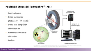
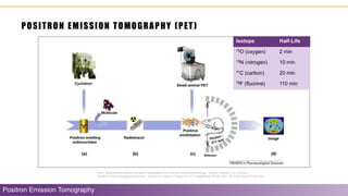
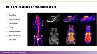
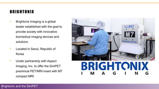
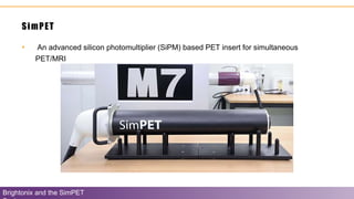
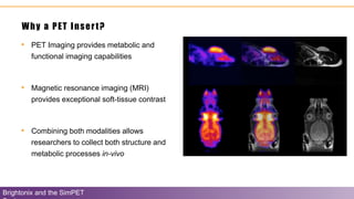
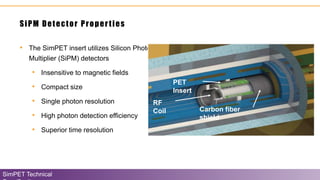
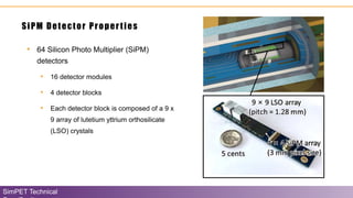
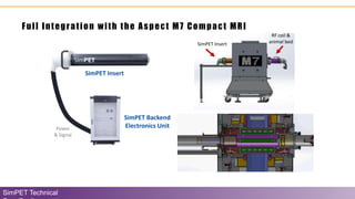
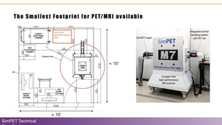
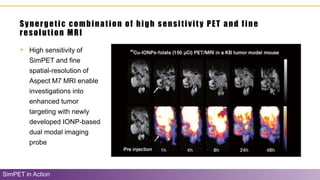
The document discusses the Simpet system, a preclinical positron emission tomography (PET) device developed by Brightonix that integrates with MRI technology for advanced imaging in research. It highlights the technical specifications, performance capabilities, and applications of the Simpet system, including its role in oncology, neuroimaging, and cardiology. Key features include high image quality, low maintenance, and integration with the Aspect M7 MRI system, positioning it as a leading solution for biomedical imaging.


![Capstone poster draft team 1 [Autosaved]-2-3](https://cdn.slidesharecdn.com/ss_thumbnails/28a50426-fc3b-4306-86ca-277632b72254-160530072646-thumbnail.jpg?width=640&height=640&fit=bounds)


