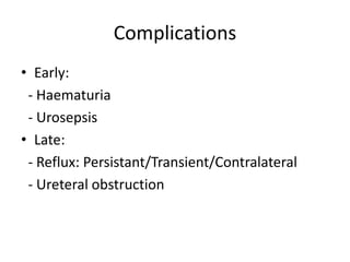
VUR
Resident: Dr SD Sanyal
Moderator: Lt Col MS Vinod
Cl Spl Surg & Paediatric Surgeon
Introduction
• Vesicoureteral reflux (VUR) represents the retrograde flow
of urine from the bladder to the upper urinary tract
• Galen and da Vinci:
- First references to VUR by Western medicine
- UVJ as a mediator of unidirectional flow of urine
from the kidneys to the bladder
• Hutch(1952): Relationship between VUR and chronic
pyelonephritis in paraplegic patients
• Hodson(1959):UTI and renal scarring carried a high
likelihood of VUR in children
Inheritance & Genetics
• The prevalence of reflux is higher in siblings
• There is a tendency for an autosomal
dominant pattern of inheritance
• Probably many genes are involved:
- PAX2
- GDNF-RET
- UPK3
- AGTR2-ACE
Anti-Reflux Mechanism
• Functional integrity of the ureter:
- Antegrade peristalsis
• Anatomic composition of the UVJ:
- 5:1 ratio of tunnel length to ureteral diameter
in nonrefluxing junctions ( Paquin, 1959 )
• Functional compliance of the bladder
Etiology
• Primary Reflux:
- fundamental deficiency in the function of the
UVJ
- bladder and ureter remain normal
- reflux occurs despite an adequately low-pressure
urine storage profile in the bladder
- length-diameter ratio is almost always less than
that described by Paquin
- inadequate tunnel length has greater implication
Etiology
• Secondary reflux:
- normal function of the UVJ being
overwhelmed
- bladder dysfunction : congenital, acquired, or
behavioral
- considered secondary if absence was
documented at some point before its
detection
Etiology
• Anatomical causes:
- PUV’s
- Prostatomegaly
- Ureteroceles
- Ureteral duplication
• Neuro-functional causes:
- Neurogenic bladder – Spina bifida
- Infant voiding patterns
- Dysfunctional voiding
- Uninhibited bladder contractions
- Constipation
International classification(VCUG)
Radionuclide Classification(DRCG)
• grade 1 = grade I of the international grading
system
• grade 2 = grade II-III
• grade 3 = grade IV-V
Clinical features
• Features of recurrent UTI:
- Fever
- Flank pain
- probability of finding VUR in children with UTI
is 29% to 50%
• Renal failure
• Palpable renal mass
Diagnosis & Evaluation
• Urine microscopy & culture:
- Infant: placement of an adhesive urine collection bag
over the genitalia
- Patients who void spontaneously : clean voided
midstream catch
- Topical cleansing of the area to reduce contamination
and false-positive culture
- Repeated catheterization for the acquisition of
specimens
- SPC
Diagnosis & Evaluation
• Radiographic evaluation:
- Indications:
1. children younger than 5 years
2. all children with a febrile UTI
3. any male with a UTI regardless of age or
fever, unless sexually active
Diagnosis & Evaluation
• Imaging modalities :
- USG
- DRCG
- VCUG
- Scintigraphy
Diagnosis & Evaluation
• Other modalities:
- Uroflowmetry & urodynamic studies
- Cystoscopy & PI Cystogram
Sonography
• Quantitative assessment of renal dimensions :
- used to monitor renal growth
- impact of any intercurrent febrile episode on renal
growth
- need for further assessment of renal
function by scintigraphy or the need for
correction of reflux
• Degree of corticomedullary differentiation
• Imaging of perfusion abnormalities :
- Renal resistive index measurements
- Contrast-enhanced harmonic ultrasound
VCUG
• Provides information on :
- functional dynamics
- structural anatomy
• Parameters observed:
A. Static films
- bladder contour - presence of diverticula
- ureteroceles - grade of reflux
- configuration & blunting of calyces - bladder neck anatomy
- urethral patency
- intrarenal reflux
VCUG
B. Dynamic films:
- Passive/active reflux
C. Delayed or postvoid films:
- Crucial in documenting clearance of contrast
from the upper tracts
- Dilated PCS + Retained contrast = PUJO
• Contraindicated in active cystitis
- Exceptions: In children with a h/o recurrent
pyelonephritis and repeatedly negative voiding
studies in the intercurrent periods
DRCG
• Radiation exposure 1% of VCUG
• Little anatomic detail is afforded
• Ideal for:
- screening
- monitoring the natural history
- surgical follow-up of reflux
• Greater sensitivity in grades II to V reflux
• Grade I reflux into distal ureter  poorly
detected
Scintigraphy
• DMSA:
- detection of reflux-associated renal damage
- acute pyelonephritic changes
- follow-up of reflux
• SPECT:
- 3D images
Uroflowmetry & Urodynamic study
• For establishing bladder functioning
• Lack of smoothness of the flow-velocity curve =
incomplete relaxation of the bladder outlet
during voiding
delays the natural history of reflux resolution or
even
perpetuate reflux
• Increased PVRU may be a risk factor for UTI
Cystoscopy
• Routine use is not mandated
• Role immediately prior to surgery for confirming:
- orifice position
- duplication
- proximity of diverticula to the orifice
- urethral patency
- endoscopic Mx(DEFLUX)
• PIC:
- to detect reflux under GA in pts with febrile UTIs
but a normal VCUG
Associated anomalies
1. PUJ Obstruction
- incidence of VUR associated with PUJO = 9% -
18%
- the incidence of PUJO in patients with reflux =
0.75% to 3.6%
- incidence with high-grade reflux = five times
more likely than lower grades of reflux
Associated anomalies
2. Ureteral duplication:
- VUR is the most common
abnormality associated
- reflux occurs most commonly
into the lower pole
Associated anomalies
3. Bladder diverticulae:
- outpouching of mucosa between detrusor
muscle bundles without any true muscle backing
itself
- Cause of reflux:
1. paraureteral diverticulum
2. large paraureteral diverticulum could expand
within Waldeyer's fascia and cause ureteral
obstruction/ project forward into the bladder
and obstruct the bladder outlet
Associated anomalies
4. Renal anomalies:
- Renal agenesis: 46% association
- MCDK: 28% association
- Presence mandates VCUG
5. Megacystis-Megaureter syndrome:
- More common in males
- Differentiation from PUV
Associated anomalies
6. Pregnancy associated reflux:
- Women with hypertension and an element of
renal failure are particularly at risk
7. Other anomalies:
- VACTERL anomalies
- CHARGE syndrome
- Imperforate anus
Natural history
• Spontaneous resolution:
- At birth, the probability of spontaneous
resolution of primary reflux is inversely
proportional to the initial grade
- If a patient is encountered at a later age,
resolution from any point in time forward will
depend on the initial grade of reflux
Natural history
• Resolution by grade:
- Most cases of low-grade reflux (grade I and II)
will resolve : 63-85%
- Grade III reflux will resolve in approximately
50% of cases
- Higher-grade reflux (grades IV and V and
bilateral grade III) : 9-25%
Natural history
• Resolution with age :
- Age has greater significance than grade
- Most prevalent in neonates and young
children and will demonstrate the greatest
tendency to resolve in this group
Management
• Principles of management:
1. Spontaneous resolution of reflux is very
common
2. High-grade reflux is less likely to resolve
spontaneously
3. Extended use of prophylactic antibiotics &
“ Watchful waiting”
4. The success rate with surgical correction is very
high
Medical management
• Watchful waiting
• Antibiotic prophylaxis:
- Amoxycillin < 2mths
- Co-trimoxazole > 2mths
- Alternatives: Nitrofurantoin/ Pro-biotics
• Breakthrough pyelonephritis
- indication for termination of medical mgt
Indications for surgery
ABSOLUTE INDICATIONS :
 Breakthrough urinary tract infections
 Failure of medical management
– - patient noncompliance
– - persistance of reflux with prolonged medical management.
– - progressive deterioration in renal function.
 Ureteral obstruction assoc with VUR
 Refluxing ureter opening into bladder diverticulum
 Cystoscopic observation of golf hole orifice
RELATIVE INDICATIONS :
– Presence of massive reflux – gr IV & V
– Reflux associated with paraureteral diverticulum
– In girls whose reflux persists after they have reached the full somatic
growth potential at puberty.
– Parental preference
Surgical management
The principles of surgical correction :
- Exclude secondary reflux
- Adequate ureteral mobilization and protection of the
ureteral blood supply
- A generous submucosal tunnel should be fashioned
- Attention should be directed to angulation and twisting
- Bladder tissues must be handled gently
- Always consider bladder function preoperatively, as
well as in all cases of persistent or recurrent reflux
- Indications for correction of reflux are the same
regardless of whether the planned approach is open,
endoscopic, or laparoscopic.
Surgical modalities
• Endoscopic management
• Ureteric reimplantation
 open
 laparoscopic
SUCCESS RATE > 90% for all open surgical
procedures
Classification
According to approach :
• Intravesical
• Extravesical
• Combined
According to the position of the sub mucosal
tunnel in relation to the original hiatus :
• Suprahiatal
• Infrahiatal
Cohen’s Transtrigonal
• Intravesical, infrahiatal procedure
• Simple, safe and most commonly used
• Avoids complications of neo-hiatus formation
• Good for small capacity bladder
• Success > 95%
Problem :
• Difficult retrograde catheterization of ureters.
Cohen’s Transtrigonal
Surgical procedures
• Politano-Leadbetter:
- Intravesical-Suprahiatal
• Glenn Anderson:
- Intravesical-Infrahiatal
• Lich-Gregoir:
- Extra-vesical
Endoscopic management
• Injection of a bio- compatible bulking agent
beneath intravesical portion of ureter in sub-
mucosal tunnel
• Elevates the intra-vesical ureter  narrowing
of lumen
• Prevents regurgitation of urine & allows
antegrade flow
Technique
Endoscopic management
ADVANTAGES
• OPD based treatment
• less morbidity, no mortality
• No surgical scar
• Success rate almost equivalent to open surgery for primary
reflux.
DISADVANTAGES
• Cost
• Lower success rate compared to surgery for high grade reflux
Indications
• Primary reflux
• Secondary reflux
- Dysfn voiding
- Neurogenic bladder
- Duplex system
• Failed open re-implant
Surgical outcomes
Success rates:
• Open - 98%
• Endoscopic – 80-89%
Complications
• Early:
- Haematuria
- Urosepsis
• Late:
- Reflux: Persistant/Transient/Contralateral
- Ureteral obstruction
Follow up
• Discharged on uro-prophylaxis
• Monitoring of pt’s
- BP
- renal function
- urine analysis
• Follow up USG and urine c/s after 1 mth
• VCUG after 3 mnths
• Discontinuation of uroprophylaxis on resolution
of reflux
• DMSA after 1 yr
Thank You

Vesicouretric reflux

  • 1.
    VUR Resident: Dr SDSanyal Moderator: Lt Col MS Vinod Cl Spl Surg & Paediatric Surgeon
  • 2.
    Introduction • Vesicoureteral reflux(VUR) represents the retrograde flow of urine from the bladder to the upper urinary tract • Galen and da Vinci: - First references to VUR by Western medicine - UVJ as a mediator of unidirectional flow of urine from the kidneys to the bladder • Hutch(1952): Relationship between VUR and chronic pyelonephritis in paraplegic patients • Hodson(1959):UTI and renal scarring carried a high likelihood of VUR in children
  • 3.
    Inheritance & Genetics •The prevalence of reflux is higher in siblings • There is a tendency for an autosomal dominant pattern of inheritance • Probably many genes are involved: - PAX2 - GDNF-RET - UPK3 - AGTR2-ACE
  • 4.
    Anti-Reflux Mechanism • Functionalintegrity of the ureter: - Antegrade peristalsis • Anatomic composition of the UVJ: - 5:1 ratio of tunnel length to ureteral diameter in nonrefluxing junctions ( Paquin, 1959 ) • Functional compliance of the bladder
  • 6.
    Etiology • Primary Reflux: -fundamental deficiency in the function of the UVJ - bladder and ureter remain normal - reflux occurs despite an adequately low-pressure urine storage profile in the bladder - length-diameter ratio is almost always less than that described by Paquin - inadequate tunnel length has greater implication
  • 7.
    Etiology • Secondary reflux: -normal function of the UVJ being overwhelmed - bladder dysfunction : congenital, acquired, or behavioral - considered secondary if absence was documented at some point before its detection
  • 8.
    Etiology • Anatomical causes: -PUV’s - Prostatomegaly - Ureteroceles - Ureteral duplication • Neuro-functional causes: - Neurogenic bladder – Spina bifida - Infant voiding patterns - Dysfunctional voiding - Uninhibited bladder contractions - Constipation
  • 9.
  • 10.
    Radionuclide Classification(DRCG) • grade1 = grade I of the international grading system • grade 2 = grade II-III • grade 3 = grade IV-V
  • 12.
    Clinical features • Featuresof recurrent UTI: - Fever - Flank pain - probability of finding VUR in children with UTI is 29% to 50% • Renal failure • Palpable renal mass
  • 13.
    Diagnosis & Evaluation •Urine microscopy & culture: - Infant: placement of an adhesive urine collection bag over the genitalia - Patients who void spontaneously : clean voided midstream catch - Topical cleansing of the area to reduce contamination and false-positive culture - Repeated catheterization for the acquisition of specimens - SPC
  • 14.
    Diagnosis & Evaluation •Radiographic evaluation: - Indications: 1. children younger than 5 years 2. all children with a febrile UTI 3. any male with a UTI regardless of age or fever, unless sexually active
  • 15.
    Diagnosis & Evaluation •Imaging modalities : - USG - DRCG - VCUG - Scintigraphy
  • 16.
    Diagnosis & Evaluation •Other modalities: - Uroflowmetry & urodynamic studies - Cystoscopy & PI Cystogram
  • 17.
    Sonography • Quantitative assessmentof renal dimensions : - used to monitor renal growth - impact of any intercurrent febrile episode on renal growth - need for further assessment of renal function by scintigraphy or the need for correction of reflux • Degree of corticomedullary differentiation • Imaging of perfusion abnormalities : - Renal resistive index measurements - Contrast-enhanced harmonic ultrasound
  • 18.
    VCUG • Provides informationon : - functional dynamics - structural anatomy • Parameters observed: A. Static films - bladder contour - presence of diverticula - ureteroceles - grade of reflux - configuration & blunting of calyces - bladder neck anatomy - urethral patency - intrarenal reflux
  • 19.
    VCUG B. Dynamic films: -Passive/active reflux C. Delayed or postvoid films: - Crucial in documenting clearance of contrast from the upper tracts - Dilated PCS + Retained contrast = PUJO • Contraindicated in active cystitis - Exceptions: In children with a h/o recurrent pyelonephritis and repeatedly negative voiding studies in the intercurrent periods
  • 20.
    DRCG • Radiation exposure1% of VCUG • Little anatomic detail is afforded • Ideal for: - screening - monitoring the natural history - surgical follow-up of reflux • Greater sensitivity in grades II to V reflux • Grade I reflux into distal ureter  poorly detected
  • 21.
    Scintigraphy • DMSA: - detectionof reflux-associated renal damage - acute pyelonephritic changes - follow-up of reflux • SPECT: - 3D images
  • 22.
    Uroflowmetry & Urodynamicstudy • For establishing bladder functioning • Lack of smoothness of the flow-velocity curve = incomplete relaxation of the bladder outlet during voiding delays the natural history of reflux resolution or even perpetuate reflux • Increased PVRU may be a risk factor for UTI
  • 23.
    Cystoscopy • Routine useis not mandated • Role immediately prior to surgery for confirming: - orifice position - duplication - proximity of diverticula to the orifice - urethral patency - endoscopic Mx(DEFLUX) • PIC: - to detect reflux under GA in pts with febrile UTIs but a normal VCUG
  • 24.
    Associated anomalies 1. PUJObstruction - incidence of VUR associated with PUJO = 9% - 18% - the incidence of PUJO in patients with reflux = 0.75% to 3.6% - incidence with high-grade reflux = five times more likely than lower grades of reflux
  • 25.
    Associated anomalies 2. Ureteralduplication: - VUR is the most common abnormality associated - reflux occurs most commonly into the lower pole
  • 26.
    Associated anomalies 3. Bladderdiverticulae: - outpouching of mucosa between detrusor muscle bundles without any true muscle backing itself - Cause of reflux: 1. paraureteral diverticulum 2. large paraureteral diverticulum could expand within Waldeyer's fascia and cause ureteral obstruction/ project forward into the bladder and obstruct the bladder outlet
  • 28.
    Associated anomalies 4. Renalanomalies: - Renal agenesis: 46% association - MCDK: 28% association - Presence mandates VCUG 5. Megacystis-Megaureter syndrome: - More common in males - Differentiation from PUV
  • 29.
    Associated anomalies 6. Pregnancyassociated reflux: - Women with hypertension and an element of renal failure are particularly at risk 7. Other anomalies: - VACTERL anomalies - CHARGE syndrome - Imperforate anus
  • 30.
    Natural history • Spontaneousresolution: - At birth, the probability of spontaneous resolution of primary reflux is inversely proportional to the initial grade - If a patient is encountered at a later age, resolution from any point in time forward will depend on the initial grade of reflux
  • 31.
    Natural history • Resolutionby grade: - Most cases of low-grade reflux (grade I and II) will resolve : 63-85% - Grade III reflux will resolve in approximately 50% of cases - Higher-grade reflux (grades IV and V and bilateral grade III) : 9-25%
  • 32.
    Natural history • Resolutionwith age : - Age has greater significance than grade - Most prevalent in neonates and young children and will demonstrate the greatest tendency to resolve in this group
  • 34.
    Management • Principles ofmanagement: 1. Spontaneous resolution of reflux is very common 2. High-grade reflux is less likely to resolve spontaneously 3. Extended use of prophylactic antibiotics & “ Watchful waiting” 4. The success rate with surgical correction is very high
  • 35.
    Medical management • Watchfulwaiting • Antibiotic prophylaxis: - Amoxycillin < 2mths - Co-trimoxazole > 2mths - Alternatives: Nitrofurantoin/ Pro-biotics • Breakthrough pyelonephritis - indication for termination of medical mgt
  • 36.
    Indications for surgery ABSOLUTEINDICATIONS :  Breakthrough urinary tract infections  Failure of medical management – - patient noncompliance – - persistance of reflux with prolonged medical management. – - progressive deterioration in renal function.  Ureteral obstruction assoc with VUR  Refluxing ureter opening into bladder diverticulum  Cystoscopic observation of golf hole orifice RELATIVE INDICATIONS : – Presence of massive reflux – gr IV & V – Reflux associated with paraureteral diverticulum – In girls whose reflux persists after they have reached the full somatic growth potential at puberty. – Parental preference
  • 37.
    Surgical management The principlesof surgical correction : - Exclude secondary reflux - Adequate ureteral mobilization and protection of the ureteral blood supply - A generous submucosal tunnel should be fashioned - Attention should be directed to angulation and twisting - Bladder tissues must be handled gently - Always consider bladder function preoperatively, as well as in all cases of persistent or recurrent reflux - Indications for correction of reflux are the same regardless of whether the planned approach is open, endoscopic, or laparoscopic.
  • 38.
    Surgical modalities • Endoscopicmanagement • Ureteric reimplantation  open  laparoscopic SUCCESS RATE > 90% for all open surgical procedures
  • 39.
    Classification According to approach: • Intravesical • Extravesical • Combined According to the position of the sub mucosal tunnel in relation to the original hiatus : • Suprahiatal • Infrahiatal
  • 40.
    Cohen’s Transtrigonal • Intravesical,infrahiatal procedure • Simple, safe and most commonly used • Avoids complications of neo-hiatus formation • Good for small capacity bladder • Success > 95% Problem : • Difficult retrograde catheterization of ureters.
  • 41.
  • 42.
    Surgical procedures • Politano-Leadbetter: -Intravesical-Suprahiatal • Glenn Anderson: - Intravesical-Infrahiatal • Lich-Gregoir: - Extra-vesical
  • 43.
    Endoscopic management • Injectionof a bio- compatible bulking agent beneath intravesical portion of ureter in sub- mucosal tunnel • Elevates the intra-vesical ureter  narrowing of lumen • Prevents regurgitation of urine & allows antegrade flow
  • 44.
  • 45.
    Endoscopic management ADVANTAGES • OPDbased treatment • less morbidity, no mortality • No surgical scar • Success rate almost equivalent to open surgery for primary reflux. DISADVANTAGES • Cost • Lower success rate compared to surgery for high grade reflux
  • 46.
    Indications • Primary reflux •Secondary reflux - Dysfn voiding - Neurogenic bladder - Duplex system • Failed open re-implant
  • 47.
    Surgical outcomes Success rates: •Open - 98% • Endoscopic – 80-89%
  • 48.
    Complications • Early: - Haematuria -Urosepsis • Late: - Reflux: Persistant/Transient/Contralateral - Ureteral obstruction
  • 49.
    Follow up • Dischargedon uro-prophylaxis • Monitoring of pt’s - BP - renal function - urine analysis • Follow up USG and urine c/s after 1 mth • VCUG after 3 mnths • Discontinuation of uroprophylaxis on resolution of reflux • DMSA after 1 yr
  • 50.