
Recommended
More Related Content
What's hot
What's hot (20)
Similar to Skeletal Muscle Physiology of Muscle Contraction _RDP
Similar to Skeletal Muscle Physiology of Muscle Contraction _RDP (20)
More from rishi2789
More from rishi2789 (20)
Recently uploaded
Recently uploaded (20)
Skeletal Muscle Physiology of Muscle Contraction _RDP
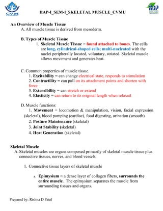
- 1. HAP-I_SEM-I_SKELETAL MUSCLE_CVMU Prepared by: Rishita D Patel An Overview of Muscle Tissue A. All muscle tissue is derived from mesoderm. B. Types of Muscle Tissue 1. Skeletal Muscle Tissue = found attached to bones. The cells are long, cylindrical-shaped cells; multi-nucleated with the nuclei peripherally located, voluntary, striated. Skeletal muscle allows movement and generates heat. C. Common properties of muscle tissue. 1. Excitability = can change electrical state, responds to stimulation 2. Contractility = can pull on its attachment points and shorten with force 3. Extensibility = can stretch or extend 4. Elasticity = can return to its original length when relaxed D. Muscle functions: 1. Movement = locomotion & manipulation, vision, facial expression (skeletal), blood pumping (cardiac), food digesting, urination (smooth) 2. Posture Maintenance (skeletal) 3. Joint Stability (skeletal) 4. Heat Generation (skeletal) Skeletal Muscle A. Skeletal muscles are organs composed primarily of skeletal muscle tissue plus connective tissues, nerves, and blood vessels. 1. Connective tissue layers of skeletal muscle a. Epimysium = a dense layer of collagen fibers, surrounds the entire muscle. The epimysium separates the muscle from surrounding tissues and organs.
- 2. HAP-I_SEM-I_SKELETAL MUSCLE_CVMU Prepared by: Rishita D Patel b. Perimysium = a fibrous layer that divides the skeletal muscle into a series of bundles called fascicles. Perimysium surrounds the fascicles. In addition to possessing collagen and elastic fibers, the perimysium contains blood vessels and nerves. c. Endomysium = a delicate connective tissue that surrounds the individual skeletal muscle cells (muscle fibers), and loosely interconnects adjacent muscle fibers. d. At the ends of a skeletal muscle, collagen fibers of the connective tissue layers merge to form either a bundle, known as a tendon, or a broad sheet, known as an aponeurosis, to anchor the muscle to the bone.
- 3. HAP-I_SEM-I_SKELETAL MUSCLE_CVMU Prepared by: Rishita D Patel 2. Anatomy of Skeletal Muscles. a. Muscle fiber = a skeletal muscle cell b. Sarcoplasm = cytoplasm. The sarcoplasm of a skeletal muscle cell has more mitochondria than the average body cell and large amounts of myoglobin, a protein important in the storage of oxygen. c. Sarcolemma = cell membrane = plasma membrane
- 4. HAP-I_SEM-I_SKELETAL MUSCLE_CVMU Prepared by: Rishita D Patel Transverse tubules (TT) o A set of membranous channels extend from the sarcolemma into the sarcoplasm as invaginations continuous with muscle cell membrane. o TTs are filled with extracellular fluid and extend deep into the cell. o Each TT runs between two enlarged portions of sarcoplasmic reticulum called terminal cisternae (TC). ▪ These structures form a triad A triad = two terminal cisternae + one transverse tubule). d. Sarcoplasmic reticulum (SR) = specialized smooth endoplasmic reticulum. o Network of membranous channels that surrounds each myofibril and runs parallel to it. o SR stores high concentrations of calcium ions compared to the sarcoplasm (maintained by active transport calcium pump). o When stimulated by muscle impulse, membranes become more permeable to calcium ions and calcium diffuses out of SR and into sarcoplasm.
- 5. HAP-I_SEM-I_SKELETAL MUSCLE_CVMU Prepared by: Rishita D Patel o Enlarged portions of sarcoplasmic reticulum = terminal cisternae (TC). e. Each muscle fiber is composed of myofibrils = long, cylindrical organelles that run parallel within the muscle fiber and contains the sarcomeres . f. Each myofibril is composed of two contractile proteins called myofilaments. g. The thick myofilaments are called myosin and the thin myofilaments are called actin. h. The myofilaments of myosin and actin are arranged in an overlapping pattern creating hundreds to thousands of functional units called sarcomeres. 3. Anatomy of a Sarcomere = the functional or contractile unit of skeletal muscle tissue. a. Each myofibril consists of approximately 10,000 sarcomeres Each sarcomere is composed of a very specific arrangement of the myofilaments of actin and myosin. b. A band = dense region of the sarcomere that contains overlapping thick and thin filaments (both myosin and actin). c. I band = on either side of the A band is an area that contains only thin filaments (actin) d. H band = middle of each A band, an area that contains only thick filaments (myosin) e. M Line = located in the middle of each H band, anchors the center of each thick filament.
- 6. HAP-I_SEM-I_SKELETAL MUSCLE_CVMU Prepared by: Rishita D Patel f. Z Disk (Lines) = mark the boundary between adjacent sarcomeres. Z lines anchor the thin filaments of adjacent sarcomeres. 4. Anatomy of the Myofilaments (actin and myosin) a. Each thin filament of actin is composed of several interacting proteins.
- 7. HAP-I_SEM-I_SKELETAL MUSCLE_CVMU Prepared by: Rishita D Patel i. Actin molecule of protein that possesses an active site where one myosin molecule can bind. ii. Tropomyosin is the protein that covers the active sites of actin in a resting muscle. This prevents the actin and myosin fibers from interacting. iii. Troponin is a complex of three protein subunits: 1) binds to calcium ions 2) binds to actin 3) binds to tropomyosin. When actin and myosin bind, they form a crossbridge. b. Each thick filament of myosin is composed of roughly 300 myosin molecules. i. Myosin molecules are a pair of myosin subunits twisted around one another. ii. The myosin subunits have a tail portion connected to a free head portion. iii. The tail and free head are connected in a way that allows the head to pivot
- 8. HAP-I_SEM-I_SKELETAL MUSCLE_CVMU Prepared by: Rishita D Patel
- 9. HAP-I_SEM-I_SKELETAL MUSCLE_CVMU Prepared by: Rishita D Patel Skeletal Muscle Contraction and Relaxation A. Neuromuscular junction and nerve stimulation 1. Skeletal muscle cells are stimulated by a motor neuron. 2. The axon of each motor neuron branches extensively to form numerous cellular extensions called synaptic terminals. 3. The synaptic terminals then interact with the sarcolemma of the muscle fiber at a specialized site called the neuromuscular junction. 4. When the electrical impulse (action potential) reaches the synaptic terminals, calcium channels with the synaptic terminal begin to open, causing calcium to rush into the axon terminal. 5. As a result of the influx of calcium, synaptic vesicles containing the neurotransmitter acetylcholine (Ach) fuse with the axon membrane releasing the neurotransmitter into the synaptic cleft by exocytosis. 6. Acetylcholine then diffuses across the synaptic cleft and attaches to receptors (also known as ion channels) on a highly folded region of the sarcolemma called the motor end plate. 7. The binding of acetylcholine to the channels causes the channel to open and influx of sodium ions into the muscle cell resulting in depolarization of the sarcolemma and eventually leading to an action potential and contraction. 8. As the action potential spreads away from the motor end plate and across the sarcolemma, acetylcholine is swiftly broken down by acetylcholinesterase. The destruction of Ach prevents continued muscle contraction in the absence of nervous stimulation.
- 10. HAP-I_SEM-I_SKELETAL MUSCLE_CVMU Prepared by: Rishita D Patel
- 11. HAP-I_SEM-I_SKELETAL MUSCLE_CVMU Prepared by: Rishita D Patel
- 12. HAP-I_SEM-I_SKELETAL MUSCLE_CVMU Prepared by: Rishita D Patel B. Generating the action potential of the muscle 1. Electrical conditions of a resting sarcolemma, also called the resting membrane potential, is said to be polarized. That is, the extracellular environment is more positive with respect to the inside of the membrane. The predominant extracellular ion is Na+ while the predominant intracellular ion is K+. The sarcolemma is relatively impermeable to both ions while at rest but more so to the Na+. To get the muscle to contract, the membrane potential must depolarize. 2. Step 1: Depolarization. The binding of acetylcholine to the Na+ ion channels on the motor end plate cause the channels to open. Sodium then rushes rapidly across the sarcolemma into the cytoplasm. As sodium begins to accumulate on the inside of the muscle cell, the resting potential is decreased and localized depolarization occurs. That is, the patch of sarcolemma immediately around the Na+ channel becomes more positive inside with respect to the outside. 3. Step 2: Propagation of the action potential. The positive charge inside the initial patch of sarcolemma changes the permeability of an adjacent patch, opening sodium channels there. Consequently, the sodium begins to rush into the membrane there causing the membrane potential in that region to decrease and depolarization occurs in that area. Thus, the action
- 13. HAP-I_SEM-I_SKELETAL MUSCLE_CVMU Prepared by: Rishita D Patel potential begins to travel rapidly away from the motor end plate, across the entire sarcolemma and down into the T tubules. 4. Step 3: Repolarization of the sarcolemma. Immediately after the depolarization wave passes, the sarcolemma’s permeability changes once again. Acetylcholine is removed from the synaptic cleft by acetylcholinesterase causing the Na+ ion channels to close while the K+ channels to finally open in a delayed response. The influx of sodium now stops but K+ begins to leak out so that the outside of the membrane switches back to positive and the inside of the membrane becomes more negative. This restores the charge across the membrane (repolarization) however, because more K+ ions eventually leave than is necessary, the membrane becomes hyperpolarized. 5. Step 4: Sodium-Potassium Pump restores ion concentrations in the hyperpolarized membrane to reach the polarized conditions of the resting membrane potential. For each ATP, 3 Na+ ions are pumped out and 2 K+ ions are pumped in. Because the ion exchange is so close to equal, there is no change in the charge across the membrane as the ions are being redistributed. 6. During repolarization, muscle fibers are in a refractory period when they are insensitive to further stimulation. 7. Action potentials are considered all or none responses. C. Excitation-Contraction Coupling 1. As the action potential propagates along the sarcolemma it moves down the T tubules. 2. Transmission of the action potential past the triads causes the terminal cisternae of the SR to release Ca+ into the sarcoplasm, it now becomes available to the myofilaments of the sarcomere. 3. The presence of calcium on the sarcomere causes myosin to bind to actin in a process commonly referred to as the sliding filament mechanism.
- 14. HAP-I_SEM-I_SKELETAL MUSCLE_CVMU Prepared by: Rishita D Patel D. Sliding Filament Mechanism 1. Attachment of myosin cross bridges within a resting muscle is inhibited by the presence of tropomyosin which covers the active sites on actin. If calcium ions are released from the terminal cisternae by the action potential, they bind to troponin changing its shape. 2. The troponin then pulls on the tropomyosin so that the binding sites on actin are exposed. 3. Once the active site is exposed, the high-energy myosin heads attach to the actin for the cross-bridge formation. 4. The release of ADP + P from the high-energy myosin head causes the head to pivot and bend as it pulls on the actin filament, sliding it towards the midline. This is called the power stroke and is equivalent to the contraction of the muscle. 5. As a new ATP attaches to the now low-energy myosin head, the myosin head detaches from the actin. 6. ATP is split to form ADP + P and the bond energy is transferred to the myosin head causing it to move in the high-energy position (cocked or reactivated) so that its ready to bind to the actin binding sites once again. 7. When the action potential dissipates, the calcium is reabsorbed back into the terminal cisternae. When this occurs, tropomyosin moves back over the active sites on the actin so that crossbridge formation can no longer occur and muscle relaxes. https://youtu.be/UZNPv86y7Fg https://youtu.be/sIH8uOg8ddw
- 15. HAP-I_SEM-I_SKELETAL MUSCLE_CVMU Prepared by: Rishita D Patel Contraction Cycle Begins - Increase in cytosolic Ca2+ Active Sites Expose - Calcium interacts with troponin causing a conformation change in tropomyosin, which exposes actin’s active site.
- 16. HAP-I_SEM-I_SKELETAL MUSCLE_CVMU Prepared by: Rishita D Patel Cross-Bridges Form - myosin heads attach to actin at active site.
- 17. HAP-I_SEM-I_SKELETAL MUSCLE_CVMU Prepared by: Rishita D Patel Myosin Heads Pivot - resulting in the movement of the actin filaments towards the H-disc. Then the attached ADP and phosphate group are released. Cross-Bridges Detach - A new molecule of ATP attaches to the myosin head, causing the cross-bridge to detach
- 18. HAP-I_SEM-I_SKELETAL MUSCLE_CVMU Prepared by: Rishita D Patel Reactivate the Myosin Head - The myosin head hydrolyzes ATP to ADP and phosphate, which returns the myosin to the cocked position.