
Recommended
More Related Content
What's hot
What's hot (20)
Similar to Joints THEORY_RDP
Similar to Joints THEORY_RDP (20)
More from rishi2789
More from rishi2789 (20)
Recently uploaded
Recently uploaded (20)
Joints THEORY_RDP
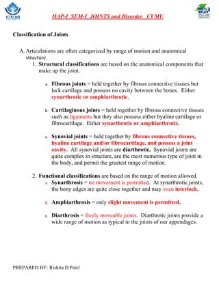
- 1. HAP-I_SEM-I_JOINTS and Disorder _CVMU PREPARED BY: Rishita D Patel Classification of Joints A. Articulations are often categorized by range of motion and anatomical structure. 1. Structural classifications are based on the anatomical components that make up the joint. a. Fibrous joints = held together by fibrous connective tissues but lack cartilage and possess no cavity between the bones. Either synarthrotic or amphiarthrotic. b. Cartilaginous joints = held together by fibrous connective tissues such as ligaments but they also possess either hyaline cartilage or fibrocartilage. Either synarthrotic or amphiarthrotic. c. Synovial joints = held together by fibrous connective tissues, hyaline cartilage and/or fibrocartilage, and possess a joint cavity. All synovial joints are diarthrotic. Synovial joints are quite complex in structure, are the most numerous type of joint in the body, and permit the greatest range of motion. 2. Functional classifications are based on the range of motion allowed. a. Synarthrosis = no movement is permitted. At synarthrotic joints, the bony edges are quite close together and may even interlock. b. Amphiarthrosis = only slight movement is permitted. c. Diarthrosis = freely moveable joints. Diarthrotic joints provide a wide range of motion as typical in the joints of our appendages.
- 2. HAP-I_SEM-I_JOINTS and Disorder _CVMU PREPARED BY: Rishita D Patel Fibrous joints i. Sutures = a synarthrotic joint located only between the bones of the skull. The edges of the bones are interlocked and bound together at the suture by dense fibrous connective tissue. ii. Syndesmosis = bones are connected by an interosseous ligament and are amphiarthrotic. The most common example is the distal articulation between the tibia and fibula called the tibiofibular joint. iii. Gomphosis = a synarthrotic joint sometimes called a “peg-in-socket” joint. A gomphosis joint is found on the maxillae and mandible where the teeth are fixed securely in the sockets of the alveolar margins. The fibrous connective tissue between a tooth and its socket is a periodontal ligament.
- 3. HAP-I_SEM-I_JOINTS and Disorder _CVMU PREPARED BY: Rishita D Patel Cartilaginous joints A. Synchondrosis = a rigid, hyaline cartilage bridge unites the bones of a synchondrosis joint. Ends of the first pair of ribs and the manubrium of the sternum (all other ribs form synovial joints). Epiphyseal plate found holding the epiphysis of a long bone to the diaphysis Both are synarthrotic joints. B. Symphysis = articulating bones are separated by a wedge of fibrocartilage. The articulation between the two pubic bones (called the pubic symphysis) is another joint typical of this category. a. fibrous cartilage between opposing bones within a synovial joint. Synchondroses
- 4. HAP-I_SEM-I_JOINTS and Disorder _CVMU PREPARED BY: Rishita D Patel - joined by hyaline cartilage Symphyses - joined by fibrocartilage 9.3 Synovial joints A. Structural features of a Synovial Joint 1. Joint cavity = possess a space between the articulating bones, called the synovial cavity. 2. Articular cartilages = line the surfaces of the articulating bones; composed of hyaline cartilage 3. Synovial fluid = this fluid is largely derived from blood and has a clear, viscous, consistency. a. Lubrication b. Nutrient distribution c. Shock absorption
- 5. HAP-I_SEM-I_JOINTS and Disorder _CVMU PREPARED BY: Rishita D Patel 4. Joint capsule = dense fibrous connective tissues enclose the synovial cavity a. Fibrous capsule = thick outer layer continuous with the periosteum. b. Synovial membrane = inner soft tissue with network of capillaries 5. Accessory structures of a typical synovial joint = in complex synovial joints, such as the knee, a variety of accessory structures provide support and additional stability. b. Ligaments = support, strengthen, and reinforce synovial joints c. Bursa = a small, fluid-filled pocket that forms in a connective tissue. d. Fat pads = localized masses of adipose tissue covered by a layer of synovial membrane.
- 6. HAP-I_SEM-I_JOINTS and Disorder _CVMU PREPARED BY: Rishita D Patel e. Meniscus = pad of fibrous cartilage between opposing bones within a synovial joint. 9.4 types of body movements A. the greater the range of motion at a joint, the weaker it becomes. B. types of motion 1. Gliding=bones slide across the surface of one another 2. Angular=changing the angle between two bones 3. Circumduction=draw around; conical shape or circular motion 4. Rotation=turning movement of a bone around its own axis
- 7. HAP-I_SEM-I_JOINTS and Disorder _CVMU PREPARED BY: Rishita D Patel 9.5 Anatomy of Selected Synovial Joints A. Ball-and-socket joints = most freely movable joints; all angular movement; 1. The head of one bone fits into the socket of another; 2. Examples = hip and shoulder. B. Condyloid joints = permit all angular motion, except rotation. Examples = wrists and knuckles, C. Gliding joints/plan joint = cartilaginous joints; flat bones glide/slide over one another Example = intervertebral discs. D. Hinge joints = permit flexion & extension only; Examples = elbow and knee. E. Pivot joints = permit rotation; Example = first intervertebral joint (atlantoaxial joint)
- 8. HAP-I_SEM-I_JOINTS and Disorder _CVMU PREPARED BY: Rishita D Patel F. Saddle joints = thumb; Common Joint Injuries A. Sprain = stretching or tearing of a ligament across the joint capsule. B. Dislocation = also known as a luxation; when reinforcing structures cannot protect a joint from extreme stresses, the articulating surfaces may be forced out of position. The displacement may damage the articular cartilages, tear ligaments, or distort the joint capsule. Although the inside of a joint has no pain receptors, nerves that monitor the capsule, ligaments, and tendons are quite sensitive, so dislocations are very painful. A partial dislocation is called a subluxation. C. Bursitis = inflammation of the bursa
- 9. HAP-I_SEM-I_JOINTS and Disorder _CVMU PREPARED BY: Rishita D Patel D. Tendonitis = inflammation of the tendon E. Synovitis =inflammation of the synovial membrane F. Osteopenia and osteoporosis=inadequate ossification of bone is called osteopenia and begins between the ages of 30 and 40 years of age when osteoblast activity declines while osteoclast activity continues at previous levels. Thereafter, women begin to lose roughly 8% of their bone mass every decade (men lose 3% per decade). When the reduction in bone mass is sufficient to compromise normal function, the condition is known as osteoporosis. G. Arthritis = inflammatory or degenerative disease of the joint where synovial membranes thicken (called pannus) and fluid production decreases resulting in friction and pain. 1. Osteoarthritis = also known as degenerative arthritis or degenerative joint disease, generally affects individuals age 60 or older. It can result from the cumulative effects of wear and tear on the joint surfaces or from genetic factors affecting collagen formation. In the U.S. population, 25% of women and 15% of men over age 60 show signs of this condition. 2. Rheumatoid arthritis = an autoimmune disease. RA can occur at any age but is more common in middle age and women get RA more often than men. Infection, genes, and hormone changes may be linked to the disease. RA usually affects joints on both sides of the body equally. Wrists, fingers, knees, feet, and ankles are the most common affected body parts. The disease often begins slowly with only minor pain but progressively becomes debilitating. 3. Gouty arthritis = Gout is caused by too much uric acid in the blood. Most of the time, having too much uric acid is not harmful. Many people with high levels in their blood never get gout. But when uric acid levels in the blood are too high, the uric acid may form hard crystals in your joints. It can cause an attack of sudden burning pain, stiffness, and swelling in a joint, usually a big toe. These attacks can happen over and over unless gout is treated. More common in men.