The document provides information about the anatomy and functions of the cerebellum. It can be summarized as follows:
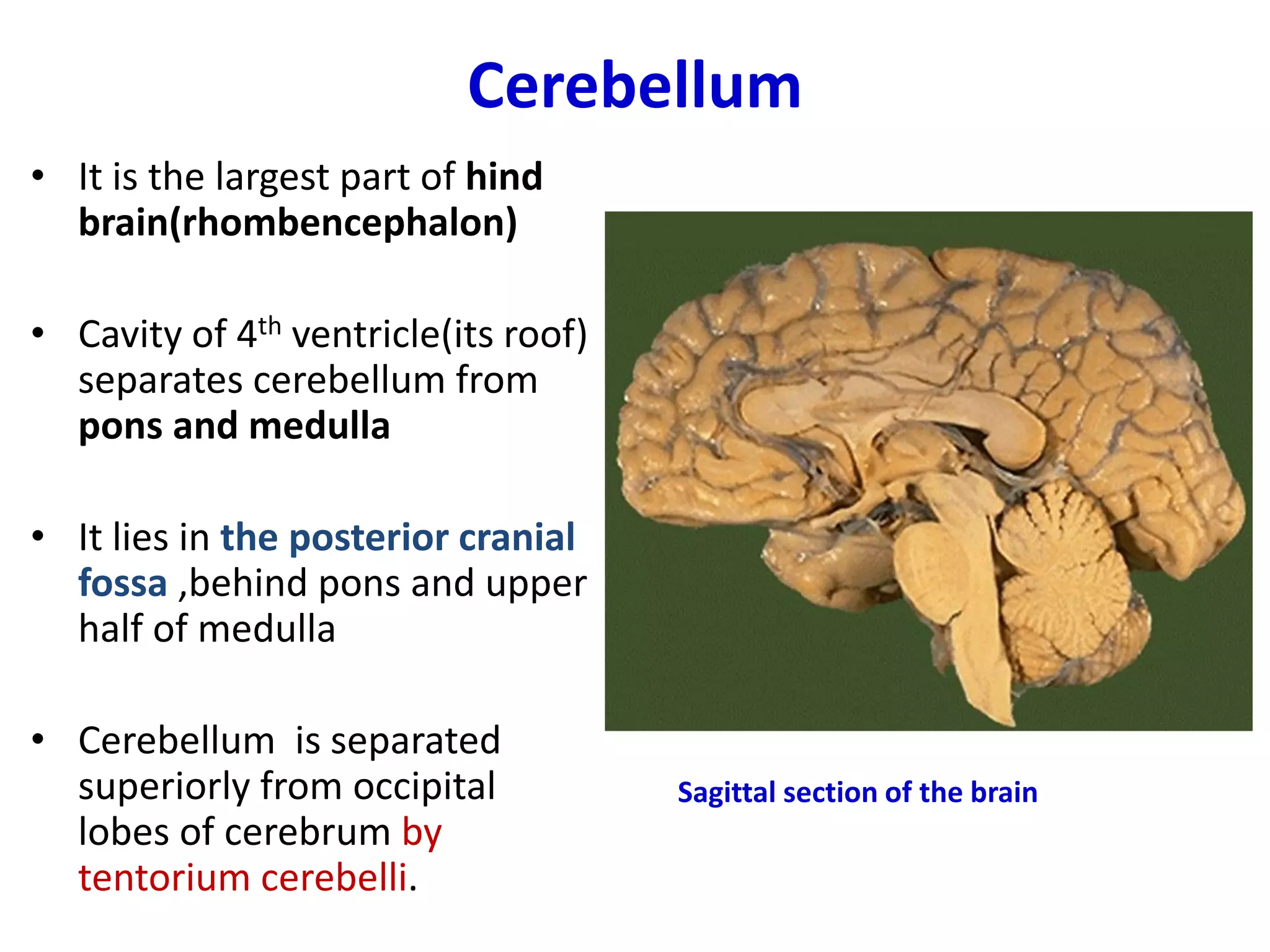
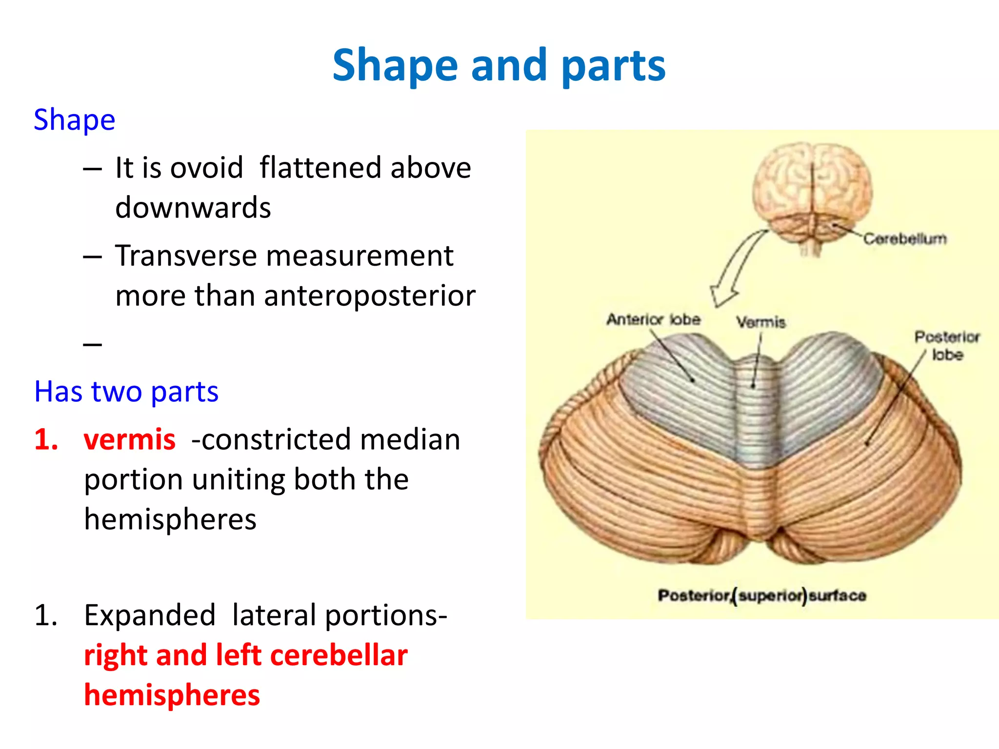
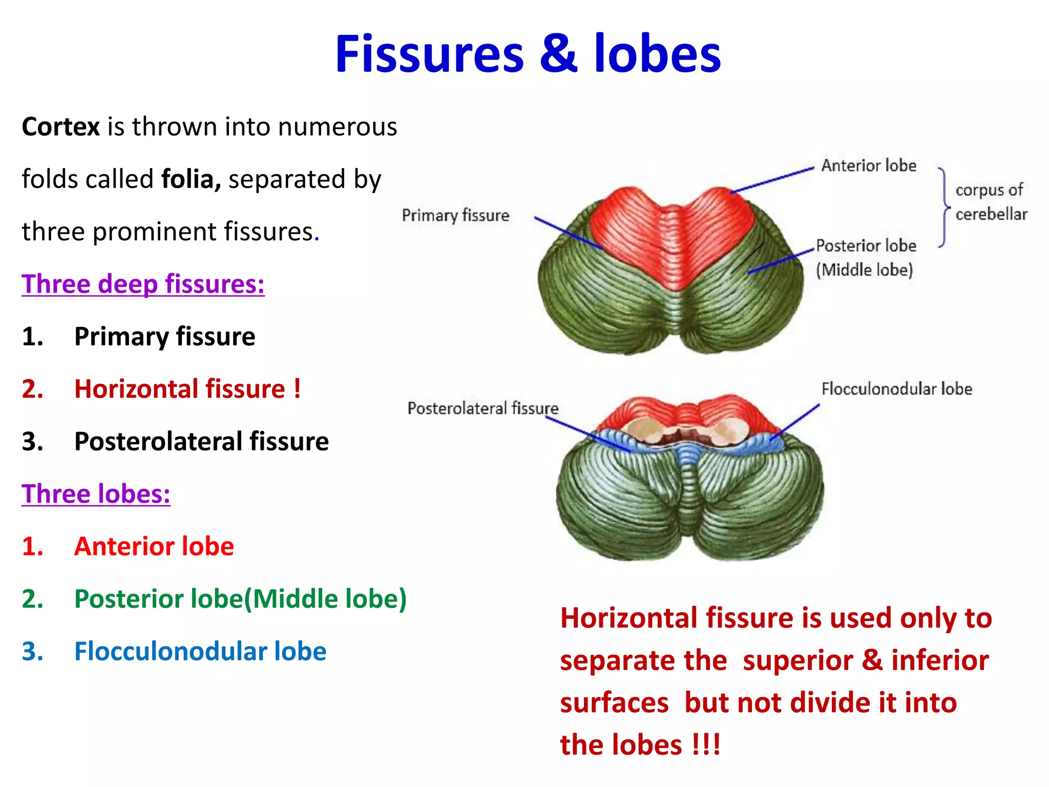
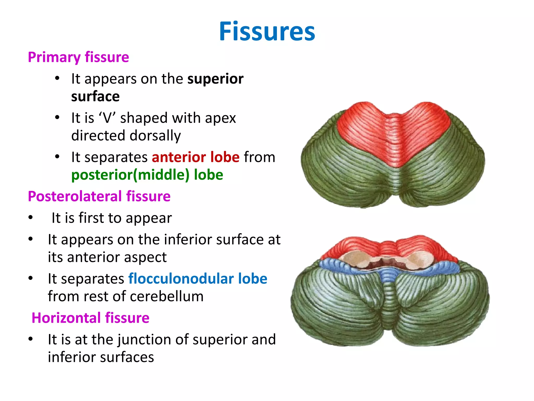
1. The cerebellum is located in the posterior cranial fossa and is separated into two hemispheres and a median vermis. It has three lobes and three functional subdivisions that control balance, coordination of movements, and muscle tone.
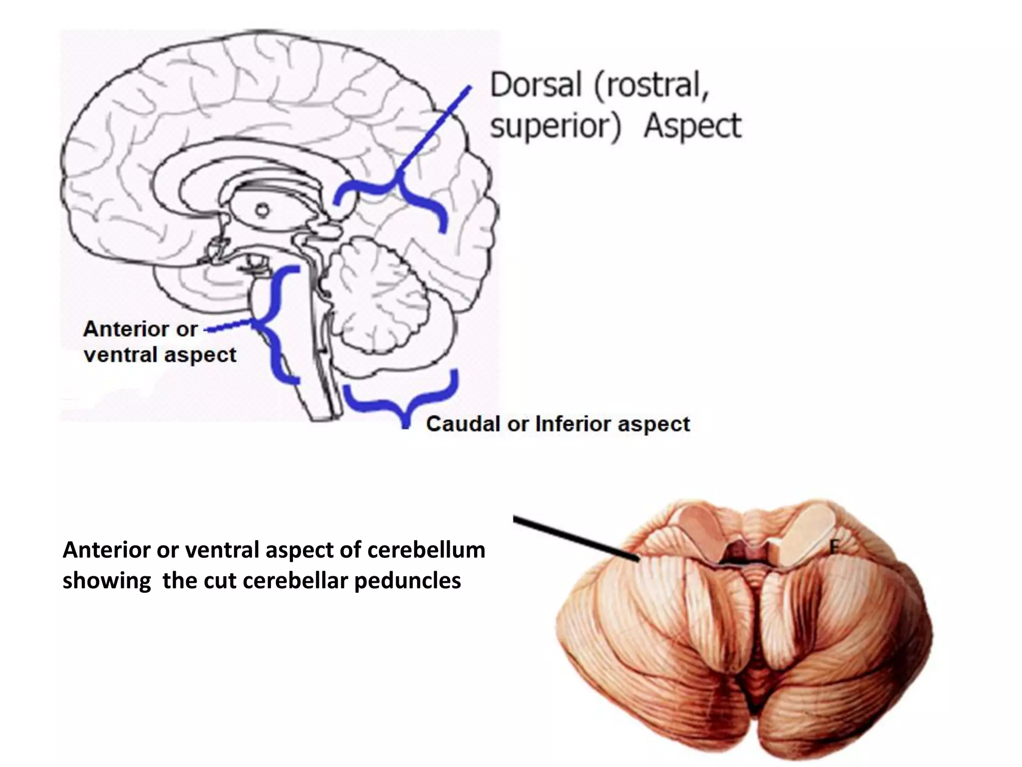
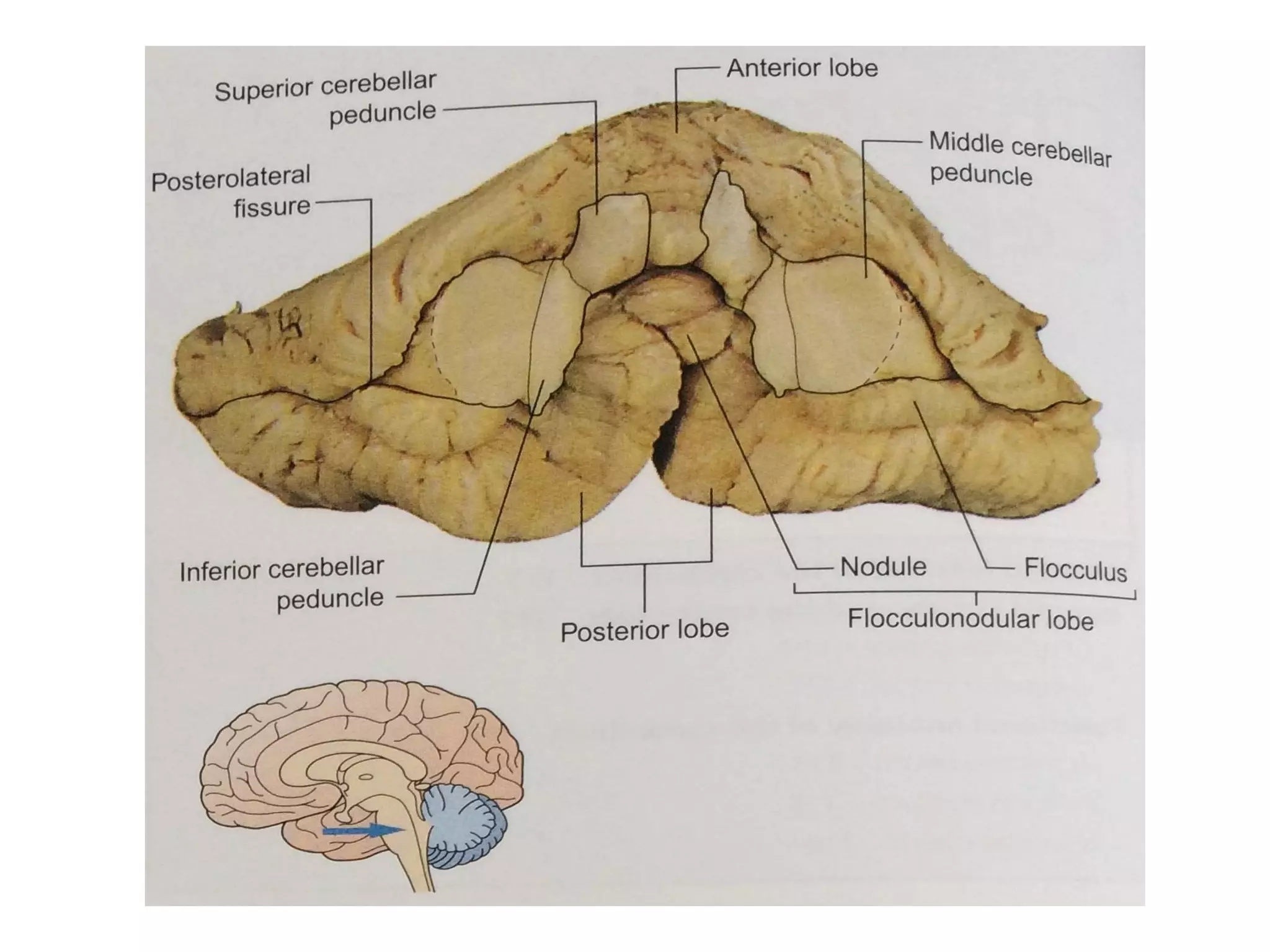
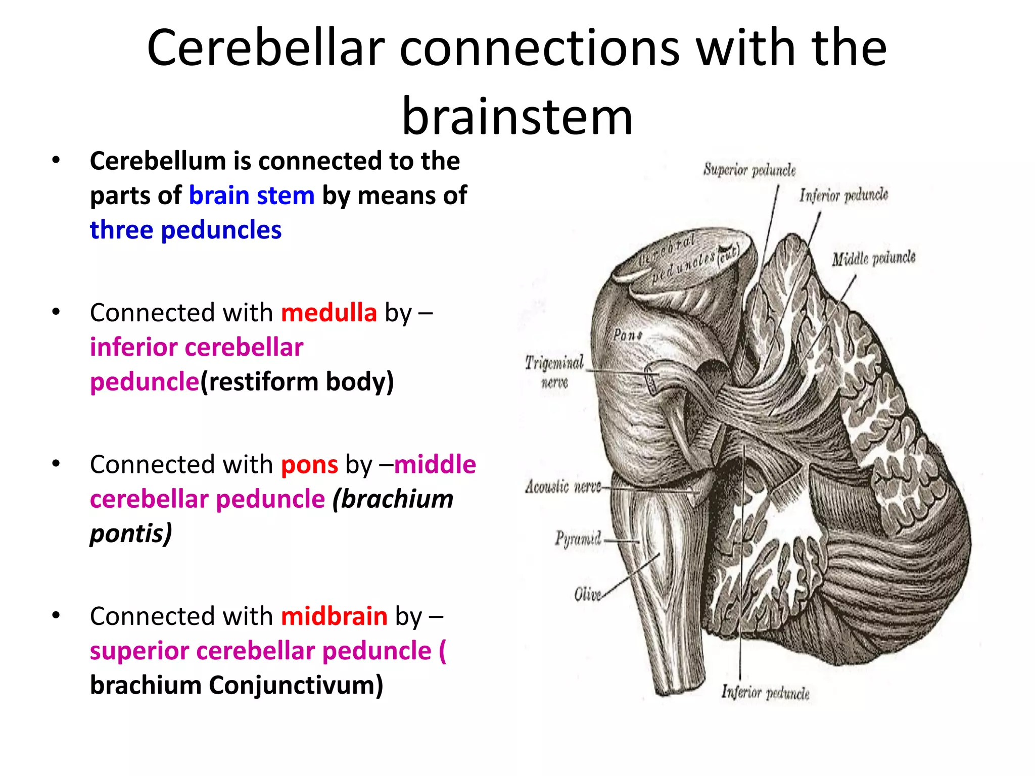
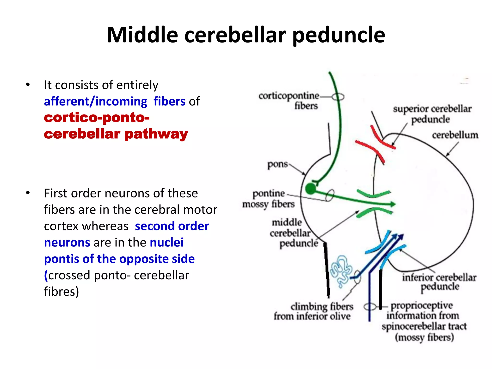
2. The cerebellum connects to the brainstem via three cerebellar peduncles - superior, middle, and inferior - which carry afferent and efferent fibers. Deep within the cerebellum are four nuclei - dentate, globose, emboliformis, and fastigius.
3. The cerebell