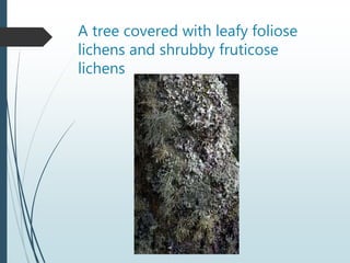
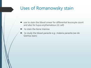
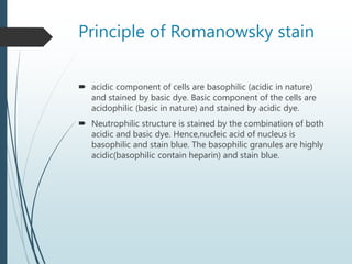
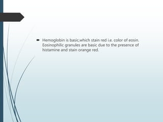
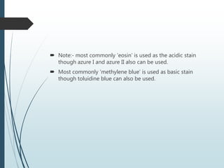
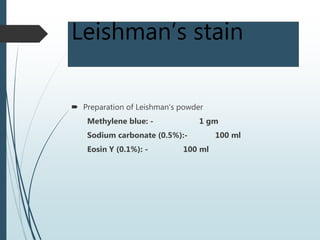
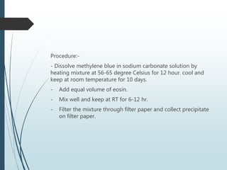
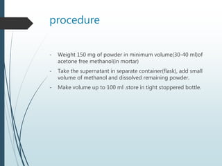
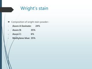
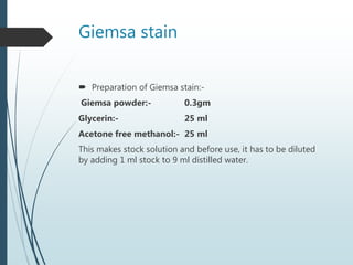
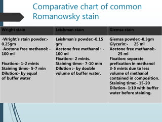
Dyes and stains
Introduction
 All the biological stains are prepared from
dyes,which have been manufactured for
special purpose that means to study the
anatomical details of blood cells and
tissue microscopically.
 The dye used to denoted powder and the stain to denote
the solution of one or more dyes. The dyes are classified
into two groups:-
1) Natural dyes
2) Synthetic dyes
1) Natural dyes
they are prepared or extracted from natural sources
such as hematoxylin(It is a compound extracted from
the heartwood of the logwood tree (Haematoxylum
campechianum), litmus(Litmus is a water-
soluble mixture of different dyes extracted from lichens),
orcein(Orcein, also archil, orchil, lacmus and C.I. Natural
Natural Red 28, are names for dyes extracted from
several species of lichen, commonly known as "orchella
weeds", found in various parts of the world), carmine,
safranin.
A tree covered with leafy foliose
lichens and shrubby fruticose
lichens
Caramine forming things
2) Synthetic dyes
These are manufactured or prepared or sometimes referred as
coal tar dyes.
stain can further be divided into 3 parts:-
1) Acidic dyes/stains:- acidic dyes stains basic component of
the cell such as cytoplasm.
the coloring substance present in the acidic component and
basic part is colorless. E.g. eosin, safranine, phylozine.
2) Basic dyes/stains:-
coloring substance is present in the basic part of the
component and acidic part is colorless.e.g. methylene blue,
crystal violet, gentian violet, methyl violet, brilliant crysal blue.
So basic dyes stain the acidic component of the cell(nucleus).
3) Neutral dyes/stains:-
in these days, both acidic and basic components are colored.
These dyes are also called amphoteric dyes e.g. Romanowsky
dyes.
AMPHOTERIC DYES
These dyes stain both acidic and basic component of the cell
at suitable PH. In acidic PH,it behaves like acidic dye and in
basic PH,it behaves like basic dye.
Acidic and basic dyes are usually prepared in aqueous
solution. But, neutral dyes are prepared in organic
solvent(methanol) and differentiated by aqueous solution.
Acidic/basic dyes are differentiated by organic media.
ROMANOWSKY’S STAIN
In 1819,Romanowsky discovered that when alkali is added to
methylene blue, it produce oxidation of methylene blue and
forms polychrome methylene blue which further combine
with eosin to form methylene blue azure. These dyes are not
soluble in water but dissolve in methanol(acetone free
methanol). Acetone acts as strong dehydrating agent and
discoloring agent. So, not used in above stain.
Purpose of staining
 To study morphological structure of the cells by stain.
Uses of Romanowsky stain
 use to stain the blood smear for differential leucocyte count
and also for lupus erythematous (LE cell)
 to stain the bone marrow
 to study the blood parasite e.g.: malaria parasite.(we do
Giemsa stain)
Principle of Romanowsky stain
 acidic component of cells are basophilic (acidic in nature)
and stained by basic dye. Basic component of the cells are
acidophilic (basic in nature) and stained by acidic dye.
 Neutrophilic structure is stained by the combination of both
acidic and basic dye. Hence,nucleic acid of nucleus is
basophilic and stain blue. The basophilic granules are highly
acidic(basophilic contain heparin) and stain blue.
 Hemoglobin is basic,which stain red i.e. color of eosin.
Eosinophilic granules are basic due to the presence of
histamine and stain orange red.
Types of Romanowsky stain
1) Leishman’s stain
2) Wright’s stain
3) Giemsa’s stain
4) Jenner stain
5) May Grunwald Giemsa stain
6) Field’s stain
7) JSB stain( Jaswanta singh bhattacharya stain)
 Note:- most commonly ‘eosin’ is used as the acidic stain
though azure I and azure II also can be used.
 Most commonly ‘methylene blue’ is used as basic stain
though toluidine blue can also be used.
Leishman’s stain
 Preparation of Leishman’s powder
Methylene blue: - 1 gm
Sodium carbonate (0.5%):- 100 ml
Eosin Y (0.1%): - 100 ml
Procedure:-
- Dissolve methylene blue in sodium carbonate solution by
heating mixture at 56-65 degree Celsius for 12 hour. cool and
keep at room temperature for 10 days.
- Add equal volume of eosin.
- Mix well and keep at RT for 6-12 hr.
- Filter the mixture through filter paper and collect precipitate
on filter paper.
- Wash precipitate with several changes of distilled water
until no more colour is extracted.
- Dry the precipitate at 37 c in incubator and grind to a
powder in glass mortar.
Preparation of
Leishman’s stain
 Leishman’s powder : - 0.15 gm
 Acetone free methanol : - 100 ml
procedure
- Weight 150 mg of powder in minimum volume(30-40 ml)of
acetone free methanol(in mortar)
- Take the supernatant in separate container(flask), add small
volume of methanol and dissolved remaining powder.
- Make volume up to 100 ml .store in tight stoppered bottle.
- Label (date of manufacturing, name of stain or reagent)
- Keep the stain at room temperature for 24 hours before
use( Ripeing time)
- If stain is needed urgently, it can be warmed at 50 c for 15-
20 minutes with occasional shaking.
Wright’s stain
 Composition of wright stain powder:-
- Azure A Eosinate: 24%
- Azure B: 35%
- Azure C: 6%
- Methylene blue: 35%
Preparation of wright’s stain
- Wright’s stain powder: - 0.25gm
- Acetone free methanol: - 100 ml
Dissolve powder in methanol. Keep for few days(for
ripening) before using.
Giemsa stain
 Preparation of Giemsa stain:-
Giemsa powder:- 0.3gm
Glycerin:- 25 ml
Acetone free methanol:- 25 ml
This makes stock solution and before use, it has to be diluted
by adding 1 ml stock to 9 ml distilled water.
How to make buffer water
 Composition of buffer water which used in Romanowsky
stain.
- Anhydrous disodium hydrogen
phosphate(Na2HPO4)=0.47 gm
- Anhydrous potassium dihydrogen
phosphate(KH2PO4)=0.46 gm
- Distilled water = up to 1 liter
- Dissolve the phosphate in distilled water and check the PH.
The PH should be 6.8.
Staining method for wright’s
stain
1) Prepare smear and air dry it.
2) Cover the smear with wright’s stain for 1-2 minutes(
fixation time), taking care that it should not dry on the
slide. Main aim is fixation.
3) Dilute with an equal volume of buffer water,PH 6.8.
4) Keep the diluted stain for 3-5 minute(staining time)
5) Wash the smear with buffer water or tape water.
6) Clean the back side of slide by using tissue paper.
7) See under microscope for proper staining i.e. to check the
smear under or over stained.
8) Let it dry at RT by keeping upright position in slide rack for
drainage and examine under microscope.
Results:-
Nucleus:- Purple
Eosinophilic granules:- Orange red
Basophilic granules:- dark blue
Lymphocytes:- dark purple nuclei
with pale blue cytoplasm
Platelets:- violet
RBCs:- salmon pink
Comparative chart of common
Romanowsky stain
Wright stain Leishman stain Giemsa stain
-Wright’s stain powder:-
0.25gm
-Acetone free methanol: -
100 ml
Fixation:- 1-2 mints
Staining time:- 5-7 min
Dilution:- by equal
of buffer water
Leishman’s powder:-0.15
gm
Acetone free methanol : -
100 ml
Fixation:- 2 mints.
Staining time:- 7-10 min
Dilution :- by double
volume of buffer water.
Giemsa powder:-0.3gm
Glycerin:- 25 ml
Acetone free methanol:-
25 ml
Fixation: separate
prefixation in methanol
3-5 mints due to less
volume of methanol
contained in composition.
Staining time:- 15-20
Dilution- 1:10 with buffer
water before staining.
Staining method with
Leishman’s stain.
1) Prepare smear and air dry it.
2) Cover the smear with stain and wait for 2 mints for
fixation.
3) Dilute with double volume of buffer water and wait for 7-
10 minutes for staining.
4) Wash the smear with tap water.
5) Clean the back side of slide and then drain and air dry the
smear
6) Examine under microscope.
Giemsa staining technique
1) Prepare the smear and air dry at RT.
2) Fix in methanol for 3-5 mints.
3) Take out and dry it.
4) Cover the smear with diluted stain(1 in 10 dilutions), 1 part
Giemsa stain + 9 part distilled water or buffer water.
5) Keep for 15-20 mints or longer, depending up on the
potency of stain.
6) Wash with tap water and dry at RT.
Field’s stain
 Field stain is a histological method for staining of blood
smears. It is used for staining thick blood films in order to
discover malarial parasites. Field's stain is a version of
a Romanowsky stain, used for rapid processing of the
specimens.
MGG stain
 Most commonly used for bone marrow. First, fix in
methanol and then stain with may Grunwald stain(0.3 gm
powder+100ml methanol).after that ,stain with Giemsa
stain.
Jenner’s stain
 Most commonly used for DLC(differential leucocyte count)
JSB stain
 It is a method of rapid staining of malaria parasites by a
water soluble dye/stain.
Dyes and stain

Dyes and stain

  • 1.
  • 2.
    Introduction  All thebiological stains are prepared from dyes,which have been manufactured for special purpose that means to study the anatomical details of blood cells and tissue microscopically.
  • 3.
     The dyeused to denoted powder and the stain to denote the solution of one or more dyes. The dyes are classified into two groups:- 1) Natural dyes 2) Synthetic dyes
  • 4.
    1) Natural dyes theyare prepared or extracted from natural sources such as hematoxylin(It is a compound extracted from the heartwood of the logwood tree (Haematoxylum campechianum), litmus(Litmus is a water- soluble mixture of different dyes extracted from lichens), orcein(Orcein, also archil, orchil, lacmus and C.I. Natural Natural Red 28, are names for dyes extracted from several species of lichen, commonly known as "orchella weeds", found in various parts of the world), carmine, safranin.
  • 5.
    A tree coveredwith leafy foliose lichens and shrubby fruticose lichens
  • 6.
  • 7.
    2) Synthetic dyes Theseare manufactured or prepared or sometimes referred as coal tar dyes. stain can further be divided into 3 parts:- 1) Acidic dyes/stains:- acidic dyes stains basic component of the cell such as cytoplasm. the coloring substance present in the acidic component and basic part is colorless. E.g. eosin, safranine, phylozine.
  • 8.
    2) Basic dyes/stains:- coloringsubstance is present in the basic part of the component and acidic part is colorless.e.g. methylene blue, crystal violet, gentian violet, methyl violet, brilliant crysal blue. So basic dyes stain the acidic component of the cell(nucleus).
  • 9.
    3) Neutral dyes/stains:- inthese days, both acidic and basic components are colored. These dyes are also called amphoteric dyes e.g. Romanowsky dyes.
  • 10.
    AMPHOTERIC DYES These dyesstain both acidic and basic component of the cell at suitable PH. In acidic PH,it behaves like acidic dye and in basic PH,it behaves like basic dye. Acidic and basic dyes are usually prepared in aqueous solution. But, neutral dyes are prepared in organic solvent(methanol) and differentiated by aqueous solution. Acidic/basic dyes are differentiated by organic media.
  • 11.
    ROMANOWSKY’S STAIN In 1819,Romanowskydiscovered that when alkali is added to methylene blue, it produce oxidation of methylene blue and forms polychrome methylene blue which further combine with eosin to form methylene blue azure. These dyes are not soluble in water but dissolve in methanol(acetone free methanol). Acetone acts as strong dehydrating agent and discoloring agent. So, not used in above stain.
  • 12.
    Purpose of staining To study morphological structure of the cells by stain.
  • 13.
    Uses of Romanowskystain  use to stain the blood smear for differential leucocyte count and also for lupus erythematous (LE cell)  to stain the bone marrow  to study the blood parasite e.g.: malaria parasite.(we do Giemsa stain)
  • 14.
    Principle of Romanowskystain  acidic component of cells are basophilic (acidic in nature) and stained by basic dye. Basic component of the cells are acidophilic (basic in nature) and stained by acidic dye.  Neutrophilic structure is stained by the combination of both acidic and basic dye. Hence,nucleic acid of nucleus is basophilic and stain blue. The basophilic granules are highly acidic(basophilic contain heparin) and stain blue.
  • 15.
     Hemoglobin isbasic,which stain red i.e. color of eosin. Eosinophilic granules are basic due to the presence of histamine and stain orange red.
  • 16.
    Types of Romanowskystain 1) Leishman’s stain 2) Wright’s stain 3) Giemsa’s stain 4) Jenner stain 5) May Grunwald Giemsa stain 6) Field’s stain 7) JSB stain( Jaswanta singh bhattacharya stain)
  • 17.
     Note:- mostcommonly ‘eosin’ is used as the acidic stain though azure I and azure II also can be used.  Most commonly ‘methylene blue’ is used as basic stain though toluidine blue can also be used.
  • 18.
    Leishman’s stain  Preparationof Leishman’s powder Methylene blue: - 1 gm Sodium carbonate (0.5%):- 100 ml Eosin Y (0.1%): - 100 ml
  • 19.
    Procedure:- - Dissolve methyleneblue in sodium carbonate solution by heating mixture at 56-65 degree Celsius for 12 hour. cool and keep at room temperature for 10 days. - Add equal volume of eosin. - Mix well and keep at RT for 6-12 hr. - Filter the mixture through filter paper and collect precipitate on filter paper.
  • 20.
    - Wash precipitatewith several changes of distilled water until no more colour is extracted. - Dry the precipitate at 37 c in incubator and grind to a powder in glass mortar.
  • 21.
    Preparation of Leishman’s stain Leishman’s powder : - 0.15 gm  Acetone free methanol : - 100 ml
  • 22.
    procedure - Weight 150mg of powder in minimum volume(30-40 ml)of acetone free methanol(in mortar) - Take the supernatant in separate container(flask), add small volume of methanol and dissolved remaining powder. - Make volume up to 100 ml .store in tight stoppered bottle.
  • 23.
    - Label (dateof manufacturing, name of stain or reagent) - Keep the stain at room temperature for 24 hours before use( Ripeing time) - If stain is needed urgently, it can be warmed at 50 c for 15- 20 minutes with occasional shaking.
  • 24.
    Wright’s stain  Compositionof wright stain powder:- - Azure A Eosinate: 24% - Azure B: 35% - Azure C: 6% - Methylene blue: 35%
  • 25.
    Preparation of wright’sstain - Wright’s stain powder: - 0.25gm - Acetone free methanol: - 100 ml Dissolve powder in methanol. Keep for few days(for ripening) before using.
  • 26.
    Giemsa stain  Preparationof Giemsa stain:- Giemsa powder:- 0.3gm Glycerin:- 25 ml Acetone free methanol:- 25 ml This makes stock solution and before use, it has to be diluted by adding 1 ml stock to 9 ml distilled water.
  • 27.
    How to makebuffer water  Composition of buffer water which used in Romanowsky stain. - Anhydrous disodium hydrogen phosphate(Na2HPO4)=0.47 gm - Anhydrous potassium dihydrogen phosphate(KH2PO4)=0.46 gm - Distilled water = up to 1 liter - Dissolve the phosphate in distilled water and check the PH. The PH should be 6.8.
  • 28.
    Staining method forwright’s stain 1) Prepare smear and air dry it. 2) Cover the smear with wright’s stain for 1-2 minutes( fixation time), taking care that it should not dry on the slide. Main aim is fixation. 3) Dilute with an equal volume of buffer water,PH 6.8. 4) Keep the diluted stain for 3-5 minute(staining time)
  • 29.
    5) Wash thesmear with buffer water or tape water. 6) Clean the back side of slide by using tissue paper. 7) See under microscope for proper staining i.e. to check the smear under or over stained. 8) Let it dry at RT by keeping upright position in slide rack for drainage and examine under microscope.
  • 30.
    Results:- Nucleus:- Purple Eosinophilic granules:-Orange red Basophilic granules:- dark blue Lymphocytes:- dark purple nuclei with pale blue cytoplasm Platelets:- violet RBCs:- salmon pink
  • 31.
    Comparative chart ofcommon Romanowsky stain Wright stain Leishman stain Giemsa stain -Wright’s stain powder:- 0.25gm -Acetone free methanol: - 100 ml Fixation:- 1-2 mints Staining time:- 5-7 min Dilution:- by equal of buffer water Leishman’s powder:-0.15 gm Acetone free methanol : - 100 ml Fixation:- 2 mints. Staining time:- 7-10 min Dilution :- by double volume of buffer water. Giemsa powder:-0.3gm Glycerin:- 25 ml Acetone free methanol:- 25 ml Fixation: separate prefixation in methanol 3-5 mints due to less volume of methanol contained in composition. Staining time:- 15-20 Dilution- 1:10 with buffer water before staining.
  • 32.
    Staining method with Leishman’sstain. 1) Prepare smear and air dry it. 2) Cover the smear with stain and wait for 2 mints for fixation. 3) Dilute with double volume of buffer water and wait for 7- 10 minutes for staining. 4) Wash the smear with tap water. 5) Clean the back side of slide and then drain and air dry the smear 6) Examine under microscope.
  • 33.
    Giemsa staining technique 1)Prepare the smear and air dry at RT. 2) Fix in methanol for 3-5 mints. 3) Take out and dry it. 4) Cover the smear with diluted stain(1 in 10 dilutions), 1 part Giemsa stain + 9 part distilled water or buffer water. 5) Keep for 15-20 mints or longer, depending up on the potency of stain. 6) Wash with tap water and dry at RT.
  • 34.
    Field’s stain  Fieldstain is a histological method for staining of blood smears. It is used for staining thick blood films in order to discover malarial parasites. Field's stain is a version of a Romanowsky stain, used for rapid processing of the specimens.
  • 35.
    MGG stain  Mostcommonly used for bone marrow. First, fix in methanol and then stain with may Grunwald stain(0.3 gm powder+100ml methanol).after that ,stain with Giemsa stain.
  • 36.
    Jenner’s stain  Mostcommonly used for DLC(differential leucocyte count)
  • 37.
    JSB stain  Itis a method of rapid staining of malaria parasites by a water soluble dye/stain.

Editor's Notes

  • #4 Denoted ;mark
  • #8 Coal tar is a thick dark liquid which is a by-product of the production of coke and coal gas from coal( a substance that can be burned to provide heat or power)black in color.
  • #11 Aqueous; produced by the action of water.
  • #21 Grind; grinds; past tense: ground
  • #24 Ripeing:- fully developed
  • #36 May Grunwald Giemsa stain