Download to read offline


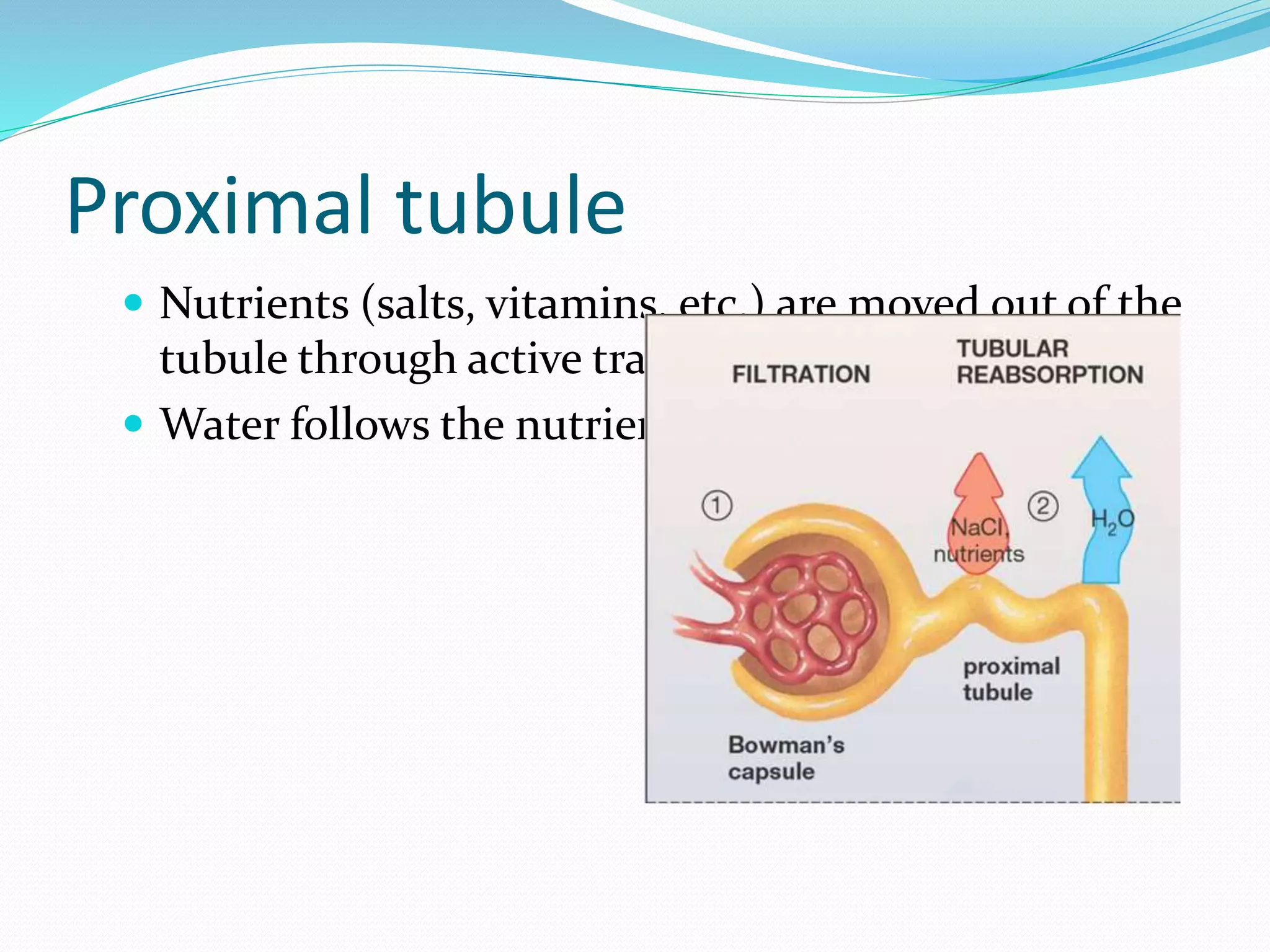

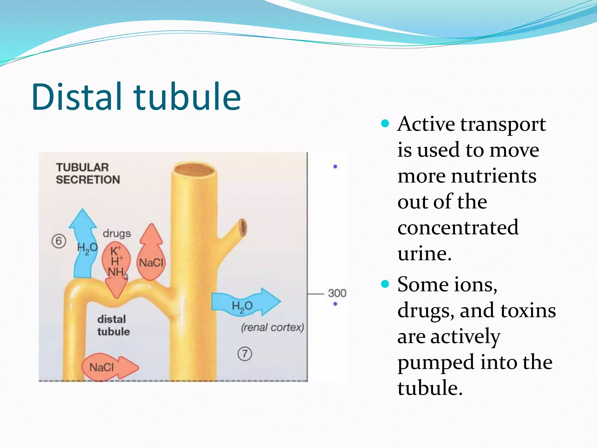


The document summarizes the urinary system of goats. It describes how the system maintains homeostasis through processes like removing waste, regulating water and salt balance, and controlling blood pressure. It then outlines the key structures of the urinary system including the kidneys, nephrons, ureters, bladder, and urethra. The document provides detailed descriptions of kidney anatomy and the role of different parts of the nephron in filtering and processing blood to form urine.


