Downloaded 513 times

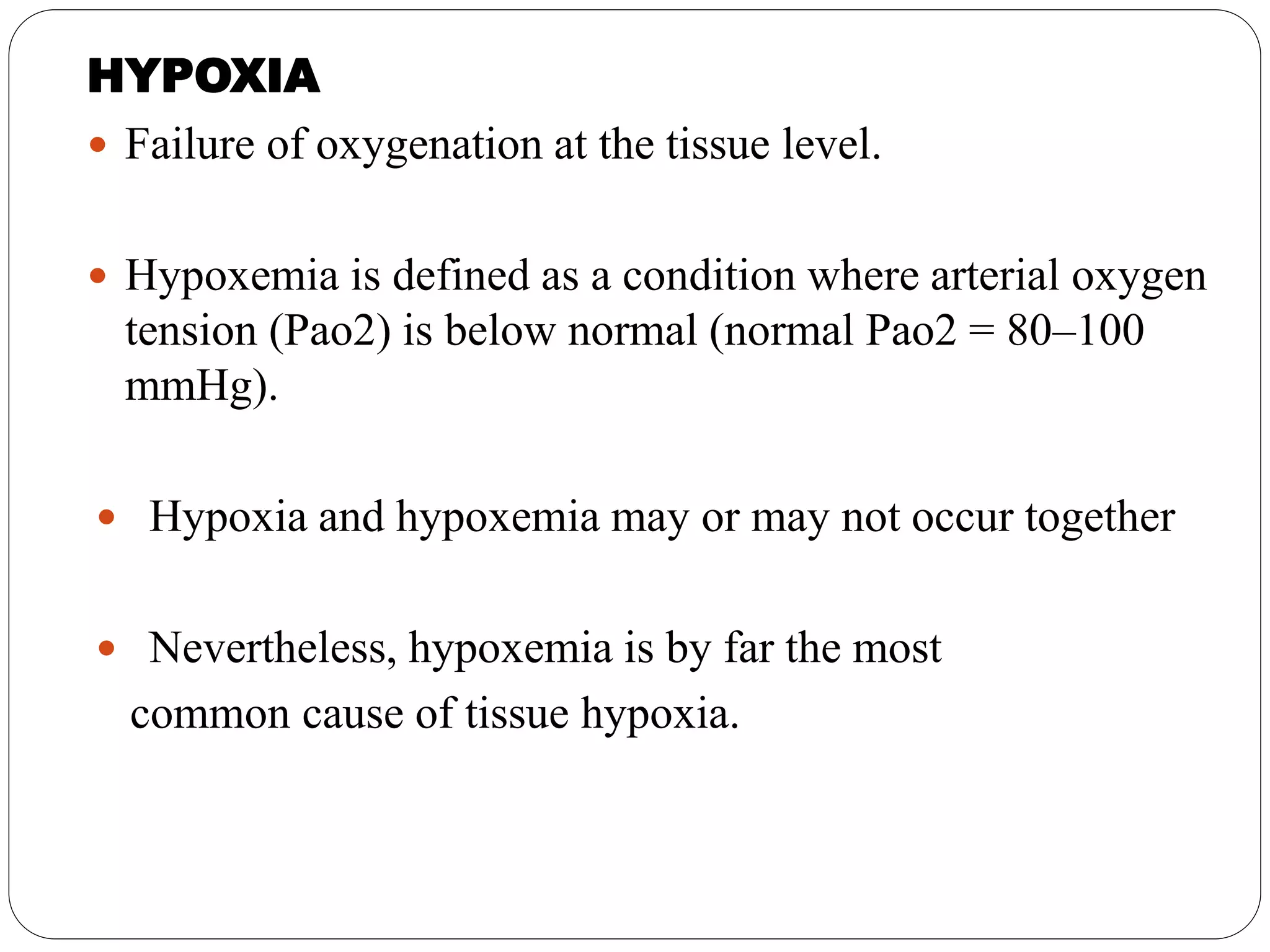

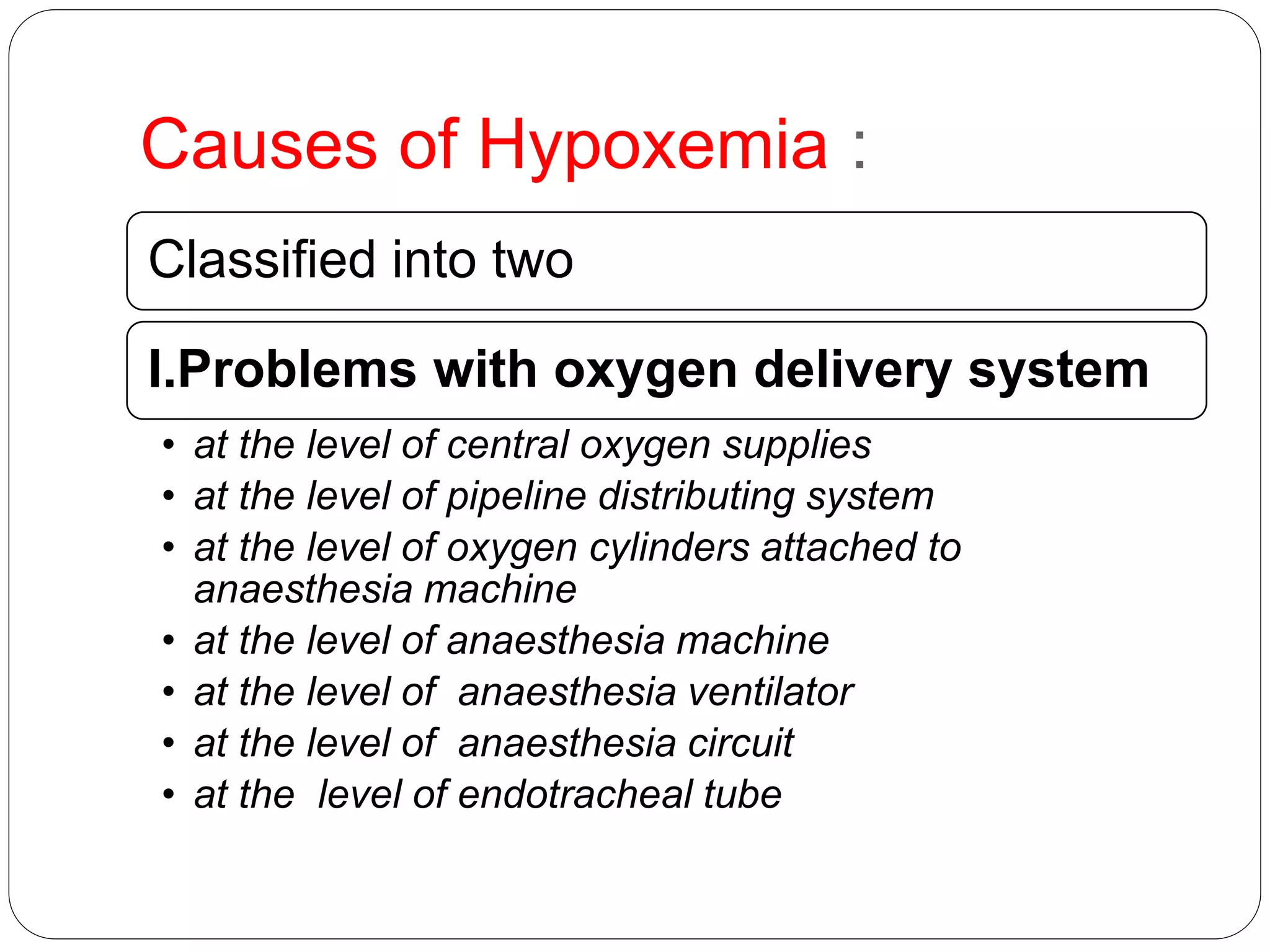
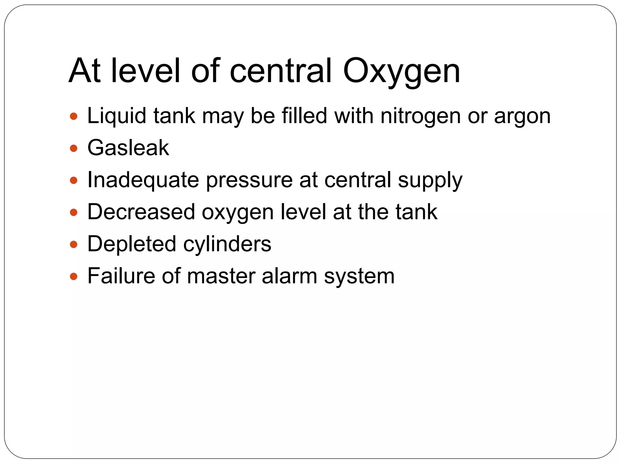


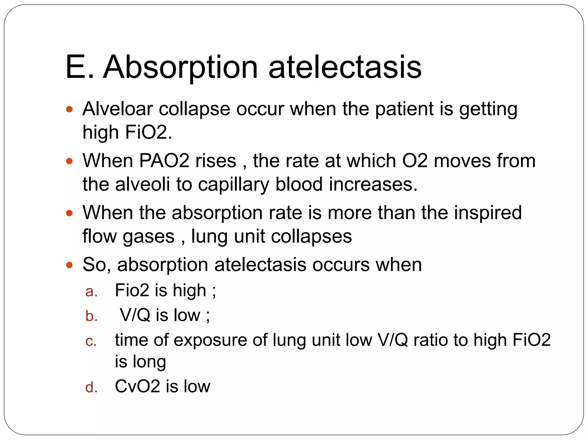


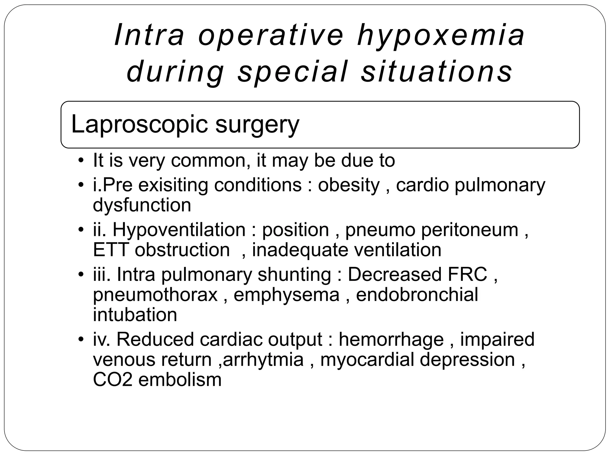


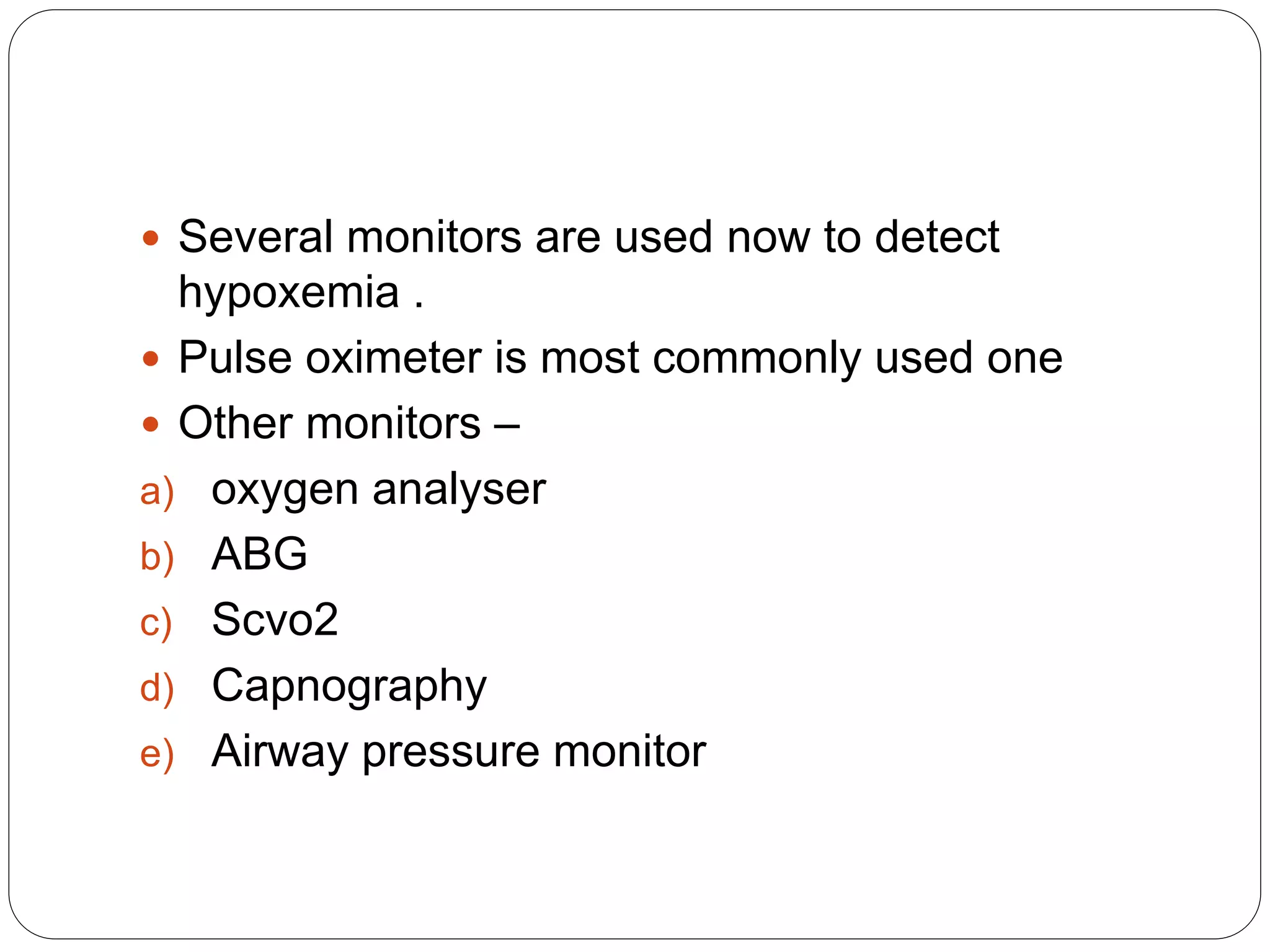


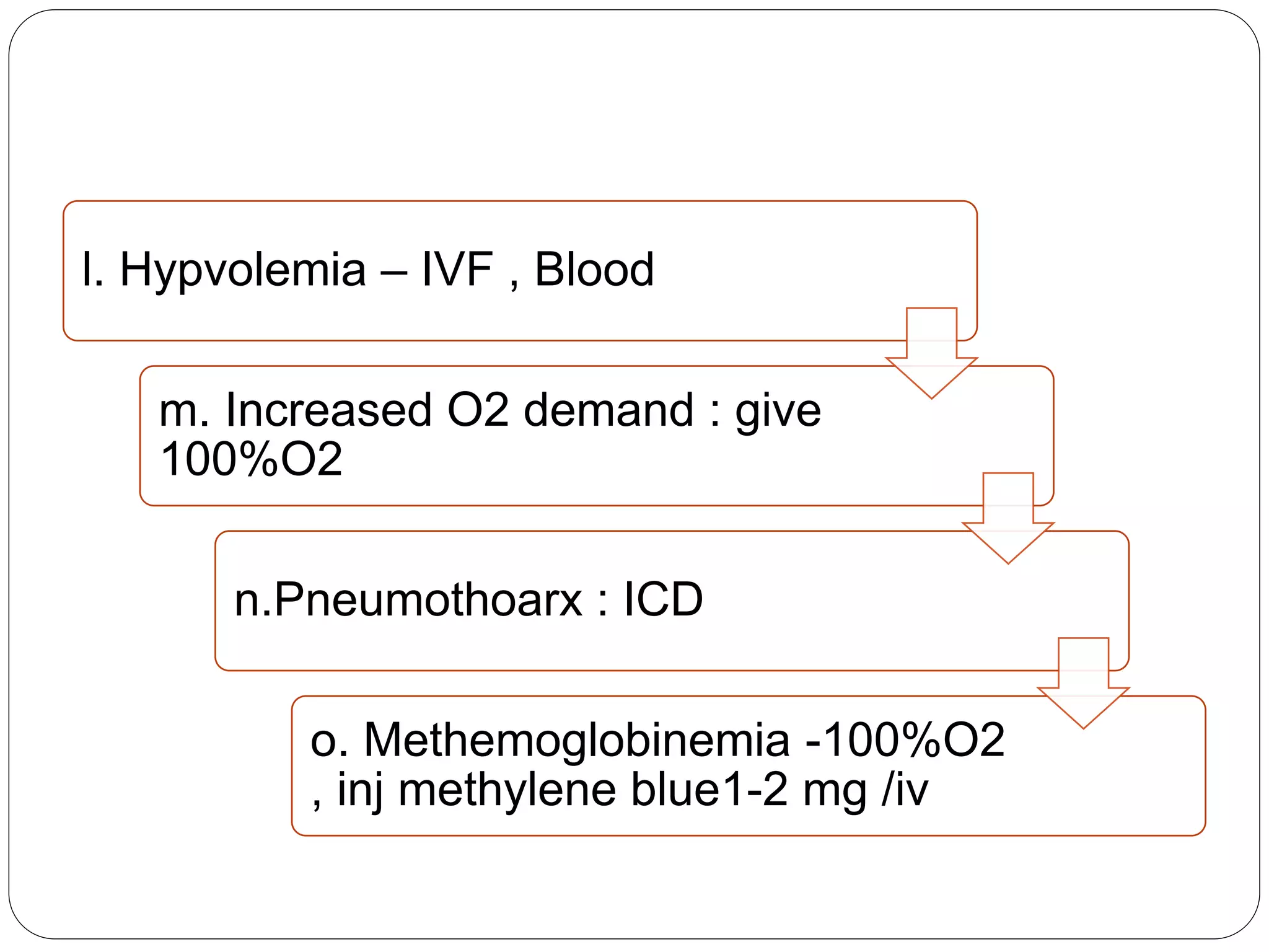
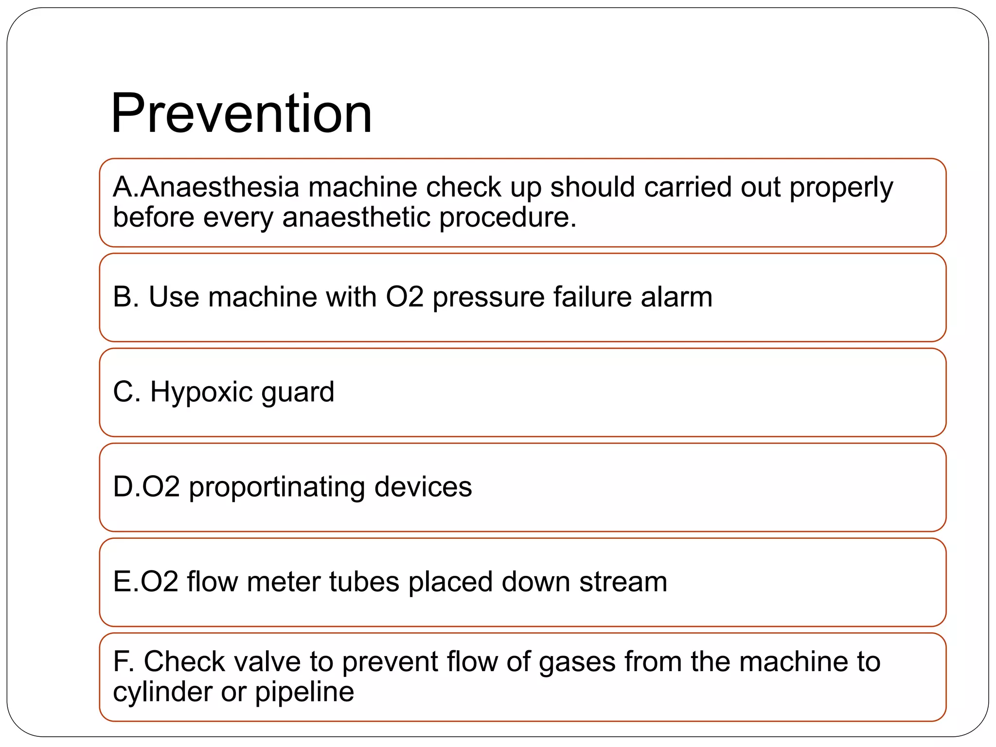


This document discusses intraoperative hypoxemia. It defines hypoxemia and classifies its causes. Causes are problems with oxygen delivery systems like ventilators, circuits or endotracheal tubes. Or problems with patients like reduced lung volumes, atelectasis or increased oxygen demand. Specific risk factors are discussed like obesity, pregnancy, elderly and one lung ventilation. Diagnosis involves monitoring like pulse oximetry. Management focuses on giving high oxygen, ventilation support and treating underlying causes. Prevention emphasizes machine checks and safety features.


