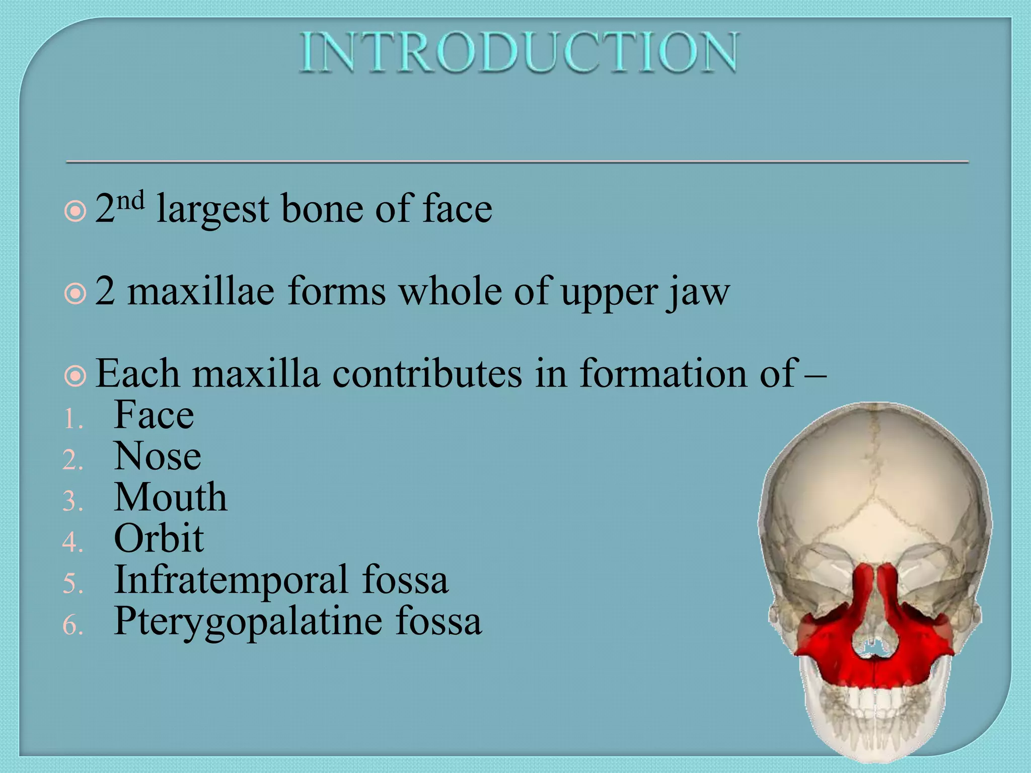
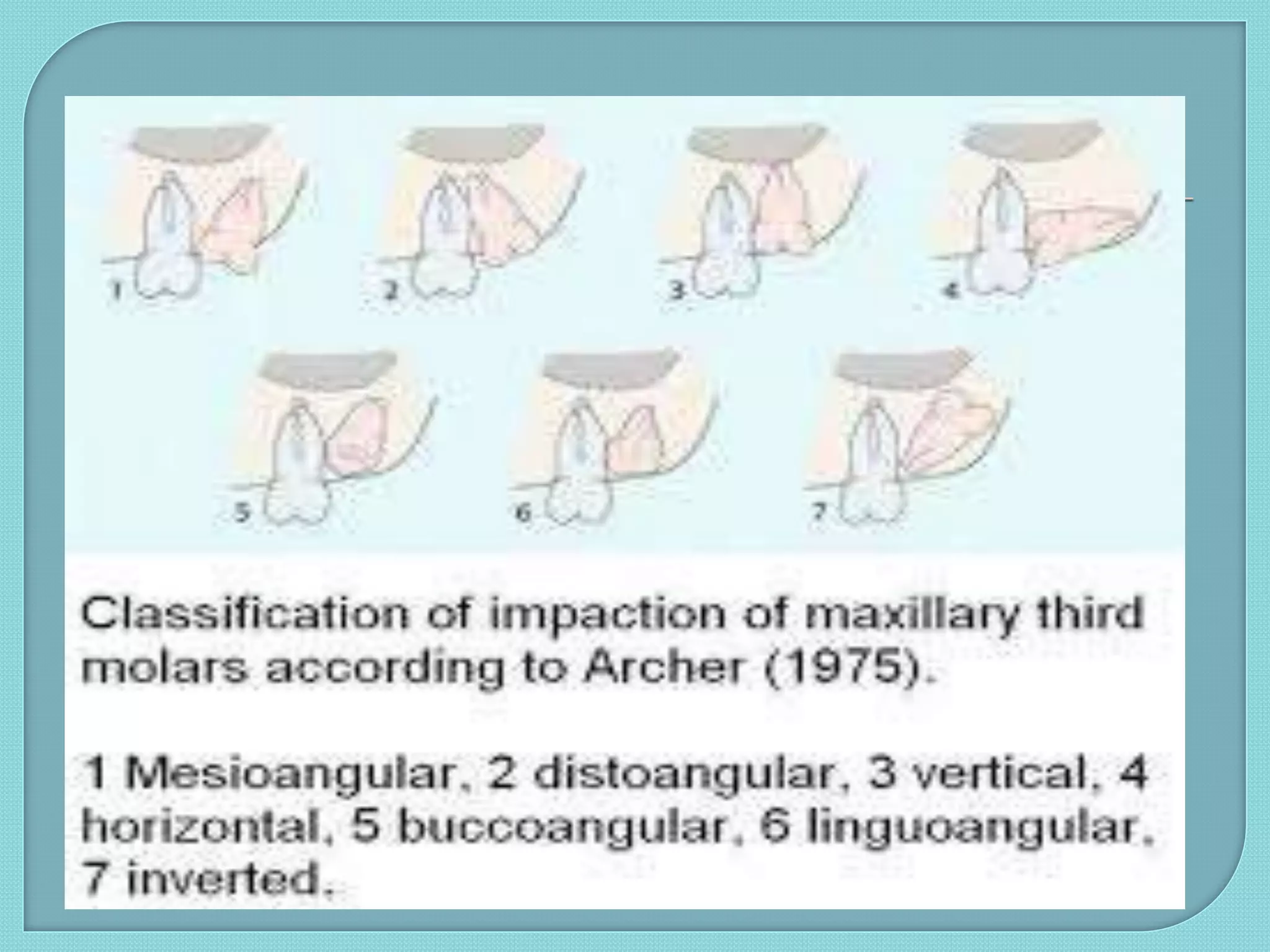
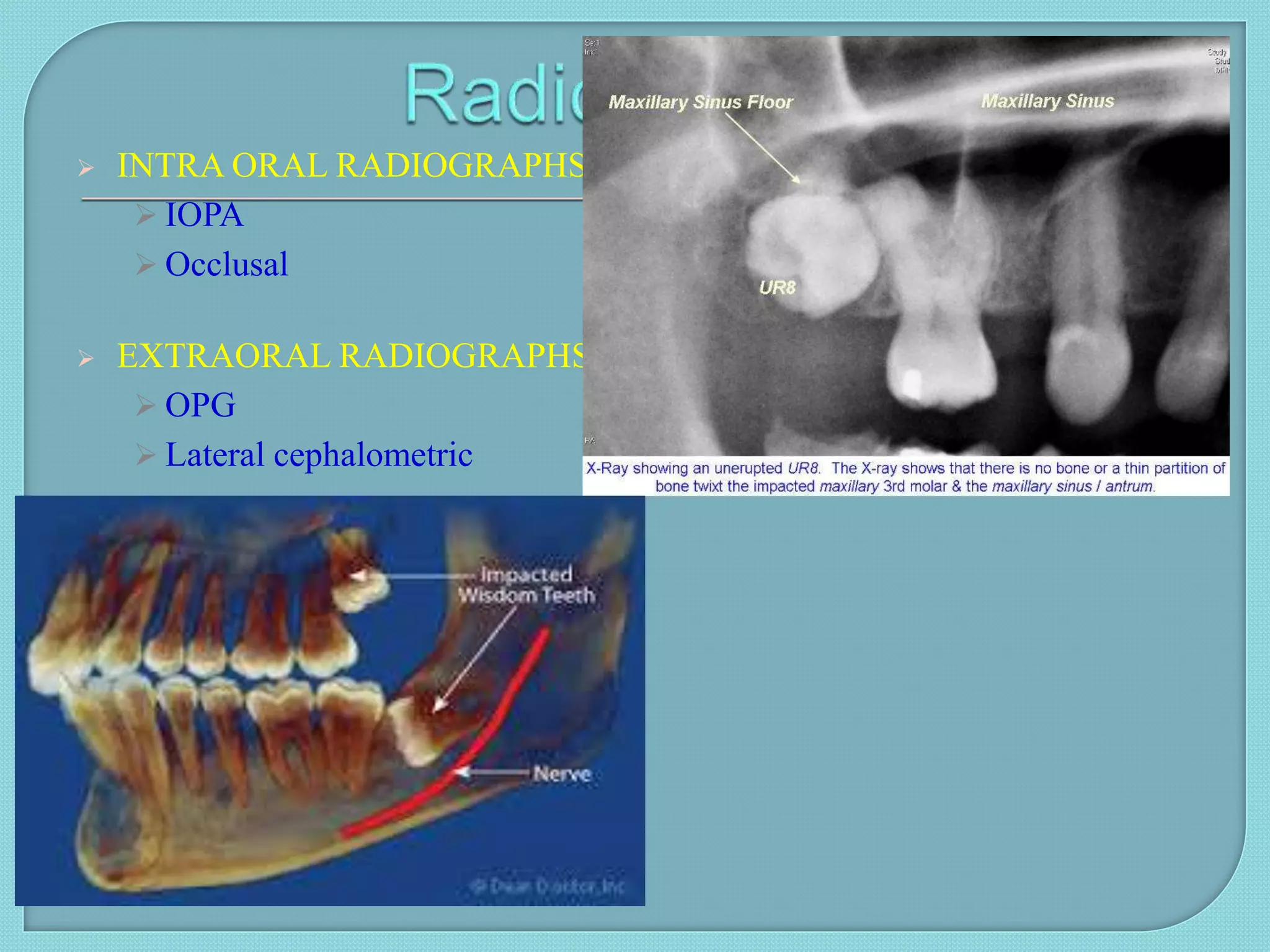
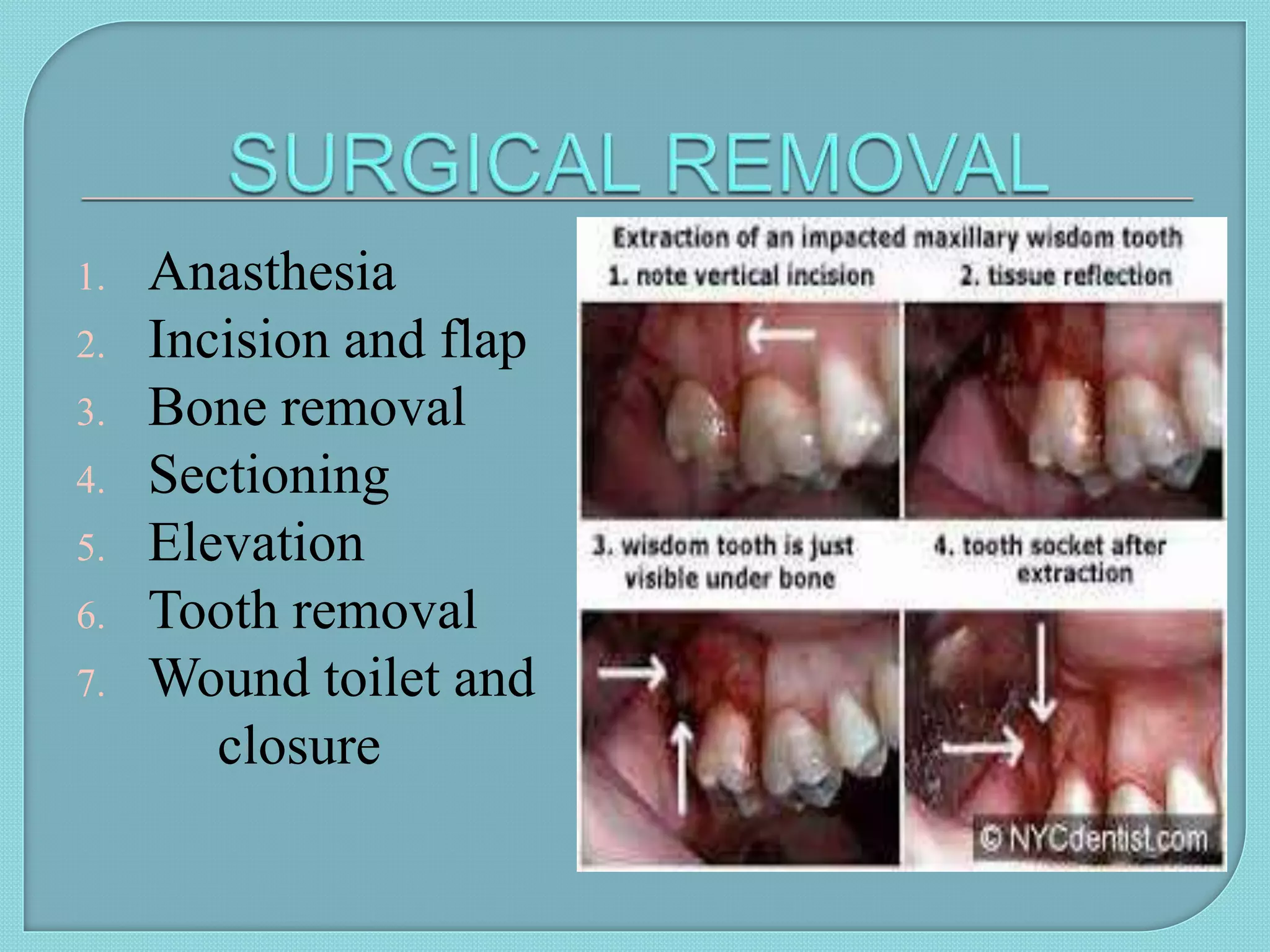
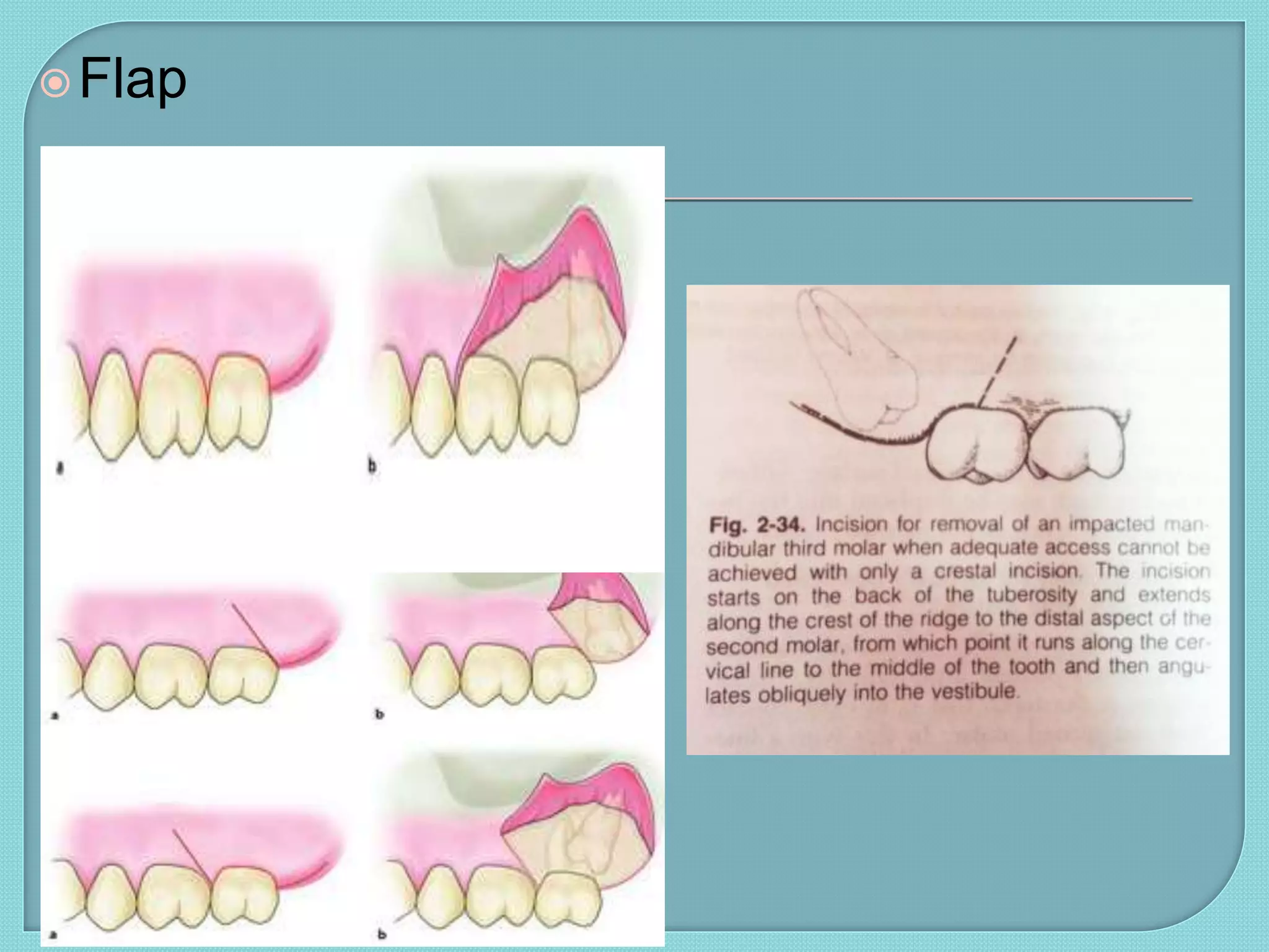
2. 2nd largest bone of face
2 maxillae forms whole of upper jaw
Each maxilla contributes in formation of –
1. Face
2. Nose
3. Mouth
4. Orbit
5. Infratemporal fossa
6. Pterygopalatine fossa
3. Shape – pyramidal
Each maxilla has –
1. A body
2. 4 processes – frontal
zygomatic
alveolar
palatine
It has –
1. Base – directed medially at nasal surface
2. Apex - directed laterally at zygomatic process
3. 4 surfaces – anterior / facial
posterior / infratemporal
medial / nasal
superior / orbital
Encloses a cavity – maxillary sinus
4. Unerupted teeth: tooth in the process of
eruption & is likely to erupt.
Impacted teeth: that has failed to erupt
completely or partially to its correct position in
the dental arch &its eruption potential is lost.
More common in females than males, in maxilla
than in mandible and on right side than left.
6. Local causes:
1. Density of overlying bone
2. Lack of space due to undedeveloped jaws
3. Obstruction-
a. Over-retained deciduous tooth
b. Thick fibrous alveolar mucosa
c. Cyst/tumors
4. Dialceration
5. Ectopic tooth eruption
7. Systemic causes:
1. Pre-natal: heredity
2. Post-natal: factors that interfer with
development-
a. Rickets
b. Malnutrition
c. anaemia
3. Rare conditions:
a. Cleft lip
b. osteoporosis
9. Pericoronitis was found to
be common in vertical
(23.0%) followed by
mesioangular (15.0%),
distoangular(8.0%) and
horizontal
angulatio(3.0%).
Common in females than
males
Streptococcus Viridans is
the most common
facultative isolate.
11. Teeth which can be used as abutments of
FPD
Age (Longer recovery periods
Difficult - more densely calcified bone
Bone removal more due to reduced PDL
space
Longer healing time )
Medical history
Deeply impacted teeth; in relation with vital
structures (vessels, sinus)
12. Based on nature of overlying tissue
o Soft tissue
o Hard tissue
SIGNIFICANCE - Each
type of impaction has
some definite path of
withdrawal of
the teeth.
17. o General
Systemic disorders
Radiation therapy
Age
Extra oral:
Signs of swelling & redness of the cheek.
Lymph node - enlargment & tenderness.
Anesthesia or paraesthesia of lower lip.
Intra oral:
State of eruption of tooth, signs of pericoronitis
Condition of 1st & 2nd molars
Elasticity of oral tissues
24. Bone removal
To remove the bone obstructing the pathway for
removal of the impacted tooth.
Bone should be removed till we reach below the
height of contour, where we can apply the
elevator.
25. Tooth sectioning
Rationale of tooth sectioning is to create a space
into which impacted tooth can be displaced & then
removed.
Impacted maxillary teeth are rarely sectioned,
because the overlying bone is relatively thin &
elastic.
Also, in no case should a chisel be used, for it may
cause displacement of tooth in the sinus.
Sectioning when done, is done at the cervical
line.
26. Periapical curettage.
Smoothening of sharp bony margins by Bone file / burs.
Thorough irrigation of the socket with Betadine solution
+ Saline .
Suturing
27.
28.
29. • Amount of space available in
dental arch for impacted canine is
assessed in model.
Study model
analysis
• Gives clue of position of impacted
tooth.
Morphology of
adjacent tooth
• Canine bulge present buccally or
palatally.
Contours of
adjacent
alveolar bone
• Root resorption.
Mobility of
adjacent tooth
30. Age of patient
Suitability of 1st premolar to replace a permanent canine
Patient motivation for orthodontic applainces
General dental health and oral hygiene
Radiographic position of canine
Availability of space
34. Flap is elevated
Attachment placed on impacted
tooth
Ligature or chain placed over the
attachment to activate after a
week
Raised flap is repositioned in its
original location
Permit eruption of impacted
canine in normal direction
35. 1. During incision
a. Injury to facial artery
b. Injury to lingual nerve
2. During bone removal
a. Damage to second molar
b. Slipping of bur- soft tissue injury
c. Extra oral/ mucosal burns
d. Fracture of the mandible when using chisel &
mallet
e. Subcutaneous emphysema
36. 3. During elevation or tooth removal
a. Luxation of neighbouring tooth/ fractured
restoration
b. Soft tissue injury due to slipping of elevator
c. Forcing tooth root INTO MAXILAARY
SINUS or BUCCAL SPACE
d. Breakage of instruments
38. patient complains of mild pain
and heaviness in the left
maxillary sinus area and the
left maxillary sinus was tender
on palpation. maxillary sinus
was exposed through a
Caldwell-Luc approach. The
sinus was irrigated with sterile
saline solution under pressure
and the tooth was removed
only by negative pressure of
the suction pump
39. CT image of the case
depicting the displaced tooth
between the buccinator and
masseter muscle in the
buccal space..
3D CT image of the displaced
maxillary third molar seen as
localized obliquely in front of the
anterior border of the ramus of the
mandible in the buccopalatine
direction.